Neonatal aortic thrombus in the proximal descending aorta is a rare but critical condition that can present mimicking interrupted aortic arch or coarctation of the aorta.Reference Neal, Mattishent and Reynolds1,Reference Knadler, Zobeck, Masand, Sartain and Kyle2 Management of a neonatal aortic thrombus is challenging because there is a higher risk of bleeding with anticoagulation and thrombolytic therapy, while surgical thrombectomy may not be suitable due to its invasiveness in critically ill neonates. Transcatheter thrombectomy has been reported scarcely. We report a case of a premature neonate with an occlusive thrombus in the proximal descending aorta, mimicking coarctation of the aorta. This thrombus was successfully treated with transcatheter mechanical thrombectomy using an Amplatzer Piccolo PDA occluder (Abbott, North Chicago, IL, USA).
Case report
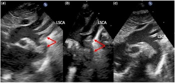
A newborn male was born at 35 weeks gestation due to non-reassuring foetal heart tones with a birth weight of 3.0 kg. Initial echocardiogram was performed due to a failed pulse oximetry screening along with a significant upper to lower extremity pressure gradient. Echocardiogram was concerning for possible critical coarctation versus interrupted aortic arch. He was started on prostaglandin E1 infusion and was then transferred to the tertiary care centre. Repeat echocardiogram showed a large occlusive thrombus in the proximal descending aorta extending to the left subclavian artery with the distal descending aorta dependent on patent ductus arteriosus flow (Fig 1a and b, Video 1). Furthermore, the left ventricular systolic function was severely reduced. Due to these findings, he was urgently taken to the cardiac catheterisation laboratory for transcatheter mechanical thrombectomy. Prior to the catheterisation, brain MRI showed a small centrum semi-ovale infarction.
(a and b) Transthoracic echocardiography, suprasternal notch view at the presentation and post-thrombectomy (c). There was echo-bright structure at the aortic isthmus, extending to the left subclavian artery. Post-thrombectomy, there was no obvious obstructive structure observed in the proximal descending aorta or LSCA.

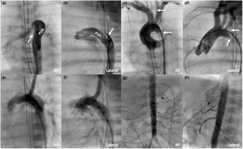
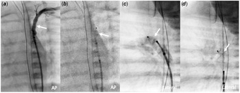
Cardiac catheterisation was performed within 12 hours of arrival. Through a low-profile 4-Fr Prelude sheath in the right femoral artery, a 4-Fr Glide catheter was advanced to the proximal descending aorta. The first aortography showed the occlusive thrombus in the descending aorta proximal to the insertion of the patent ductus arteriosus (Fig 2a–d). There was a significant systolic pressure gradient of 42 mmHg across the occluded isthmus. Angiography showed complete occlusion of the proximal descending aorta at the isthmus and partial occlusion of the left subclavian artery due to the thrombus. The diameter of the left subclavian artery and proximal descending aorta measured 3 mm and 5.5 mm, respectively. A 54 mm Amplatzer Piccolo PDA occluder was deemed suitable for mechanical thrombectomy based on the measured vessel sizes. First, the device was deployed in the left subclavian artery superior to the thrombus using an Amplatzer TorqVue LP catheter and the device was then gently withdrawn to the descending aorta along with suctioning of the thrombus through the Piccolo delivery catheter (Fig 3, Video 2). This manoeuvre was then repeated from the aortic arch to the distal descending aorta (Video 3). A small thrombus was found on the withdrawn device. Post-thrombectomy, angiography showed no evidence of residual thrombus in the left subclavian artery, proximal and abdominal descending aorta along with no pressure gradient across the aortic arch (Fig 2e–h). Prostaglandin E1 infusion was discontinued and intravenous heparin infusion was initiated to prevent recurrent thrombosis. Haematology was consulted to investigate the cause of the neonatal thrombosis but there has been no identifiable cause to date. Within 24 hours of the procedure, the left ventricular systolic function improved significantly with no evidence of residual thrombus on a follow-up echocardiography (Fig 1c). Repeat brain MRI showed no evidence of cerebral infarction. He was discharged home with subcutaneous enoxaparin therapy at 14 days of life. At 2 months of age, he is clinically well with no recurrent thrombosis.
(a and d) Cardiac catheterisation before and after (e–h) the transcatheter thrombectomy. (a–d) The proximal descending aorta is completed occluded by thrombus (arrow), which extends into the origin of the left subclavian artery. The descending aorta flow is dependent on the right-to-left shunt through the large patent ductus arteriosus. (e–f) Post-thrombectomy, there is no residual thrombus seen in the proximal descending aorta or left subclavian artery. (g– h) Abdominal descending aortography shows no filling defects in the abdominal arterial system. AP = anteriorposterior.

Transcatheter mechanical thrombectomy using a 5–4 mm Amplatzer Piccolo PDA occluder (Abbott, North Chicago, IL, USA). (a) and (b) The device (arrow) is deployed at the left subclavian artery distal to the thrombus and gently withdrawn inferiorly. (c) and (d) The device is deployed at the aortic arch and gently withdrawn inferiorly.

Discussion
Aortic thrombus in a neonate is a rare phenomenon. The most common causes include an associated umbilical artery catheter, inherited thrombophilia, or infection.Reference Rashish, Paes, Nagel, Chan and Thomas3,Reference Wieland, Jack and Seidemann4 There was no identifiable cause found in our patient. When an aortic thrombus is occlusive, its presentation mimics that of severe coarctation or interrupted aortic arch.Reference Neal, Mattishent and Reynolds1,Reference Knadler, Zobeck, Masand, Sartain and Kyle2,Reference Gerardin, Anderson, Armstrong and Grifka5,Reference Francis, Monagle, Hope and Sehgal6 Our case exhibited the same presentation and prostaglandin E infusion was immediately started due to lower body dependence on the right-to-left flow through the PDA.
Management of a neonatal aortic thrombus is challenging. Medical management including anticoagulation and/or thrombolysis therapy can be considered in a clinically stable neonate with partially occlusive thrombus.Reference Sharathkumar, Lamear and Pipe7,Reference Metsvaht, Hermlin, Kern, Kahre and Starkopf8 However, the risk of intracranial bleeding associated with thrombolysis therapy is not negligible, especially in premature infants. Surgical thrombectomy is a definitive therapy, albeit more invasive, and therefore not deemed suitable for a critically ill neonate. Transcatheter mechanical thrombectomy is a less invasive alternative option. In our case, the aortic thrombus was completely occlusive with severe left ventricular systolic dysfunction. Urgent recanalization of the occluded aorta was crucial so the patient was taken to the cardiac catheterisation laboratory without a trial of initial medical management.
Transcatheter thrombectomies for aortic thrombi have been described scarcely in neonates, and are limited only to abdominal aortic thrombi. Transcatheter thrombectomy using the Amplatzer Vascular Plug II and IV has been described in four infants, where the technique to “scrape” the thrombus was well described.Reference McGovern, Qureshi and Goldstein9 Similarly, the use of a 54 mm Amplatzer Piccolo PDA occluder was described for successful removal of an abdominal aorta thrombus in a 5-month-old infant.Reference Ferraro, Marini and Agnoletti10 The benefit of this device is its softer design to prevent vascular injury associated with thrombectomy and the use of a very low-profile system (4-Fr). The same size of a Piccolo occluder was used for the thrombus in the left subclavian artery and proximal descending aorta in our case. The selected device (Supplemental Table) was oversized compared to the left subclavian artery and proximal descending aorta diameter to prevent distal thrombus dislodgement. Fortunately, there was no evidence of distal thromboembolic phenomenon post-thrombectomy clinically or on repeat brain MRI.
Conclusion
Transcatheter mechanical thrombectomy was successfully performed for neonatal aortic thrombus using an Amplatzer Piccolo PDA occluder. This method would be a good alternative to surgical thrombectomy in a critically ill neonate with acute and occlusive aortic thrombus.
Supplementary material
To view supplementary material for this article, please visit https://doi.org/10.1017/S1047951121003267
Financial support
This research received no specific grant from any funding agency, commercial, or not-for-profit sectors.
Conflicts of interest
None.
Ethical standards
All procedures performed in studies involving human participants were in accordance with the ethical standards of the institutional and/or national research committee and with the 1964 Helsinki declaration and its later amendments or comparable ethical standards. Informed consent was obtained from all individual participants included in this case report.