Introduction
In asthenoteratozoospermia patients there is a specific syndrome named multiple morphological abnormalities of the sperm flagella (MMAF), which is characterized by the conjoint presence of sperm whose flagella are absent, short, irregular in width, bent and coiled (Ben Khelifa et al., Reference Ben Khelifa, Coutton, Zouari, Karaouzène, Rendu, Bidart, Yassine, Pierre, Delaroche, Hennebicq, Grunwald, Escalier, Pernet-Gallay, Jouk, Thierry-Mieg, Touré, Arnoult and Ray2014). The denomination of MMAF was established to include former descriptions of sperm with dysplasia of the fibrous sheath (DFS), short tails, stump tails or non-specific flagellar anomalies (Chemes et al., Reference Chemes, Brugo, Zanchetti, Carrere and Lavieri1987; Barthelemy et al., Reference Barthelemy, Tharanne, Lebos, Lecomte and Lansac1990; Stalf et al., Reference Stalf, Sánchez, Köhn, Schalles, Kleinstein, Hinz, Tielsch, Khanaga, Turley and Gips1995; Dávila Garza and Patrizio, Reference Dávila Garza and Patrizio2013).
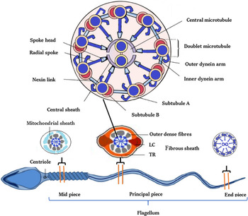
The flagellum contains the axoneme, a highly evolutionarily conserved structure responsible for sperm motion. The flagellum is divided into the mid piece and principal piece. In the mid piece, the axoneme is surrounded by outer dense fibres (ODF) and a mitochondrial sheath. In the principal piece, the ODF are surrounded by the fibrous sheath (FS). The axoneme is made up of nine peripheral doublets of microtubules and a central complex of a pair of microtubules (CPC). The central pair is connected to the peripheral doublets by the radial spokes (RS). Doublets are linked by nexin bridges that contain the dynein regulatory complex (DRC). The complete and inner microtubule A from each doublet contains two complex expansions made of dyneins, the outer dynein arm (ODA) and the inner dynein arm (IDA). Chemical signalling derived from the CPC is carried through the RS to the RDC, triggering dynein arm binding to the more external and incomplete microtubule B of the neighbouring doublet, causing microtubule sliding and sperm motion (Afzelius et al., Reference Afzelius, Dallai, Lanzavecchia and Bellon1995; Mortimer, Reference Mortimer2018) (Figure 1).
Structure of human sperm flagellum. Sperm flagella are structurally divided into three parts: mid piece, principal piece and end piece. the cross-section of each part is shown. The mid piece consists of a helical mitochondrial sheath (MS) surrounding nine outer dense fibres (ODF; there are seven ODF in the principal piece) and the axoneme. MS is replaced by the fibrous sheath (FS) in the principal piece. The FS is composed of two longitudinal columns (LC) which are connected by transverse ribs (TR). The end piece is devoid of any peri-axonemal structures. The axoneme is a highly evolutionarily conserved structure present in the whole flagellum. The axoneme is enlarged and the offset shows: the nine outer microtubule doublets of the axoneme (MTs) with associated inner dynein arms (IDA), outer dynein arms (ODA), radial spokes (RS), nexin-dynein regulator complex (N-DRC), nexin links (NL) and the central pair (CP) of microtubule doublets.

In a recent report of 78 patients with MMAF (46 North African, 10 Middle East, 22 Caucasian), 50% with consanguinity, semen analysis revealed 98% abnormal morphology and 4% sperm motility, showing sperm with short flagella (44%), irregular width of the flagella (32%), absent flagella (21%), coiled flagella (13%) and bent flagella (4%). Ultrastructurally, all sperm also showed abnormal periaxonemal structures (FS, ODF, and mitochondrial sheath). Of these, 95% had an abnormal axoneme structure, 67–82% with absent central pair complex associated in 14–19% with disruption of the peripheral doublet organization. The authors found MMAF associated pathogenic variants (22/78, 28%) not only in the gene DNAH1, but also in two further genes (Coutton et al., Reference Coutton, Vargas, Amiri-Yekta, Kherraf, Ben Mustapha, Le Tanno, Wambergue-Legrand, Karaouzène, Martinez, Crouzy, Daneshipour, Hosseini, Mitchell, Halouani, Marrakchi, Makni, Latrous and Kharouf2018). Recently, more new genes have been reported to be associated with MMAF. Pathogenic variants in these genes have led to axoneme and periaxonemal defects (Yang et al., Reference Yang, Gao, Wang, Ding, Xu and Li2018; Shen et al., Reference Shen, Zhang, Li, Jiang, Yang, Li, Li, Wang, Cheng, Liu, Zhang, Yuan, Pei, Cai, Hu, Sun, Yan and Tang2019; Huang et al., Reference Huang, Yin, Liu, Li, Yu, Wang, Zhang, Muhammad, Gao, Li, Chen, Liu and Ma2020; Liu et al., Reference Liu, Miyata, Gao, Sha, Tang, Xu, Whitfield, Patrat, Wu, Dulioust, Tian, Shimada, Cong, Noda, Li, Morohoshi, Cazin and Kherraf2020b; Sha et al., Reference Sha, Wei, Ding, Mei, Huang, Lin, Su, Kong, Zhang and Ji2020b). Although sharing the same axoneme structure, patients with MMAF do not have a clinic suggestive of primary ciliary dyskinesia (PCD), i.e. chronic rhinosinusitis or bronchiectasis (Nsota Mbango et al., Reference Nsota Mbango, Coutton, Arnoult, Ray and Touré2019; Wang et al., Reference Wang, Tu and Tan2020).
At present, intracytoplasmic sperm injection (ICSI) is the only available technique to achieve fertilization in those patients with MMAF. In previous MMAF patients, healthy children were delivered following ICSI with ejaculated sperm, as reported by other authors (McLachlan et al., Reference McLachlan, Ishikawa, Osianlis, Robinson, Merriner, Healy, de Kretser and O’Bryan2012; Wambergue et al., Reference Wambergue, Zouari, Fourati Ben Mustapha, Martinez, Devillard, Hennebicq, Satre, Brouillet, Halouani, Marrakchi, Makni, Latrous, Kharouf, Amblard, Arnoult, Ray and Coutton2016; Sha et al., Reference Sha, Wei, Ding, Ji, Mei, Huang, Su, Wang, Zhang and Lin2020a). Here, we report a pregnancy with delivery of a healthy child after vitrified blastocyst transfer from vitrified oocytes injected with immotile testicular spermatozoa.
Case presentation
Patient
The couple presented with 4 years of primary infertility. The 33-year-old woman was assessed by a gynaecologist, and no contributing female factors were detected. The body mass index was 20 kg/m2. The 34-year-old male partner had a normal clinical history and physical examination. Serum follicle-stimulating hormone (FSH; 4.73 IU/L, 1.5–12.5 IU/L), luteinizing hormone (LH; 4.86 IU/L, 1.7–8.6 IU/L), and testosterone (T; 10.62 nmol/L, 8.36–39.59 nmol/L) were within normal ranges, and his karyotype was 46,XY.
Ovarian stimulation and oocyte retrieval
A long protocol for ovulation induction was performed with daily administration of recombinant FSH (200 IU/day) (Metrodine® HP; Serono, France) following pituitary desensitization with a gonadotropin-releasing hormone (GnRH) agonist (Enantone®; Takeda, France). Ovulation trigger was achieved with 10,000 IU of human recombinant chorionic gonadotrophin (rHCG, Ovidrel, Merck-Serono) as soon as 50% of the follicles of >10 mm reached a diameter of ≥18 mm. After 36 h, cumulus–oocyte complexes (COC) were retrieved transvaginally under ultrasound guidance using a 17-gauge single lumen needle (K-OPS-7035-RWH-ET; Cook Australia) under general anaesthesia. COC were cultured in four-well dishes (Nunc; Thermo Scientific) with each well containing 0.6 ml of Fertilization Medium (SAGE) covered with 0.35 ml of oil (SAGE) in a standard incubator at 37°C (Astec, Japan; 5% O2, 5% CO2, 90% N2).
Oocyte vitrification and warming
Follicular cells from COC were removed by brief exposure to 10 IU/ml hyaluronidase (hyaluronidase, SAGE) and by gentle mechanical aspiration using plastic pipettes (Denuding Flexipet™ Cook, Eight Mile Plains Queensland, Australia) 3 h after retrieval. Vitrified oocytes were performed a few minutes later using the Cryotop method (Kitazato Corporation, Shizuoka, Japan) with open devices (Kitazato Biopharma, Tokyo, Japan) as described elsewhere (Nagy et al., Reference Nagy, Varghese and Agarwal2019). Oocytes were loaded into the equilibration solution for 12–15 min, and sequentially transferred to the vitrification solution for 60 s. Oocytes were immediately placed on the Cryotop® strip with a minimal amount of the vitrification solution and quickly immersed into liquid nitrogen (LN2). A maximum of three oocytes were placed by each Cryotop. Oocytes were warmed in thawing solution for 1 min and sequentially transferred to the dilution solution for 3 min. Oocytes were then washed in washing solution for 5 min. Oocytes were thereafter transferred to culture dishes (Falcon) and placed in the incubator for 2 h before ICSI.
Semen analysis and sperm preparation
Semen samples were evaluated according to World Health Organization (WHO) guidelines (World Health Organization, 2010) to measure sperm concentration, motility, morphology and vitality. Sperm morphology was assessed with Diff-Quick staining (Diff-Quick stain kit, Guangzhou, China), and at least 200 spermatozoa were examined. Sperm vitality of 39% was performed by using the one-step eosin–nigrosin staining technique. Spermatozoa were separated from seminal plasma by washing in Fertilization Medium (1020, SAGE, USA). Following centrifugation at 200 g for 5 min, the sperm pellet was resuspended in 0.3 ml Fertilization Medium. After routine assessment of sperm morphology, the stained sperm smear was examined for the shape of the sperm tail, which was categorized into five types: (i) no tail, (ii) short tail, (iii) bent tail, (iv) coiled tail, and (v) irregularly shaped tail (World Health Organization, 2010; Wang et al., 2019). Diagnosis of MMAF demands that >5% of sperm exhibit simultaneously at least four of the five characteristics of the syndrome (Ben Khelifa et al., Reference Ben Khelifa, Coutton, Zouari, Karaouzène, Rendu, Bidart, Yassine, Pierre, Delaroche, Hennebicq, Grunwald, Escalier, Pernet-Gallay, Jouk, Thierry-Mieg, Touré, Arnoult and Ray2014).
Testicular sperm retrieval
Testicular sperm aspiration (TESA) (Craft and Tsirigotis, Reference Craft and Tsirigotis1995) was performed under local anaesthesia with intravenous sedation, as described elsewhere (Esteves et al., Reference Esteves, Miyaoka, Orosz and Agarwal2013). The testicular tissue fragments were flushed into a Falcon tube (Becton Dickinson, USA) containing sperm medium (1023, SAGE, USA), and gently crushed with microneedles in a Petri dish (Becton Dickinson, USA) containing the same medium. When sperm cells were identified, the sperm suspension was then centrifuged for 5 min at 200 g in a Falcon tube. The supernatant was removed and the final pellet was incubated at 35°C in 5% CO2 until the ICSI procedure was performed.
ICSI procedure, culture, and transfer
We performed ICSI (Tesarik and Sousa, Reference Tesarik and Sousa1995; Palermo et al., Reference Palermo, Schlegel, Sills, Veeck, Zaninovic, Menendez and Rosenwaks1998) at least 1 h after removing follicular cells (Hyun et al., Reference Hyun, Cha, Son, Yoon, Kim and Lim2007). Metaphase II oocytes were microinjected with the help of a hypo-osmotic swelling test (HOST) (Casper et al., Reference Casper, Meriano, Jarvi, Cowan and Lucato1996; Stanger et al., Reference Stanger, Vo, Yovich and Almahbobi2010). Each immotile spermatozoon was transferred individually (i.e. one at a time) into a microdroplet of hypo-osmotic medium prepared by diluting the HEPES-buffered culture medium with an equal amount of Milli-Q water, as described by Sallam et al. (Reference Sallam, Farrag and Aganeya2005). A viable spermatozoon was recognized by its curved or swollen tail after a maximum of 10 s. It was then transferred into another microdroplet of HEPES-buffered medium where it was washed three times for osmotic re-equilibration, before being transferred to the PVP microdroplet. In our experience, in most cases, coiling or swelling of the sperm tail was observed within 1 or 2 s of putting the spermatozoon in the hypo-osmotic solution. The tails of all selected spermatozoa were crushed before the injection step.
Fertilization was assessed 17–19 h after insemination for the appearance of two distinct pronuclei and two polar bodies. Each fertilized oocyte was cultured in a 25-μl pre-equilibrated droplet of Quinn’s Advantage Cleavage medium (1026, SAGE, USA) under oil (SAGE, USA) in a standard incubator at 37°C (Astec, Japan; 5% O2, 5% CO2, 90% N2) (Sciorio and Smith, Reference Sciorio and Smith2019). Embryonic development was assessed on day 2 (41–43 h) and on day 3 (65–67 h), according to the requirements for blastomeres, the percentage and pattern of anucleate fragments, and all dysmorphic characteristics of the embryos (Vandervorst et al., Reference Vandervorst, Liebaers, Sermon, Staessen, De Vos, Van de Velde, Van Assche, Joris, Van Steirteghem and Devroey1998). After day 3, the embryos were cultured in groups of two or three in oil-covered droplets of 25 μl Blastocyst Medium (1029, SAGE, USA). The morphologic features of the blastocysts were assessed on day 5 according to published criteria (Gardner et al., Reference Gardner, Lane, Stevens, Schlenker and Schoolcraft2000).
On day 3 or day 5, high quality embryos were transferred using embryo transfer catheters (Cook, Cook Incorporated, Indiana, USA) under transabdominal ultrasound guidance. For luteal supplementation, progesterone vaginal gel (Crinone 8%, Merck-Serono SA, Geneva, Switzerland) was used from the day of embryo transfer until the day of implantation and confirmation by a rise in β-human chorionic gonadotrophin (β-HCG) serum (12 days). Clinical pregnancy was determined by a fetal heartbeat on ultrasound screening after 35 days.
Results
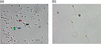
Semen analysis showed sperm densities of 11–21 million/ml, volumes of 2.6–3.6 ml, and sperm vitality of 39% with normal pH 7.2. All spermatozoa presented complete immotility and abnormal tails after repeated semen analyses under a light microscope (Figure 2a). The sperm flagellar phenotype was in accordance with MMAF (Yang et al., Reference Yang, Li, Wang, Shi, Cheng, Wang, Li, Hou and Wen2015), showing bent (64%), short (7%), irregular (4%), absent (5%) and coiled (20%) flagella (Table 1).
(a) Light microscopy observation (×200 magnification) of the ejaculated sperm with MMAF shows bent (green arrow), short (red arrow), absent (blue arrow), coiled (black arrow) and irregular sperm flagella (white arrow). (b) Most of the testicular sperm flagella (×400 magnification) are bent.

Semen parameters of the patient and ICSI outcomes of the two cycles

ICSI: intracytoplasmic sperm injection; 2PN 2PB: normal fertilized oocytes (two pronuclei and two polar bodies).
The couple was counselled regarding the potential negative outcomes of abnormal sperm on fertilization and the risk of genetic defects in the offspring. The couple opted to have some of the mature oocytes cryopreserved pending a decision on future treatment and the appropriate consent forms were signed. Of the 24 COC retrieved, 22 presented as mature oocytes (MII), which gave a maturation rate of 90.9% (20/22). Of the mature oocytes, 10 were microinjected with ejaculated sperm and 12 prepared for cryopreservation in case of fertilization failure. Of the injected 10 oocytes, six were normally fertilized, which gives a 60% fertilization rate (6/10). All fertilized oocytes cleaved, which gives an embryo cleavage rate of 100%. At day 3, only 2 (33%) embryos showed high quality, whereas the remaining embryos became arrested in development up to day 5. The two embryos of high quality were transferred at day 3, but no pregnancy was achieved (Table 1).
Seven months later, the couple returned to the clinic to try again with their vitrified oocytes. Testicular sperm were obtained by needle aspiration because of ejaculation failure on the day of oocyte retrieval. Most of the testicular retrieved sperm were bent at the tip and fully immotile (Figure 2b), showing a sperm vitality of 43%. The sperm flagellar phenotype exhibited bent (56%), short (9%), irregular (2%), absent (7%), coiled (25%) and normal flagella (1%) (Table 1).
All 12 vitrified oocytes survived successfully after thawing. After ICSI with testicular sperm, nine MII normally fertilized, which gave a fertilization rate of 75% (9/12). Of these, eight embryos were formed at day 2, which gives an embryo cleavage rate of 88.8% (8/9). At day 3, four of the eight embryos were of high quality. Two 8-cell embryos of high quality were transferred at day 3. At 12 days after transfer, the β-HCG concentration was positive (80 mIU/dl), but a gestational sac with fetal heartbeat was not observed 7 weeks later. The remaining six embryos were cultured up to day 5, and one developed to blastocyst with a grade of 5BB at day 5, being vitrified (Table.1). After 5 months, a third attempt was scheduled, with transfer of the vitrified blastocyst. A healthy female child was delivered, weighing 3050 g and 53 cm in length.
Discussion
Several studies have reported the birth of normal children following ICSI with ejaculated sperm showing a MMAF phenotype (Inaba, Reference Inaba2007; Coutton et al., Reference Coutton, Vargas, Amiri-Yekta, Kherraf, Ben Mustapha, Le Tanno, Wambergue-Legrand, Karaouzène, Martinez, Crouzy, Daneshipour, Hosseini, Mitchell, Halouani, Marrakchi, Makni, Latrous and Kharouf2018; Dong et al., Reference Dong, Amiri-Yekta, Martinez, Saut, Tek, Stouvenel, Lorès, Karaouzène, Thierry-Mieg, Satre, Brouillet, Daneshipour, Hosseini, Bonhivers, Gourabi, Dulioust, Arnoult and Touré2018; Lorès et al., Reference Lorès, Coutton, El Khouri, Stouvenel, Givelet, Thomas, Rode, Schmitt, Louis, Sakheli, Chaudhry, Fernandez-Gonzales, Mitsialis, Dacheux, Wolf, Papon, Gacon and Escudier2018; Yang et al., Reference Yang, Gao, Wang, Ding, Xu and Li2018; Shen et al., Reference Shen, Zhang, Li, Jiang, Yang, Li, Li, Wang, Cheng, Liu, Zhang, Yuan, Pei, Cai, Hu, Sun, Yan and Tang2019; Huang et al., Reference Huang, Yin, Liu, Li, Yu, Wang, Zhang, Muhammad, Gao, Li, Chen, Liu and Ma2020; Liu et al., Reference Liu, Lv, He, Zhu, Amiri-Yekta, Li, Wu, Kherraf, Liu, Zhang, Tan, Tang, Zhu, Zhong, Li, Tian, Zhang and Jin2020a; Sha et al., Reference Sha, Wei, Ding, Ji, Mei, Huang, Su, Wang, Zhang and Lin2020a; Talreja et al., Reference Talreja, Gupta, Pai and Palshetkar2020; Wang et al., Reference Wang, Tu and Tan2020). To our knowledge, the present case is the first report, in a patient with MMAF, of a successful birth that ensued after microinjection of vitrified oocytes with immotile testicular spermatozoa and using a vitrified blastocyst.
The present results showed that both ejaculated and testicular sperm were fully immotile and presented total abnormal flagella morphology (Figure 1). With ejaculated sperm and ICSI, the fertilization rate of 60% obtained using fully immotile sperm with MMAF, compares with those previously presented in other MMAF cases, being 70.8% (Wambergue et al., Reference Wambergue, Zouari, Fourati Ben Mustapha, Martinez, Devillard, Hennebicq, Satre, Brouillet, Halouani, Marrakchi, Makni, Latrous, Kharouf, Amblard, Arnoult, Ray and Coutton2016), and 63% (Chemes, Reference Chemes and Schatten2012; Dávila Garza and Patrizio, Reference Dávila Garza and Patrizio2013). With testicular sperm and ICSI, the fertilization rate of 75% obtained using 100% immotile sperm from a patient with MMAF, compared with those previously presented in other MMAF cases, being 44% (Wambergue et al., Reference Wambergue, Zouari, Fourati Ben Mustapha, Martinez, Devillard, Hennebicq, Satre, Brouillet, Halouani, Marrakchi, Makni, Latrous, Kharouf, Amblard, Arnoult, Ray and Coutton2016), 42.9% (Yang et al., Reference Yang, Gao, Wang, Ding, Xu and Li2018) and 66.7% (McLachlan et al., Reference McLachlan, Ishikawa, Osianlis, Robinson, Merriner, Healy, de Kretser and O’Bryan2012). Overall, fertilization achieved by ICSI with ejaculated or testicular sperm are effective in patients with MMAF, regardless of severe flagellar defects, although the fertilization rate is variable.
Studies using ejaculated sperm with MMAF revealed a fertilization rate of 40% (Stalf et al., Reference Stalf, Sánchez, Köhn, Schalles, Kleinstein, Hinz, Tielsch, Khanaga, Turley and Gips1995), 49% (Fauque et al., Reference Fauque, Albert, Serres, Viallon, Davy, Epelboin, Chalas, Jouannet and Patrat2009), 53% (Nijs et al., Reference Nijs, Vanderzwalmen, Vandamme, Segal-Bertin, Lejeune, Segal, van Roosendaal and Schoysman1996; Barros et al., Reference Barros, Sousa, Oliveira, Silva, Almeida and Beires1997), 67% (Peeraer et al., Reference Peeraer, Nijs, Raick and Ombelet2004; Sha et al., Reference Sha, Xu, Mei, Li, Su, He and Li2017), 71–77% (Wambergue et al., Reference Wambergue, Zouari, Fourati Ben Mustapha, Martinez, Devillard, Hennebicq, Satre, Brouillet, Halouani, Marrakchi, Makni, Latrous, Kharouf, Amblard, Arnoult, Ray and Coutton2016) and 80% (Sha et al., Reference Sha, Wei, Ding, Ji, Mei, Huang, Su, Wang, Zhang and Lin2020a). A few cases used testicular spermatozoa from MMAF cases, which revealed a fertilization rate of 67% (McLachlan et al., Reference McLachlan, Ishikawa, Osianlis, Robinson, Merriner, Healy, de Kretser and O’Bryan2012), 65% (Nijs et al., Reference Nijs, Vanderzwalmen, Vandamme, Segal-Bertin, Lejeune, Segal, van Roosendaal and Schoysman1996), 44% (Wambergue et al., Reference Wambergue, Zouari, Fourati Ben Mustapha, Martinez, Devillard, Hennebicq, Satre, Brouillet, Halouani, Marrakchi, Makni, Latrous, Kharouf, Amblard, Arnoult, Ray and Coutton2016) and 40% (Yang et al., Reference Yang, Gao, Wang, Ding, Xu and Li2018), with the three first using TESE and the latter TESA for sperm retrieval. In one study, the fertilization rate of ejaculated sperm (53%) was compared with that of epididymal sperm (60%) and testicular sperm (65%) in patients with MMAF, with results showing significant better fertilization rates with testicular sperm (Nijs et al., Reference Nijs, Vanderzwalmen, Vandamme, Segal-Bertin, Lejeune, Segal, van Roosendaal and Schoysman1996), whereas another study revealed a lower fertilization rate with testicular sperm (40%) compared with ejaculated sperm 71–77%) (Wambergue et al., Reference Wambergue, Zouari, Fourati Ben Mustapha, Martinez, Devillard, Hennebicq, Satre, Brouillet, Halouani, Marrakchi, Makni, Latrous, Kharouf, Amblard, Arnoult, Ray and Coutton2016).
One ultrastructural study of spermatogenesis revealed that flagellar anomalies in MMAF occurred during late spermiogenesis, when flagella are elongating in spermatids (Barthelemy et al., Reference Barthelemy, Tharanne, Lebos, Lecomte and Lansac1990; Wang et al., Reference Wang, Tu and Tan2020). The presence of MMAF in the testis from patients with MMAF in the ejaculates further suggested that flagellar anomalies arose in the testis, and were not secondary to an epididymal hostile environment (Wambergue et al., Reference Wambergue, Zouari, Fourati Ben Mustapha, Martinez, Devillard, Hennebicq, Satre, Brouillet, Halouani, Marrakchi, Makni, Latrous, Kharouf, Amblard, Arnoult, Ray and Coutton2016; Wang et al., Reference Wang, Tu and Tan2020). Comparisons between ejaculated and testicular sperm in patients who are non-azoospermic revealed no significant differences regarding clinical outcomes, except in the presence of severe sperm abnormalities and compromised motility, whereas testicular sperm gave better results (Awaga et al., Reference Awaga, Bosdou, Goulis, Chatzimeletiou, Salem, Roshdy, Grimbizis, Tarlatzis and Kolibianakis2018). The present report suggested that testicular sperm is an alternative when ejaculated sperm presents MMAF. Nonetheless, these better clinical outcomes obtained with testicular sperm need to be further assessed in a large multicentre study.
To date, most patients with MMAF have been reported to have 100% immotile sperm (Wang et al., Reference Wang, Tu and Tan2020). Consistent with that report, the present results showed that both ejaculated and testicular sperm were fully immotile in our case. Therefore, the key to success in ICSI for patients with MMAF is the selection of viable sperms. HOST is a simple and valuable tool for selecting spermatozoa with a functionally intact plasma membrane with improved fertilization rates (Casper et al., Reference Casper, Meriano, Jarvi, Cowan and Lucato1996; Stanger et al., Reference Stanger, Vo, Yovich and Almahbobi2010). In our case, when viable testicular sperm identified by HOST were used for ICSI, fertilization rates of 75% were achieved. Yang L et al. (Reference Yang, Banerjee, Cao, Bai, Peng, Chen, Huang, Han, Feng, Yi, Song and Wu2018) also report a successful fertilization outcome by ICSI by HOST from a patient with PCD (Yang L et al., Reference Yang, Banerjee, Cao, Bai, Peng, Chen, Huang, Han, Feng, Yi, Song and Wu2018). While, the use of unselected immotile sperm from patients with MMAF was usually associated with low fertilization rates of 42.9% due to the high chance of selecting non-viable sperm for ICSI (Yang et al., Reference Yang, Gao, Wang, Ding, Xu and Li2018).
The main mechanisms described to explain the development of MMAF include structural defects in the axoneme and peri-axoneme region, defects in intramanchette transport (IMT) or intraflagellar transport (IFT), and defects in centriole assembly (Wang et al., Reference Wang, Tu and Tan2020). It has been reported that the success rates of ICSI may be correlated with the type of ultrastructural flagellar defect carried by patients. Lower implantation (8%) and clinical pregnancy rates (15%) in patients without axonemal central structures (9+0 axoneme) have been reported (Mitchell et al., Reference Mitchell, Rives, Albert, Peers, Selva, Clavier, Escudier and Escalier2006). Similarly, authors reported a slower kinetics of early embryo cleavage and a lower implantation rate when the central pair was missing (Fauque et al., Reference Fauque, Albert, Serres, Viallon, Davy, Epelboin, Chalas, Jouannet and Patrat2009). In contrast, other authors, although describing the absence of the central pair as a hallmark of DNAH1-associated MMAF, observed good ICSI outcomes (Wambergue et al., Reference Wambergue, Zouari, Fourati Ben Mustapha, Martinez, Devillard, Hennebicq, Satre, Brouillet, Halouani, Marrakchi, Makni, Latrous, Kharouf, Amblard, Arnoult, Ray and Coutton2016). The difference may be explained by the molecular defects causing the 9+0 phenotype. In DNAH1 patients, it is the lack of the inner arm heavy chain that results in the abrogation of RS anchoring (Ben Khelifa et al., Reference Ben Khelifa, Coutton, Zouari, Karaouzène, Rendu, Bidart, Yassine, Pierre, Delaroche, Hennebicq, Grunwald, Escalier, Pernet-Gallay, Jouk, Thierry-Mieg, Touré, Arnoult and Ray2014). Other reports showed that the absence of the inner arm could be less damaging for embryonic development compared with other defects such as those induced by the absence of or defects in centrosomal or pericentrosomal proteins (Chemes, Reference Chemes and Schatten2012; Coutton et al., Reference Coutton, Escoffier, Martinez, Arnoult and Ray2015). The critical element in achieving fertilization after ICSI with fresh or thawed oocytes is a functional centrosome. Centrioles are microtubular organizing centres that, among other functions, give rise to axoneme and meiotic and mitotic spindles. During sperm tail development, the axoneme nucleates from the distal centriole and is surrounded by electron-dense material that forms the connecting piece. No chromosomal abnormalities were however detected in one patient with short-tailed spermatozoa (Viville et al., Reference Viville, Mollard, Bach, Falquet, Gerlinger and Warter2000) pointing out that all flagellar defects do not affect centrosomal function. Normal centrioles were seen in most patients with MMAF with good ICSI outcomes (Yang et al., Reference Yang, Li, Wang, Shi, Cheng, Wang, Li, Hou and Wen2015). Therefore, it is possible that some of the described cases of fertilization failure and abnormal embryonic development reported in patients with MMAF might be caused by defects in centriole assembly (Sathananthan, Reference Sathananthan1994; Van Blerkom, Reference Van Blerkom1996; Chemes, Reference Chemes and Schatten2012; Sha et al., Reference Sha, Xu, Mei, Li, Su, He and Li2017).
There is a risk of patients with MMAF carrying genetic defects, and therefore genetic counselling is highly significant in helping evaluate and avoid the risk of transmission of genetic defects by ICSI in these patients (Sha et al., Reference Sha, Xu, Mei, Li, Su, He and Li2017, Reference Sha, Wei, Ding, Ji, Mei, Huang, Su, Wang, Zhang and Lin2020a, 2020b; Shen et al., Reference Shen, Zhang, Li, Jiang, Yang, Li, Li, Wang, Cheng, Liu, Zhang, Yuan, Pei, Cai, Hu, Sun, Yan and Tang2019; Wang et al., Reference Wang, Tu and Tan2020). In the present report, the appropriate consent forms about genetic counselling were signed. Fortunately, as we did not perform genetic screening in this patient, the chances of genetic transmission were lessened by the fact that the newborn was female. The possibility that this female child may carry the described variant was discussed during genetic counselling. We will continue to follow up the health of the child.
In conclusion, we reported another successful delivery in patients with MMAF using immotile testicular sperm that also exhibited the MMAF phenotype. Additionally, and for the first time, we also describe a healthy delivery after vitrified blastocyst transfer derived from the microinjection of vitrified oocytes. Although several genes were revealed in recent years to be associated with MMAF, these can only explain 35–60% of the MMAF cases (Shen et al., Reference Shen, Zhang, Li, Jiang, Yang, Li, Li, Wang, Cheng, Liu, Zhang, Yuan, Pei, Cai, Hu, Sun, Yan and Tang2019). Therefore, more work should be done in the field of MMAF genetics. Our future research will involve gathering more patients with MMAF to perform full whole-exome sequencing and transmission electron microscopy assessments.
Acknowledgements
The authors thank Bo Ma for assistance in preparing this manuscript.
Authors’ contributions
Liuguang Zhang designed the research. Yuhu Li, Zongqiang Li and Yuqun Huang participated in collecting the data. All authors contributed to the drafting or revising of the manuscript, approved the final version to be published, and are willing to take public responsibility for the accuracy and integrity of its content.
Consent to participate
Not applicable.
Consent for publication
All authors have read this manuscript.
Ethics approval
This is a case study. The Ethics Committee has confirmed that no ethical approval is required.
Data availability
Not applicable.
Conflict of interest
The authors declare no competing interests.
Funding
The research was supported by no funding.