Introduction
Heat shock proteins (Hsps) are widely found in living organisms and have an evolutionarily highly conserved amino acid sequence. Hsps can be broadly categorised into five gene families based on their functional structure and relative molecular weight: Hsp100, Hsp90, Hsp70, Hsp60, Hsp40, and small heat shock proteins (sHsps) (Hoffmann et al., Reference Hoffmann, Sørensen and Loeschcke2003; Kim et al., Reference Kim, Kim and Kim1998). sHsps are 12–43 kDa and are less conserved and more diversified than other Hsp families (Feder and Hofmann, Reference Feder and Hofmann1999; Sǿrensen et al., Reference Sǿrensen, Kristensen and Loeschcke2003). sHsps possess a variable N-terminal region, which allows them to serve as molecular chaperones, participating in various physiological processes, such as the synthesis of nascent peptide chains, protein transport, and denatured protein repair (Basha et al., Reference Basha, O’Neill and Vierling2012; Poulain et al., Reference Poulain, Gelly and Flatters2010). They also have an α-crystallin domain (ACD), a conserved structure of typically 80–100 amino acid residues (Lindguist and Craig, Reference Lindguist and Craig1988). Scientific evidence highlights the presence of numerous sHsps within insect organisms. And their expression levels are significantly modulated in response to environmental stressors (Franck et al., Reference Franck, Madsen, van Rheede, Ricard, Huynen and de Jong2004; Kriehuber et al., Reference Kriehuber, Rattei, Weinmaier, Bepperling, Haslbeck and Buchner2010; Martínez-Paz et al., Reference Martínez-Paz, Morales, Martín, Martínez-Guitarte and Morcillo2013). Song et al. (Reference Song, Lu and Du2020) found that sHsp23.9 and sHsp27.3 in Chilo suppressalis enhance adult (or larvae) stress resistance through different expression patterns under extreme temperature stress. Yang et al. (Reference Yang, Meng, Zhou, Yao and Zhang2021) demonstrated that Spodoptera frugiperda could reduce ultraviolet (UV) damage by upregulating the expression of five sHsps.
As an important environmental factor, temperature can significantly affect insect growth and development, including their development duration, longevity, and flight capacity (Antolinez et al., Reference Antolinez, Moyneur, Martini and Rivera2021; Broufas et al., Reference Broufas, Pappas and Koveos2009; Fu et al., Reference Fu, Chang, He, Zhao and Wu2017; Ma et al., Reference Ma, Wang, Liu, Su and Huang2017). The developmental period and adult longevity of Brachmia macroscopa decrease with increasing temperatures, and its eggs do not hatch properly above 36°C (Ma et al., Reference Ma, Wang, Liu, Su and Huang2017). The flight ability of Diaphorina citri is significantly reduced at 40°C (Antolinez et al., Reference Antolinez, Moyneur, Martini and Rivera2021). Zhu et al. (Reference Zhu, Xu, Zhou, Wang, Chen, Hao and Zhou2018) found 21 abnormal vein types in Apis mellifera at low temperatures, including 16 types of polygenic veins and 5 types of lost veins. Chang et al. (Reference Chang, Zhang, Chen, Lu, Gong and Du2019a) demonstrated that extreme temperature stress induces the upregulation of Hsp70 mRNA expression in Liriomyza trifolii. UV-A (320–400 nm) also induces physiological and biochemical changes in insects. Excessive UV-A exposure can induce oxidative stress and affect insects’ physiological state (Ali et al., Reference Ali, Rashid, Huang and Lei2016; Rünger and Kappes, Reference Rünger and Kappes2008). Ali et al. (Reference Ali, Rashid, Huang and Lei2016) demonstrated that the total antioxidant capacity of Mythimna separata increased and then decreased with increasing UV-A treatment time. After 1 and 2 h of UV-A treatment, 103 and 444 genes were upregulated in Ostrinia furnacalis, respectively (Su et al., Reference Su, Yang, Zhou, Zhang and Meng2021). Yao et al. (Reference Yao, Liu, Meng, Yang and Zhang2022) found that the Hap38 MAPK expression in Helicoverpa armigera was significantly reduced under UV-A. Understanding the molecular mechanisms of insect response to temperature and UV could help control the occurrence and outbreak of pests.
Spodoptera frugiperda, also known as the fall armyworm (FAW), is a worldwide invasive pest with various hosts (Abbas et al., Reference Abbas, Ullah, Hafeez, Han, Dara, Gul and Zhao2022; Jeger et al., Reference Jeger, Bragard, Caffier, Candresse, Chatzivassiliou, Dehnen-Schmutz, Gilioli, Gregoire, Miret, Navarro, Niere, Parnell, Potting, Rafoss, Rossi, Urek, van Bruggen, van der Werf, West, Winter, Gardi, Aukhojee and MacLeod2017). The larvae of FAW exhibit voracious feeding behaviour and strong concealment capabilities. And its infestations can cause significant yield losses, while severe outbreaks may lead to complete crop failure. FAW adults migrate in search of hosts and lay eggs to infest them, and the occurrence of FAW is closely related to its resistance to environmental stress (Abbas et al., Reference Abbas, Ullah, Hafeez, Han, Dara, Gul and Zhao2022). Studying the stress resistance mechanism of S. frugiperda adult is crucial to understanding its response to environmental stresses. And it can help to control their migration and reduce infestation. In this study, we cloned six sHsps of S. frugiperda and detected their expression levels in different tissues and different developmental stages and under high temperature, low temperature, and UV-A stress in adults. Our findings are valuable for understanding the molecular mechanism of stress response in FAW.
Materials and methods
Insects
Spodoptera frugiperda specimens were collected from a corn field located in Puding Country (Guizhou Province, China) in 2020, and a stable laboratory population was established by captive rearing. The larvae were fed fresh corn leaves, while the adults were bred with absorbent balls containing honey water at 15%. The temperature of the climate chamber was maintained at 25°C ± 1°C, and the humidity at 70% ± 5%. The photoperiod was 16-h light/8-h dark.
Sample preparation
We collected S. frugiperda samples across varying developmental stages and diverse tissues from three-day-old adult males and females. The different developmental stages included eggs (n = 50), first-instar larvae (n = 20), second- and third-instar larvae (n = 10), fourth- to sixth-instar larvae (n = 3), pupae (n = 3), and three-day-old female and male adults (n = 3). All samples were exposed to specific stressors, including high temperature (36°C), low temperature (4°C), and UV-A radiation. Subsequently, the samples were promptly frozen in liquid nitrogen and stored at −80°C for preservation.
We randomly selected three-day-old 60 healthy adult females and 60 healthy adult males from a FAW population as one experimental group for tissue dissection. FAW were dissected to obtain antennae, compound eyes, mouthparts, heads (without antennae and compound eyes), chest, wings, feet, abdomen, midgut, hindgut, Malpighian tubule, ovary, and testis. These different tissue samples were then placed in 1.5 mL centrifuge tubes containing RNA storage reagents (Sangon Biotech Co., Ltd., Shanghai, China). Each sample was used in triplicate.
Treatment
Effect of temperature on the sHsps of S. frugiperda
The climate box was adjusted to two different temperatures, 36°C and 4°C, to study the effect of temperature on the sHsps of S. frugiperda, and the other conditions were kept constant. Three-day-old females and males were placed in a 25°C climatic chamber as a blank control (n = 30) (referred to as CK). To examine the impact of temperature exposure time on the subjects, the samples (n = 30) were separately exposed to treatments for 30, 60, 90, and 120 min at 36°C and 4°C. Each group of samples was used in triplicate. For each repetition, three males and three females were used in each replicate.
Effect of UV-A light on the sHsps of S. frugiperda
Three-day-old females and males were exposed to UV-A during the light period. The intensity of UV-A light was set at 300 μW/cm2, whereas all other environmental conditions remained unchanged. Samples (n = 30) were treated for 0, 30, 60, 90, and 120 min. Each sample was used in triplicate, and each repetition required three adults.
RNA extraction and cDNA synthesis
The total RNA of samples was extracted using the extraction steps of the Eastep Super Total RNA Extraction Kit (Promega, Shanghai, China). Quality was detected by 1% agarose gel electrophoresis. The first-strand cDNA was synthesised according to the operation method of the Hi-Fi Script cDNA Synthesis Kit (CoWin Biosciences, Beijing, China) with qualified RNA as the template.
Sequence identification
In the transcriptome data of S. frugiperda, six sHsps, SfsHsp11.2, SfsHsp15.8, SfsHsp20.2, SfsHsp21.4, SfsHsp22, and SfsHsp26.6, were identified. Specific primers were designed using Primer 6.0. And the open reading frames (ORFs) of these six genes were acquired by reverse transcription-polymerase chain reaction (RT-PCR). The thermocycling conditions were as follows: 95°C for 3 min; 35 cycles at 95°C for 30 s, 55°C (primer-dependent) for 30 s, and 72°C for 1 min; and a final extension at 72°C for 10 min. PCR products were detected and separated on a 1% agarose gel electrophoresis. The target bands were purified using a SanPrep column DNA gel recovery kit. They were ligated into the pMDTM19-T vector (TaKaRa Biotechnology [Dalian] Co., Ltd., Dalian, China) and transfected into Escherichia coli DH5α competent cells. After culture, single colonies were picked up and detected by PCR. The bacterial solution was sent to Shanghai Shenggong Bioengineering for sequencing.
Phylogenetic analysis and gene structure prediction
sHsps homologs were searched using the BLAST program (https://blast.ncbi.nlm.nih.gov/). Phylogenetic analysis was performed using DNAMAN 8.0 (Lynnon Biosoft, San Ramon, CA, USA). And ORFs were identified using ORF Finder (https://www.ncbi.nlm.nih.gov/orffinder/). Physical and chemical analyses of the SfsHsps amino acid sequence were performed using the ExPASy ProtParam tool (https://web.expasy.org/protparam/). Secondary structures were predicted using the SOPMA method of the NPS@ Web server (https://npsa-pbil.ibcp.fr) (Geourjon and Deléage, Reference Geourjon and Deléage1995). The phylogenetic relationship of sHsps was generated using the neighbour-joining (NJ) method in MEGA 11.0.
Quantitative real-time PCR
The expression levels of six sHsp genes in different developmental stages and body parts of S. frugiperda and under different exposure times to UV-A and different temperatures were detected by the CFX-96 Real-Time quantitative PCR (RT-qPCR) Detection System (Bio-Rad, Hercules, CA, USA). This experiment aimed to explore the potential role of the six sHsps in the growth and development of S. frugiperda and its response mode to environmental stress.
The reaction mixture contained 9 µL of TB Green Premix Ex Taq II (TaKaRa), 1 µL of template DNA, 1 µL of each primer, and 8 µL of nuclease-free water for a total of 20 µL. Primer-BLAST (https://www.ncbi.nlm.nih.gov/tools/primer-blast/) was used to design the primers (table 1) for the SfsHsp genes. Ribosomal proteins L27 and L7 were selected as reference genes (Karamipour et al., Reference Karamipour, Fathipour, Talebi, Asgari and Mehrabadi2018). The thermocycling conditions were as follows: 95°C for 3 min; 39 cycles at 95°C for 30 s, 55°C–58°C (primer-dependent) for 30 s, 65°C for 5 s, and 60°C for 30 s. The relative amount of SfsHsps was determined using the 2−∆∆Ct method. And all values are mean ± SD.
Primers used in this study

Data analysis
SPSS 27.0 software (IBM Corporation, Armonk, NY, USA) was used for data analysis. One-way analysis of variance (ANOVA) and Tukey’s multiple comparisons were used to analyse the data. A P-value of <0.05 was considered statistically significant. Values from qRT-PCR data were natural-log-transformed to increase homogeneity of variance. (Tissues: SfsHsp11.2 and SfsHsp22 of females and males, SfsHsp21.4 and SfsHsp26.6 of males. Developmental stages: SfsHsp11.2 and SfsHsp20.2. Treatment of 36°C: SfsHsp26.6 of females; SfsHsp11.2, SfsHsp20.2 SfsHsp21.4, SfsHsp15.8, and SfsHsp22 of males. Treatment of 4°C: SfsHsp11.2, SfsHsp21.4, and SfsHsp22 of females; SfsHsp20.2 and SfsHsp26.6 of males. Treatment of UV-A: SfsHsp11.2, SfsHsp15.8, SfsHsp21.4, and SfsHsp22 of females; SfsHsp21.4 and SfsHsp26.6 of males.) When the fluorescence data failed to meet the normality and homogeneity of variance assumptions for one-way ANOVA, the Kruskal–Wallis test of non-parametric analysis was used to analyse them. (Tissues: SfsHsp15.8 and SfsHsp20.2 of females and males; SfsHsp21.4 and SfsHsp26.6 of females. Developmental stages: SfsHsp15.8, SfsHsp21.4, SfsHsp22, and SfsHsp26.6. Treatment of 36°C: SfsHsp20.2 and SfsHsp22 of females; SfsHsp26.6 of males. Treatment of 4°C: SfsHsp26.6 of females; SfsHsp22 of males. Treatment of UV-A: SfsHsp20.2 of females.)
Results
Cloning, characterisation, and phylogenetic analysis of the six SfsHsps from FAW
Six sHsp genes, SfsHsp11.2, SfsHsp15.8, SfsHsp20.2, SfsHsp21.4, SfsHsp22, and SfsHsp26.6 (GenBank accession numbers: OR405887, OR405888, OR405884, OR405886, OR405889, and OR405890, respectively), were cloned from S. frugiperda. The ORFs were 312, 417, 531, 570, 582, and 765 bp in length, encoding 103, 138, 176, 189, 193, and 254 amino acids, respectively. The physicochemical properties of these six SfsHsps were predicted using the ExPASy ProtParam tool. The predicted molecular weights of the corresponding SfsHsp proteins were 11.2–26.6 kDa, the isoelectric points were 4.68–7.97, and the instability indices were 18.39–61.94 (table 2).
Characteristics of sHsp mRNAs in FAW

Secondary structure analyses revealed the presence of a conserved ACD and α-helix structures at various locations in the six sHsps of S. frugiperda. And four SfsHsps, SfsHsp20.2, SfsHsp21.4, SfsHsp22, and SfsHsp26.6, were found to possess distinguishing ‘V/P/I’ or ‘I/P/I’ sequences around their C-terminus (fig. 1). sHsps homologs in insects were collected from GenBank using BLAST in NCBI, and a phylogenetic tree was constructed using the NJ method. The results indicated that all SfsHsps were clustered with homologous insect sHsps (fig. 2).
The nucleotide and amino acid sequences of six SfsHsp genes from FAW were analysed. The start codon and the stop codon are represented in red, and the asterisk represents the translation termination codon. The α-crystal protein domain is represented by underlined, and the boxed portion represents the ‘V/P/I’ motif.

Phylogenetic tree of six SfsHsp genes in FAW and other insects constructed using the adjacency method in MEGA 11.0. Numbers on the branches are bootstrap values obtained from 1000 repeats. Sequence tags are indicated by species names and GenBank numbers. Red triangles represent the sequences of FAW sHsps.

Expression levels of the six SfsHsps in different FAW tissues
Sex-dependent variations were observed in SfsHsp expression in S. frugiperda tissues. In females, the SfsHsps showed tissue-specific high expression patterns. SfsHsp11.2 (F 10,22 = 747.96) and SfsHsp26.6 (W 10,33 = 31.37) were highly expressed in Malpighian tubules. SfsHsp15.8 (W 10,33 = 31.45) and SfsHsp20.2 (W 10,33 = 31.12) were highly expressed in compound eyes. SfsHsp21.4 (F 10,22 = 2067.72) and SfsHsp22 (F 10,22 = 1080.54) were highly expressed in ovary and wings, respectively (fig. 3). On the other hand, SfsHsp11.2 and SfsHsp22 exhibited the lowest expression in the ovary and SfsHsp15.8, SfsHsp20.2, SfsHsp21.4, and SfsHsp26.6 in the wing, thorax, hindgut, and foot, respectively (fig. 3). In males, SfsHsp11.2 (F 10,22 = 701.25), SfsHsp15.8 (W 10,33 = 31.61), SfsHsp20.2 (W 10,33 = 31.57), and SfsHsp21.4 (W 10,33 = 31.64) were highly expressed in compound eyes. SfsHsp22 (F 10,22 = 1774.67) and SfsHsp26.6 (W 10,33 = 31.54) were highly expressed in the mouthparts and abdomen, respectively. SfsHsp11.2, SfsHsp15.8, SfsHsp20.2, and SfsHsp21.4 were all minimally expressed in the hindgut (fig. 3). SfsHsp22 and SfsHsp26.6 were minimally expressed in the testis and mouthparts, respectively. In different tissues, the high expression of SfsHsps indicates that they might be involved in the formation or functioning of the tissue.
Relative expression levels of six SfsHsp genes in tissues of FAW females and males. HD, head; TX, thorax; AN, abdomen; AA, antenna; CE, compound eye; WG, wing; FT, foot; MS, mouthparts; MT, mid-gut; ME, Malpighian tubule; OY/TS, ovary/testis; HT, hindgut. Error bars showed the standard errors of the means of three biological replicates. Significant differences are marked with letters (P < 0.05). All values are mean ± SD.

Expression levels of the six SfsHsps at different developmental stages in FAW
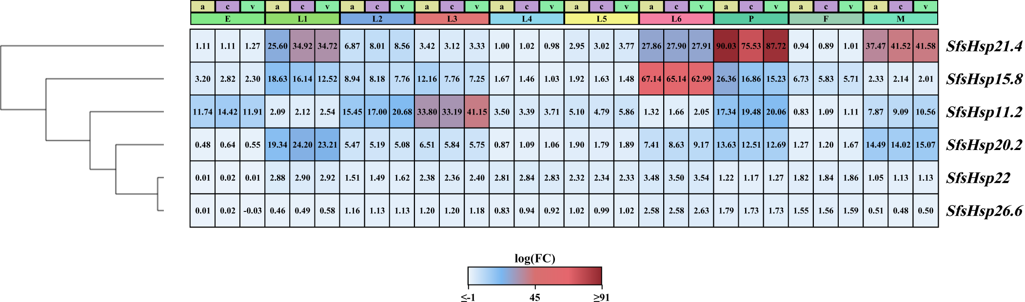
Hierarchical clustering revealed that the expression patterns of the six SfsHsp proteins varied depending on the S. frugiperda developmental stage (fig. 4). SfsHsp11.2 had the highest expression levels in the third-instar larvae, SfsHsp15.8, SfsHsp22, and SfsHsp26.6 had the highest expression in the sixth-instar larvae, SfsHsp21.4 had the highest expression in the pupal stage, and SfsHsp20.2 had the highest expression in the first-instar larvae. In addition, SfsHsp11.2 had the lowest expression in female adults, SfsHsp21.4 and SfsHsp29 in fourth-instar larvae, and SfsHsp22 and SfsHsp26.6 in eggs.
The hierarchical clustering analysis of six SfsHsp genes at the developmental stage of the FAW. The x-axis indicates the developmental stage of the FAW. E, egg; L1–L6, first- to sixth-instar larvae; P, pupa; M, male; F, female.

Expression levels of the six SfsHsps under high temperature
Under 36°C treatment (fig. 5), the expression of six SfsHsps increased in male and female adults, indicating their potential involvement in S. frugiperda response to heat. In females, the expression levels of five SfsHsps, SfsHsp11.2 (F 4,10 = 725.48), SfsHsp15.8 (F 4,10 = 20741.20), SfsHsp20.2 (W 4,15 = 12.9), SfsHsp21.4 (F 4,10 = 1786.85), and SfsHsp26.6 (F 4,10 = 19805.40), initially increased and then decreased with the maximum at 60 min and 33-, 21-, 48-, 15-, and 28-fold compared with the control, respectively. And the expression of SfsHsp22 (W 4,15 = 13.50) continued to rise with treatment time. In males, the expression levels of all six SfsHsps, SfsHsp11.2 (F 4,10 = 3503.34), SfsHsp15.8 (F 4,10 = 1645.94), SfsHsp20.2 (F 4,10 = 4803.84), SfsHsp21.4 (F 4,10 = 7793.74), SfsHsp22 (F 4,10 = 1125.76), and SfsHsp26.6 (W 4,15 = 13.50), exhibited a similar trend of initial increase and subsequent decrease with the maximum at 60 min and 13-, 17-, 273-, 229-, 279-, and 22-fold compared with the control, respectively.
Effect of high temperature (36°C) stress on the expression of six SfsHsp genes in FAW adults. Error bars showed the standard errors of the means of three biological replicates. Significant differences are marked with different letters (P < 0.05). All values are mean ± SD.

Expression levels of the six SfsHsps under low temperature
The expression levels of the six SfsHsps were influenced by exposure to 4°C (fig. 6), indicating their involvement in the response of S. frugiperda to low-temperature stress. In females, SfsHsp11.2 (F 4,10 = 906.33), SfsHsp15.8 (F 4,10 = 105.41), SfsHsp20.2 (F 4,10 = 1462.39), SfsHsp21.4 (F 4,10 = 3064.65), and SfsHsp22 (F 4,10 = 2424.58) displayed an initial increase and followed by a decrease with treatment time in expression. And they were the highest at 60 min with 13-, 6-, 16-, 12-, and 51-fold compared with the control, respectively. The expression of SfsHsp26.6 (W 4,15 = 13.50) consistently increased with treatment time. In males, SfsHsps11.2 (F 4,10 = 287.29), SfsHsp15.8 (F 4,10 = 5493.02), SfsHsp20.2 (F 4,10 = 2664.54), and SfsHsp26.6 (F 4,10 = 3066.74) first increased and then decreased with treatment time. And their expressions reached the maximum at 30, 60, 60, and 90 min with 6-, 4-, 9- and 13-fold higher than that of the control, respectively. And the expression of SfsHsp21.4 (F 4,10 = 397.96) and SfsHsp22 (W 4,15 = 13.50) increased with treatment time.
Effect of low temperature (4°C) stress on the expression of six SfsHsp genes in FAW adults. Error bars showed the standard errors of the means of three biological replicates. Significant differences are marked with letters (P < 0.05). All values are mean ± SD.

Expression levels of the six SfsHsps under UV-A stress
The six SfsHsps exhibited distinct expression patterns in males and females when exposed to UV light (fig. 7). In females (fig. 7), six SfsHsps initially increased and then decreased with treatment time. And the expression of SfsHsp21.4 (W 4,15 = 13.50) reached its maximum at 60 min (19-fold increase compared to the control), whereas that of SfsHsp11.2 (W 4,15 = 12.90), SfsHsp15.8 (F 4,10 = 4198.45), SfsHsp20.2 (W 4,15 = 12.90), and SfsHsp26.6 (F 4,10 = 1620.28) all reached their maximums at 90 min (57-, 9-, 16-, and 27-fold increases compared to the control, respectively). And the expression of SfsHsp22 (F 4,10 = 1281.09) increased with treatment time. In males (fig. 7), the expression of the six SfsHsps first increased and then decreased with treatment time. SfsHsp11.2 (F 4,10 = 301.09), SfsHsp15.8 (F 4,10 = 3307.56), SfsHsp21.4 (W 4,15 = 13.50), and SfsHsp26.6 (F 4,10 = 2545.87) exhibited the highest expression at 60 min (41-, 36-, 436-, and 16-fold increases compared to the control, respectively). And SfsHsp20.2 (F 4,10 = 5206.19) and SfsHsp22 (F 4,10 = 804.80) reached a maximum at 30 min, rising 1555- and 17-fold compared with the control, respectively.
Effect of UV-A stress on the expression of six SfsHsp genes in FAW adults. Error bars showed the standard errors of the means of three biological replicates. Significant differences are marked with letters (P < 0.05). All values are mean ± SD.

Discussion
Insects trigger a series of molecular mechanisms in response to extreme environmental conditions. As molecular chaperones, sHsps are essential in insect growth, development, and response to environmental stress (Kriehuber et al., Reference Kriehuber, Rattei, Weinmaier, Bepperling, Haslbeck and Buchner2010; Martínez-Paz et al., Reference Martínez-Paz, Morales, Martín, Martínez-Guitarte and Morcillo2013). Due to the different requirements of each environmental change, the type, tissue distribution, expression sequence, and expression levels of sHsps in different insects also differ (Sun et al., Reference Sun, Lu, Tang and Du2014). In this study, six S. frugiperda sHsp, SfsHsp11.2, SfsHsp15.8, SfsHsp20.2, SfsHsp21.4, SfsHsp22, and SfsHsp26.6, were identified and cloned. All SfsHsps contain an ACD domain. And four SfsHsps, SfsHsp20.2, SfsHsp21.4, SfsHsp22, and SfsHsp26.6, contain ‘V/P/I’ structure, which contributes to the diversity of function of the ACD structure (Basha et al., Reference Basha, O’Neill and Vierling2012). The phylogenetic tree based on different insect sHsps indicated that although the sHsps expression varied among insects, homologous sHsps were clustered into one branch (Chang et al., 2019; Chen and Zhang, Reference Chen and Zhang2015). And it indicated the evolution of sHsp is conserved and complex.
In this study, we observed distinct expression patterns of insect sHsps in different tissues. Specifically, SfsHsps15.8, SfsHsp20.2, and SfsHsp29 exhibited high expression levels in the compound eye of S. frugiperda. This finding could be explained by the crucial role of the compound eye in sensing external temperature variations, as reported by previous studies (Shen et al., Reference Shen, Gu, Huang, Zheng, Liu, Xu, Feng and Kang2011; Song et al., Reference Song, Lu and Du2020). Additionally, we discovered that these sHsps were highly transcribed in various tissues of male and female individuals, suggesting potential sex-specific roles. Similar observations have been made in studies involving other insect sHsps. For instance, OfsHsp19.9 and OfsHsp24.2 in O. furnacalis exhibited elevated transcript levels in the ovary, whereas OfsHsp21.8 was expressed in the testes and ovaries (Ruan et al., Reference Ruan, Meng, Yang, Zhou and Zhang2022). Hence, the tissue-specific expression pattern of sHsps may significantly contribute to the development and functional activity of the respective tissues. Notably, sHsp27 regulates mammalian spermatocyte prophase meiosis and displays an abundant expression in Drosophila melanogaster (Jagla et al., Reference Jagla, Dubińska-Magiera, Poovathumkadavil, Daczewska and Jagla2018; Wakayama and Iseki, Reference Wakayama and Iseki1999).
Expression patterns of insect sHsps vary in different developmental stages. For instance, LtHSP20.8 and LtHSP21.7b in L. trifolii exhibit their highest expression in the pupal stage, whereas LtHSP19.5 in the prepupal stage (Chang et al., 2019). Consistent with these findings, our study revealed that the expression levels of SfsHsps in S. frugiperda vary across different developmental stages. Additionally, we observed significant differences in the expression of the six SfsHsps between males and females, suggesting distinct roles in the growth and development of each sex. Similar findings have been reported in other insect species, including S. litura (Shen et al., Reference Shen, Gu, Huang, Zheng, Liu, Xu, Feng and Kang2011), Choristoneura fumiferana (Quan et al., Reference Quan, Duan, Fick, Kyei-Poku and Candau2018), and O. furnacalis (Ruan et al., Reference Ruan, Meng, Yang, Zhou and Zhang2022). These results highlight the importance of considering developmental stages and sex-specific differences when studying sHsp expression patterns and functions in insects.
High temperatures lead to changes in protein denaturation, fluidity of the phospholipid membrane, and intracellular homeostasis in insects (Bowler, Reference Bowler2018; King and MacRae, Reference King and MacRae2015; Ma et al., Reference Ma, Ma and Pincebourde2020). The sHsp family can help proteins fold correctly and repair denatured proteins, an essential process for insects to respond to temperature stress (Horwitz, Reference Horwitz1992; Poulain et al., Reference Poulain, Gelly and Flatters2010). In this study, the expression of the six sHsps displayed obvious changes under temperature stress. This finding agrees with the results of previous research on insect sHsps, indicating a crucial role for these heat shock proteins in conferring resistance and protecting insects against temperature-induced stress (Antolinez et al., Reference Antolinez, Moyneur, Martini and Rivera2021; Martínez-Paz et al., Reference Martínez-Paz, Morales, Martín, Martínez-Guitarte and Morcillo2013; Shen et al., Reference Shen, Gu, Huang, Zheng, Liu, Xu, Feng and Kang2011). Under 4°C and 36°C, the six sHsp genes were significantly upregulated in adults. The results demonstrated that temperature stress might trigger the synthesis of SfsHsps, similar to findings from studies in other insects. For instance, under cold and heat stress, the expression of 12 sHsps was significantly upregulated in Plutella xylostella, and Bthsp19.5, Bthsp19.2, and Bthsp21.3 in Bemisia tabaci (Chen and Zhang, Reference Chen and Zhang2015; Bai et al., Reference Bai, Liu, Lu and Du2019). In this study, the expression of SfsHsp11.2, SfsHsp15.8, SfsHsp20.2, and SfsHsp21.4 reached a maximum at different treatment times in adults. This may be due to the different protection thresholds of sHsps. For example, the expression of Hsp22, Hsp23, and Hsp27 in Myzus persicae after 10 h at 36°C was significantly higher than the control and other temperatures (Khurshid et al., Reference Khurshid, Inayat, Tamkeen, Ul Haq, Li, Boamah, Zhou and Liu2021). Under high-temperature stress, the expression of OfsHsp20.7 in O. furnacalis and sHsp27.6 in Apis cerana were also upregulated, reaching a maximum at 150 and 120 min, respectively (Liu et al., Reference Liu, Xi, Kang, Guo and Xu2012; Ruan et al., Reference Ruan, Meng, Yang, Zhou and Zhang2022). These results indicate that insects can respond to temperature stress by adjusting the expression of sHsps in the body.
As the main radiation reaching the ground, UV-A is an important environmental stress factor for insect growth and development, and can affect amino acid, sugar, and lipid metabolism in insects (Ali et al., Reference Ali, Rashid, Huang and Lei2016; Meng et al., Reference Meng, Zhang, Zhu, Wang and Lei2009; Rünger and Kappes, Reference Rünger and Kappes2008; Su et al., Reference Su, Yang, Zhou, Zhang and Meng2021). Here, UV-A induced the production of S. frugiperda SfsHsps. Their expression initially increased and decreased after a certain treatment time, which may be due to the stress intensity exceeding the protection threshold of sHsps under UV-A. Hsp70-3, Hsp70-4, Hsp70-5, and Hsp70-6 in Sogatella furcifera and Hsp26 and Hsp27 in D. melanogaster also displayed similar expression patterns (Wang et al., Reference Wang, Zhou, Zhu, Ma and Lei2014; Zhou et al., Reference Zhou, Yang, Yang, Long, Wang and Jin2019). However, the expression of sHsps reached a maximum at different treatment times, which may indicate different protective abilities against UV-A damage. Our results indicate that SfsHsps are essential for the response of S. frugiperda to UV-A stress.
In this investigation, six sHsps were identified and cloned from S. frugiperda. Subsequently, their expression patterns were studied across various tissues, diverse developmental stages, exposure to high and low temperatures, and UV-A stress conditions. The findings highlight the pivotal role of SfsHsps in the progress and maturation of FAW, in addition to their participation in the organism’s capacity to overcome extreme temperature shifts and UV-A stress. Nevertheless, the precise mechanism of action and physiological response mediated by these proteins remains unknown. Hence, further investigations are indispensable to unravel the specific molecular basis underlying these processes.
Supplementary material
The supplementary material for this article can be found at https://doi.org/10.1017/S0007485325100370.
Financial support
This study was supported by the Guizhou Provincial Department of Education Program (grant number KY[2021]057), the Guizhou Tobacco Company Program (grant numbers 2019XM37 and 2022XM09), and the Zunyi Tobacco Company Program (grant number 2022XM12).
Competing interests
The authors declare no conflicts of interest.
Ethical standards
This study did not involve any experiments with human subjects or animals.