INTRODUCTION
Trypanosoma evansi is a protozoan known as the etiologic agent of a disease called ‘Mal das Cadeiras’ or ‘Surra’ in horses (Silva et al. Reference Silva, Seidl, Ramirez and Dávila2002; Herrera et al. Reference Herrera, Dávila, Norek, Abreu, Souza, D'Andrea and Jansen2004). It is geographically distributed from the Northern Africa through the Middle East to Southeast Asia and it is often reported in the Arabian Peninsula (Desquesnes et al. Reference Desquesnes, Holzmuller, Lai, Dargantes, Lun and Jittaplapong2013). In Europe, some cases in Spain (Gutierrez et al. Reference Gutierrez, Juste, Corbera, Magnus, Verloo and Montoya2000) and France (Desquesnes et al. Reference Desquesnes, Bossard, Patrel, Herder, Patout, Lepetitcolin, Thevenon, Berthier, Pavlovic, Brugidou, Jacquiet, Schelcher, Faye, Touratier and Cuny2008) have been reported. This parasite may infect several animal species, including domestic and wild animals (Silva et al. Reference Silva, Seidl, Ramirez and Dávila2002) and it is rarely found in humans (Joshi et al. Reference Joshi, Shegokar, Powar, Herder, Katti, Salkar, Dani, Bhargava, Jannin and Truc2005). The disease is characterized by rapid weight loss, varying degrees of anaemia, intermittent fever, oedema of the hind limbs and progressive weakness (Herrera et al. Reference Herrera, Dávila, Norek, Abreu, Souza, D'Andrea and Jansen2004; Rodrigues et al. Reference Rodrigues, Fighera, Souza, Schild, Soares, Milano and Barros2005).
Pathogenicity of Trypanosoma evansi may vary according to its strain, host species and to some non-specific factors affecting simultaneously the animal, such as stress, concurrent infections or epizootiological local conditions. In the host bloodstream, T. evansi undergoes asexual multiplication by binary fission, starting at the bite site (skin) followed by parasite invasion of the bloodstream and lymphatic system, causing fever and inducing peaks of an inflammatory response (Connor and Van Den Boosche, Reference Connor, Van Den Boosche, Coetzer and Tustin2004).
Chemotherapy is probably the main form of therapeutic control of the disease and it can be used preventively in endemic areas. Currently therapy for equine trypanosomiasis is based on 4 different drugs: suramin, diminazene aceturate, quinapyramine and melarsomine (Brun et al. Reference Brun, Hecker and Lun1998), but in Brazil only diminazene aceturate and dipropionate imidocarb are officially marketed. However, the high toxicity of these drugs, the emergence of parasite resistance due to drug inappropriate use, especially in endemic regions (Silva et al. Reference Silva, Seidl, Ramirez and Dávila2002), as well as to unsatisfactory curative efficacy, some researches are still working in order to find more effective ways to treat the disease with less toxic agent.
In this sense, several studies have shown promising results using components extracted from plants in the control of parasites of different medical and veterinary importance (Machado et al. Reference Machado, Sousa, Salgueiro and Cavaleiro2010). Curcumin, the active principle of turmeric, isolated from the rhizomes of the plant Curcuma longa L. is characterized as a yellow-orange powder, insoluble in water and ether, but soluble in ethanol and acetone (Goel et al. Reference Goel, Kunnumakkara and Aggarwal2008). Its antioxidant activity (Gülçin and Ak, Reference Ak and Gülçin2008) is related to the ability to neutralize free radicals, inhibit lipid peroxidation, thus acting on cellular protection from cellular macromolecules, including DNA, from oxidative damage (Kunchandy and Rao, Reference Kunchandy and Rao1990; Subramanian et al. Reference Subramanian, Sreejayan Rao, Devasagayam and Singh1994). The effect of Curcuma sp. and its components, such as curcumin against Leishmania spp., Trypanosoma spp., Babesia spp., Toxoplasma gondii, Cryptosporidium spp., Giardia spp., Sarcoptes scabiei, Schistosoma spp., Angiostrongylus cantonensis and Toxocara canis have been reported (Haddad et al. Reference Haddad, Sauvain and Deharo2011).
The pharmacological use of curcumin is still not fully exploited due to its low aqueous solubility, chemical instability and low bioavailability (Ireson et al. Reference Ireson, Jones, Orr, Coughtrie, Boocock, Williams, Farmer, Steward and Gescher2002). Several strategies have been evaluated to increase its biological activity (Anand et al. Reference Anand, Kunnumakkara, Newman and Aggarwal2007). Lipid-core nanocapsules (LNCs) are an attractive option to circumvent these limitations, since these carriers can increase the curcumin solubility, stability, bioavailability, as well as its clinical efficacy (Frozza et al. Reference Frozza, Bernardi, Paese, Hoppe, Silva, Battastini, Pohlmann, Guterres and Salbego2010; Pohlmann et al. Reference Pohlmann, Fonseca, Paese, Detoni, Coradini, Beck and Guterres2013; Zanotto-Filho et al. Reference Zanotto-Filho, Coradini, Braganhol, Schröder, de Oliveira, Simões-Pires, Battastini, Pohlmann, Guterres, Forcelini and Beck2013). In this context, this study aimed to evaluate, through in vitro and in vivo studies, the trypanocidal effects of free curcumin (CURC) and curcumin-loaded lipid-core nanocapsules (C-LNCs) against T. evansi.
MATERIALS AND METHODS
Reagents
Curcumin, poly (ε-caprolactone) and sorbitan monostearate were purchased from Sigma-Aldrich (São Paulo, Brazil), whereas polysorbate 80 and acetone were purchased from Vetec (Rio de Janeiro, Brazil).
Preparation and characterization of LNCs
C-LNCs were prepared by interfacial deposition of preformed polymer method as described by Jäger et al. (Reference Jäger, Venturini, Poletto, Colomé, Pohlmann, Bernardi, Battastini, Guterres and Pohlmann2009) and Venturini et al. (Reference Venturini, Jäger, Oliveira, Bernardi, Battastini, Guterres and Pohlmann2011). At 40 °C, curcumin (0·1 g), poly(ε-caprolactone) (1·0 g), grape seed oil (1·65 mL) and sorbitan monostearate (0·385 g) were dissolved in acetone (270 mL). This organic phase was injected into an aqueous phase containing polysorbate 80 (0·77 g) and water (540 mL). Acetone was eliminated and the formulation was concentrated to 100 mL at 40 °C under reduced pressure. As a control, nanocapsule suspensions without curcumin, named as blank lipid-core nanocapsules (B-LNCs), were prepared. The suspensions were produced in triplicate and they were protected from light exposition.
Formulations were characterized according to their mean particle size, polydispersity index (PDI) and zeta potential using a Zetasizer Nano ZS equipment (Malvern Instruments, Malvern, UK). The analyses were performed at 25 °C after sample dilution with ultra-pure water (particle size and PDI) or 10 mm NaCl aqueous solution (zeta potential). The pH was measured using a calibrated potentiometer (VB-10, Denver Instrument, USA). The curcumin content and the encapsulation efficiency were determined by high-performance liquid chromatography (HPLC – Perkin Elmer, Shalton, USA) according to the method previously validated by Zanotto-Filho et al. (Reference Zanotto-Filho, Coradini, Braganhol, Schröder, de Oliveira, Simões-Pires, Battastini, Pohlmann, Guterres, Forcelini and Beck2013). The drug content was determined after appropriate dilution of the suspension with acetonitrile. After 30 min in the ultrasonic bath, the samples were centrifuged at 4120 g for 10 min. An aliquot of the supernatant was taken and diluted with the mobile phase for HPLC quantification. The curcumin encapsulation efficiency was determined by ultrafiltration–centrifugation technique (Ultrafree-MC 10 000 MW, Millipore, Ireland).
Trypanosoma evansi isolate
Trypanosoma evansi isolate used in this research was originally obtained from a naturally infected dog (Colpo et al. Reference Colpo, Monteiro, Stainki, Colpo and Henriques2005). Two rats (R1 and R2) were intraperitoneally (i.p.) infected with blood that was cryopreserved in liquid nitrogen. This step was carried out in order to allow the strain reactivation and achievement of a large amount of bloodstream forms of the parasite. The procedure was approved by the Animal Welfare Committee of the Universidade Federal de Santa Maria under protocol number: 095/2013.
In vitro tests
Trypanosoma evansi cultivation technique was adapted from Baltz et al. (Reference Baltz, Baltz, Giroud and Crockett1985). Briefly, the culture medium was composed of minimum essential medium without glutamine (0·376 g), glutamine (0·016 g), sodium bicarbonate (0·088 g), glucose (0·04 g), HEPES free acid (0·238 g), non-essential amino acid solution (200 μL), penicillin (1596 U mL−1) and estreptomicin (100 μg mL−1). The components were dissolved and homogenized in 30 mL of water after adjustment of the pH to 7·1 with NaOH. The volume of the solution was then raised up to 42 mL with sterile distilled water at an osmolarity of 0·30. Later, the culture medium was sterilized by filtration at 0·22 μm and stored under refrigeration. On the day of testing, 10 mL were separated into a Falcon tube to which were added 1 μL mL−1 of 50 mm hypoxanthine (dissolved in 0·1 m NaOH) and 2 μL mL−1 of 1·2 mm 2-mercaptoethanol. Subsequently, the complete culture medium was equilibrated in a CO2 incubator for 2 h (37 °C and 5% CO2).
When the animal R1 showed high parasitaemia (107 trypanosomes per μL), it was anaesthetized (isoflurane/anaesthetic chamber) for blood drawing. The sample was stored in tubes with anticoagulant (EDTA 10%). Then, 7 mL of blood was diluted (in order to separate the lymphocytes) in culture medium stabilized (1 v/v). The new solution was stored in microtubes and centrifuged at 400 g during 15 min. The supernatant was removed and placed on the culture medium. Posteriorly, the trypomastigotes count was performed in Neubauer chamber based on the methodology described by Gillingwater et al. (Reference Gillingwater, Kumar, Ismail, Arafa, Stephens, Boykin, Tidwell and Brun2010).
The culture medium with the parasites at the initial concentration of 36 240 trypomastigotes per well was distributed in microtiter plates (225 μL well−1). Formulations containing curcumin were dissolved in 1% dimethyl sulfoxide (DMSO) at final concentrations of 100, 75, 50 and 25 mg mL−1 (C-LNCs) and 100, 75, 50, 25, 12·5, 6·25, 3·12, 1·56 and 0·78 mg mL−1 (CURC) and added at each well. Control groups with B-LNCs, DMSO 1%, distilled water and diminazene aceturate (Ganazeg®: 3 μg mL−1) were used for test validation. At 1, 3, 6, 9 and 12 h after the onset of the experiment, the counting of living parasites was performed in Neubauer chambers. All tests were performed in triplicate.
In vivo tests
Animals
Fifty-four adult male Wistar rats (Rattus norvegicus), average 60 days old and 250 g in weight were housed in cages in an experimental room under controlled temperature and humidity (23 °C, 75% relative humidity, 12 h dark/light) and subjected to an adaptation period of 10 days. Throughout the experiment the animals were fed with commercial ration and received water ad libitum.
Experimental design and trypanosome infection
The animals were divided into eight groups (A–H) as follows: group A (not-infected and treated with saline/negative control/n = 4); group B (not-infected and treated with C-LNCs, n = 4); group C (infected by T. evansi and treated with B-LNCs, n = 5); group D (infected by T. evansi and treated with DMSO, n = 5); group E (infected by T. evansi and treated with saline/positive control, n = 6); group F (infected by T. evansi and treated with C-LNCs [10 mg kg−1], n = 10); group G (infected by T. evansi and treated with CURC dissolved in DMSO [10 mg kg−1], n = 10); and group H (infected by T. evansi and treated with CURC [100 mg kg−1], n = 10). Animals from groups C–H were inoculated with 0·1 mL of blood containing 3×10⁶ trypanosomes (coming from R2), whereas groups A and B remained uninfected to serve as negative controls. Twelve (12) hours post-infection (PI), curcumin treatment was delivered i.p. twice a day at 12 h interval (Table 1). This way, each treated animal received a volume of 1250 μL per application according to their body weight. This treatment was kept during 6 days with interval of 12 h between doses.
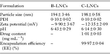
Physicochemical characteristics of B-LNCs and C-LNCs after preparation

Parasitaemia evaluation
Parasitaemia was daily estimated by blood microscopic examination. Each blood smear was prepared with blood collected from the tail vein (Da Silva et al. Reference Da Silva, Doyle and Monteiro2006) stained by the Romanowsky method and visualized at a magnification of 1000×.
Sampling
On day 7 PI the animals were anaesthetized (isoflurano/anaesthetic chamber) for blood drawing by cardiac puncture. The samples were stored in tubes without anticoagulant in order to obtain the serum. Thereafter, all the rats were euthanized, and their livers and kidneys were removed for histological examination.
Nitrite/nitrate
Seric NO levels were analysed indirectly by nitrite/nitrate (NOx) quantification according to a modified Griess method (Cobas Mira automated analyzer) described in detail by Tatsch et al. (Reference Tatsch, Bochi, Pereira, Kober, Agertt, De Campo, Gomes, Duarte and Moresco2011). The results were expressed as μmol L−1.
Advanced oxidation protein products (AOPP)
AOPP levels in serum were measured according to the techniques described by Hanasand et al. (Reference Hanasand, Omdal, Norheim, Gøransson, Brede and Jonsson2012) using a Cobas Mira automated analyzer. The results were expressed as μmol L−1.
Catalase (CAT) and superoxide dismutase (SOD) activity in whole blood
Determination of CAT activity was carried out in accordance with a modified method of Nelson and Kiesow (Reference Nelson and Kiesow1972). This assay involved the change in absorbance at 240 nm due to CAT-dependent decomposition of hydrogen peroxide (H2O2). An aliquot (0·02 mL) of blood (diluted 1:10 with saline) was homogenized in 0·910 mL of potassium phosphate buffer 50 mm, pH 7·0. The spectrophotometric determination was initiated by the addition of 0·07 mL of H2O2 0·3 mol L−1. The change in absorbance at 240 nm was measured for 2 min. CAT activity was calculated using the molar extinction coefficient (0·0436 cm2 μmol−1) and the results were expressed as nmol CAT per milligram protein.
SOD activity measurement was based on the inhibition of the radical superoxide reaction with adrenalin as described by McCord and Fridovich (Reference McCord and Fridovich1969). In this method, SOD present in the sample compeats with the detection system for radical superoxide. A unit of SOD is defined as the amount of enzyme that inhibits by 50% the speed of adrenalin oxidation. It leads to formation of the red-coloured product, adrenochrome, which is detected by spectrophotometer. SOD activity is determined by measuring the speed of adrenochrome formation, observed at 480 nm, in a reaction medium containing glycine–NaOH (50 mm, pH 10) and adrenalin (1 mm). The results were expressed as UI SOD per milligram protein.
Hepatic and renal functions
Hepatic function was evaluated by the assessment of alanine aminotransferase (ALT), aspartate aminotransferase (AST) and alkaline phosphatase (AP), whereas renal function was evaluated by the assessment of creatinine levels. All analyses were carried out in serum samples and measurement using commercial kit Bioclin® (kinetic tests), Cobas Mira® automated system (Freitas et al. Reference Freitas, Rovani, Boligon, Brum, Piana, Jesus, Veloso, Kober, Moresco, Araldi, Bauermann and Athayde2013).
Histology
Representative fragments of liver and kidney were fixed in 10% buffered formalin. Sagittal sections of every 6 μm were obtained and stained with the haematoxylin and eosin method. The slides were analysed for the presence of histopathological changes, measuring the dimensions of structures present in the cells. In each slide, 10 cells were randomly evaluated considering the mean area of hepatocytes nuclei, as well as the renal glomerular area, quantified by the number of hepatocyte nuclei.
Statistical analysis
Firstly, the data were subjected to normality test and the data that showed abnormal distribution were transformed into logarithms. The results from in vitro tests (normal data) were analysed by analysis of variance (ANOVA) (followed by Student's t-test (P>0·05). In vivo results were subjected to ANOVA followed by the Duncan's test. The histological results were analysed by the Bonferroni method followed by the Duncan's test. The results were considered significant when P<0·05.
RESULTS
Characterization of LNCs
Macroscopically, nanocapsule suspensions presented a homogeneous and opalescent appearance. After preparation, the formulations showed particle sizes about 200 nm with narrow size distributions (PDI<0·2) and negative zeta potential. The pH values were in the range of 6·1–6·5. The curcumin content was in agreement with the expected concentration (1·0 mg mL−1) and the encapsulation efficiency was close to 100% (Table 1).
In vitro tests
It was observed that all the curcumin concentrations higher than 3·12 μg mL−1 were able to kill all parasites within the first hour PI (Fig. 1A). In Fig. 1B, it is possible to observe a C-LNCs dose-dependent activity from the first hour PI, which reduced the concentration of live trypanosomes. In the third hour PI, it was possible to verify the absence of live parasites at the highest concentration (100 μg mL−1) with significant reduction of trypomastigotes in the other concentrations. Six hours PI, it was not possible to visualize live parasites in all the concentrations tested differently of the control groups (water, medium and DMSO at 1%), where they were live even after 9 and 12 h PI. These results validated our tests. In diminazene aceturate group (the control group), all the parasites died within 6 h post-treatment (Fig. 1A and B).
In vitro tests of CURC: A and C-LNCs: B against T. evansi. In order to validate the test the comparison with negative and positive control (diminazene aceturate: D.A.) was performed. Results within a circle did not differ from each other in Student's t-test (P>0·05).

In vivo tests
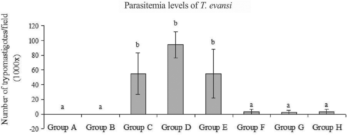
There was a significant reduction on the parasitaemia in animals treated with both concentrations of CURC and C-LNCs when compared to the positive control groups (C, D and E) on the 7th day PI (Fig. 2).
Average parasitaemia levels of rats infected by T. evansi on the 7th day PI: same letters in the same column indicate that the groups did not differ statistically among themselves at a significance level of 5% (Duncan's test). Group A (uninfected/treated with saline); B (uninfected/treated with C-LNCs); C (infected/treated with B-LNCs); D (infected/treated with DMSO); E (infected/saline-treated); F (infected/treated with C-LNCs [10 mg kg−1]); G (infected/treated with CURC [10 mg kg−1]); H (infected/treated with CURC [100 mg kg−1]).

Hepatic and renal functions
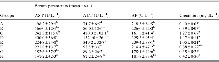
The results of the biochemical analyses are shown in Table 2. A significant decrease in the levels of AST was observed in the serum of rats treated with CURC at the highest concentration. There was a decrease in ALT levels in the groups treated with CURC (groups F and G) and C-LNCs (group H). Creatinine levels were significantly reduced in both groups treated with CURC. Serum AP levels in the animals treated with CURC did not show significant changes when compared to the infected control group.
Liver and kidney function assessment in rats infected by T. evansi and treated with CURC and C-LNCs

AST, aspartate aminotransferase; ALT, alanine aminotransferase; AP, alkaline phosphatase. Same letters in the same column indicate that the groups did not differ statistically among themselves at a significance level of 5% (Duncan's test). Group A (uninfected/treated with saline); B (uninfected/treated with C-LNCs); C (infected/treated with B-LNCs); D (infected/treated with DMSO); E (infected/saline-treated); F (infected/treated with C-LNCs [10 mg kg−1]); G (infected/treated with CURC [10 mg kg−1]); H (infected/treated with CURC [100 mg kg−1]).
Oxidative and antioxidants markers
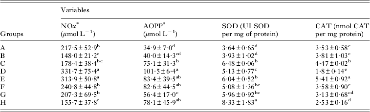
The results of oxidative and antioxidants biomarkers in serum are shown in Table 3. The serum concentration of AOPP showed a significant reduction in the group treated with CURC at the lowest concentration. Serum levels of NOx, in rats treated with CURC (in both concentrations) and in C-LNCs were significantly reduced, when compared to the control group. Blood levels of CAT were significantly reduced in groups treated with CURC when compared to the control group. On the other hand, the levels of SOD in total blood were significantly increased in animals treated with the highest concentration of CURC (the control group).
Assessment of oxidative biomarker serum (NOx and AOPP) and antioxidant enzymes (SOD and CAT) in whole blood of rats experimentally infected by T. evansi and treated with CURC and C-LNCs

Note: Same letters in the same column indicate that the groups did not differ statistically among themselves at a significance level of 5% (Duncan's Test). Groups A (uninfected/treated with saline); B (uninfected/treated with C-LNCs); C (infected/treated with B-LNCs); D (infected/treated with DMSO); E (infected/saline-treated); F (infected/treated with C-LNCs [10 mg kg−1]); G (infected/treated with CURC [10 mg kg−1]); and H (infected/treated with CURC [100 mg kg−1]). *AOPP and NOx levels did not present normal distribution, and were transformed to logarithms before comparative statistical analysis between groups.
Histology
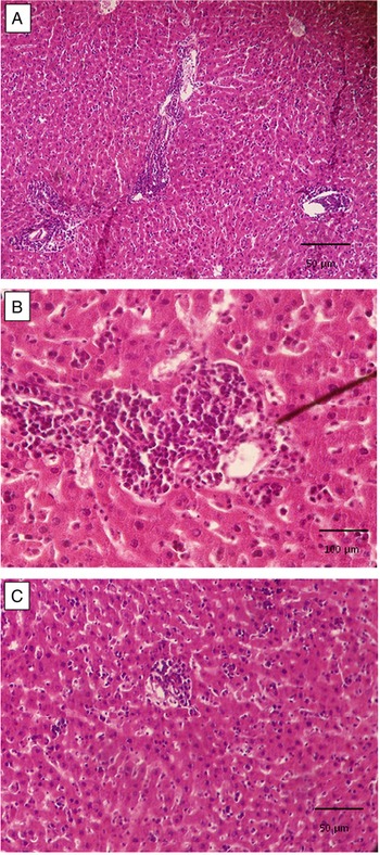
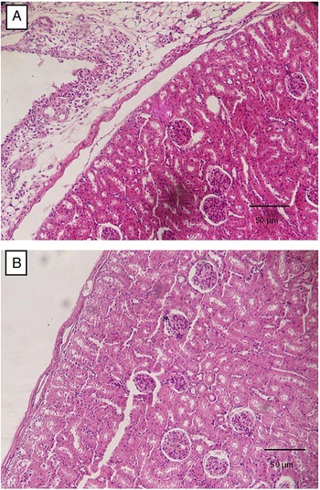
Non-infected rats did not show histological alterations (groups A and B), whereas animals infected by T. evansi (groups C, D and E) showed moderate multifocal lymphoplasmacytic inflammatory infiltrate in the liver (Fig. 3A and B), and moderate and diffuse perivascular lymphocytic inflammatory infiltration. In these animals, necrosis of hepatocytes (isolated) was also observed, characterized as mild and diffuse. In the renal capsule and pelvis, it was observed a moderate lymphocytic inflammatory infiltration in animals from groups C, D and E (Fig. 4a), which it was not observed in the animals from groups F, G and H (Fig. 4b). The animals treated with CURC (groups F, G and H) showed only mild perivascular and diffuse lymphocytic inflammatory infiltration (Fig. 3c).
Rats experimentally infected by T. evansi. In rats from group E it was observed a moderate liver tissue damage with multifocal lymphoplasmacytic inflammatory infiltration [A (obj.10×) and B (obj. 40×)] differently from animals treated with curcumin from group H, where rare and mild inflammatory infiltration was observed [C (obj.10×)].

Rats experimentally infected by T. evansi. It is observed a moderate lymphocytic inflammatory infiltration in the renal capsules of animals from group E [A], not observed in group F [B]. Obj. 10×.

Based on the statistical analysis, it was possible to detect an increase in the area of the hepatocytes nuclei in animals treated with nanocapsules with or without curcumin (groups B, C and F) when compared to group A (P<0·001); however, there was no difference between groups regarding the number of nuclei (P>0·05). In the kidneys, it was observed a significant reduction (P<0·001) in the renal glomerular area of the animals of all the groups when compared to the negative control (group A), but there was no visible morphological glomerular alteration.
DISCUSSION
The development of new therapies against trypanosomes is an important matter and it has been subject of research mainly due to the present limitations of the available therapies used currently (Baldissera et al. Reference Baldissera, Da Silva, Oliveira, Zimmermann, Vaucher, Santos, Rech, Tonin, Giongo, Mattos, Koester, Santurio and Monteiro2013; Wolkmer et al. Reference Wolkmer, da Silva, Paim, Duarte, Palma, França, Felin, Siqueira, Lopes, Schetinger, Monteiro and Mazzanti2013). This study showed the curcumin efficacy in its free or nanocapsules forms against T. evansi. This is the first study assessing the activity of nanoencapsulated curcumin against this parasite. Nanotechnology is a powerful tool to circumvent the limitations of curcumin, such as low aqueous solubility, chemical instability and low bioavailability (Anand et al. Reference Anand, Kunnumakkara, Newman and Aggarwal2007). In our study, morphological alterations in healthy rats that received B-LNCs were not found, but these animals showed increased hepatocyte nuclei area and reduction in the kidney glomerular area. These findings were similar to those observed in the groups treated with other nanocapsules with curcumin, which can be interpreted as the onset of tissue alterations, fact already described in other studies (Linkov et al. Reference Linkov, Satterstrom and Corey2008).
A significant in vitro trypanocidal activity of curcumin against T. evansi was observed 1 h PI corroborating with Nagajyothi et al. (Reference Nagajyothi, Zhao, Weiss and Tanowitz2012) and Nose et al. (Reference Nose, Koide, Ogihara, Yabu and Ohta1998) findings, who demonstrated curcumin activity (in vitro) on T. cruzi and T. brucei, respectively. Additionally, it was observed that C-LNCs treatments at doses lower than 100 μg mL−1 were not able to destroy all the parasites after 1 h PI, differently of the results observed for CURC. However, after 6 h no living parasites were observed, independently of the dose used. These results are in accordance with Zanotto-Filho et al. (Reference Zanotto-Filho, Coradini, Braganhol, Schröder, de Oliveira, Simões-Pires, Battastini, Pohlmann, Guterres, Forcelini and Beck2013), who observed that non-encapsulated curcumin was more cytotoxic than nanoencapsulated curcumin in C6 glioma cells after the first hour post-treatment, showing similar cytotoxicity at later time points. This better efficacy at later time can be related with the slow release of curcumin from LNCs (Zanotto-Filho et al. Reference Zanotto-Filho, Coradini, Braganhol, Schröder, de Oliveira, Simões-Pires, Battastini, Pohlmann, Guterres, Forcelini and Beck2013).
Considering our in vivo findings, both treatments, C-LNCs and CURC, showed antiparasitic activity on infected rats, differently of the results reported by Wolkmer et al. (Reference Wolkmer, da Silva, Paim, Duarte, Palma, França, Felin, Siqueira, Lopes, Schetinger, Monteiro and Mazzanti2013), who used oral treatment with curcumin at 20 mg kg−1. According to Pan et al. (Reference Pan, Huang and Lin1999), Dohare et al. (Reference Dohare, Garg, Jain, Nath and Ray2008) and Moon et al. (Reference Moon, Kim, Lee, Choi, Park, Heo and Kim2008) the curcumin has low absorption in the gastrointestinal tract, which justifies our choice for i.p. route, since we aimed to provide a greater absorption of this phytochemical to reach its higher biological activity.
The infiltration and dissemination of T. evansi in the central nervous system of equines have been described by Berlin et al. (Reference Berlin, Loeb and Baneth2009). This infiltration causes severe and fatal clinical disease, limiting the treatment effectiveness due to the blood–brain barrier (Wolkmer et al. Reference Wolkmer, da Silva, Paim, Duarte, Palma, França, Felin, Siqueira, Lopes, Schetinger, Monteiro and Mazzanti2013). Recent reports have shown that nanoencapsulation can increase the drug concentration into the brain (Bernardi et al. Reference Bernardi, Braganhol, Jäger, Figueiró, Edelweiss, Pohlmann, Guterres and Battastini2009, Reference Bernardi, Frozza, Meneghetti, Hoppe, Battastini, Pohlmann, Guterres and Salbego2012; Frozza et al. Reference Frozza, Bernardi, Paese, Hoppe, Silva, Battastini, Pohlmann, Guterres and Salbego2010). The formulations prepared in this study consisted of aqueous suspensions, enabling its intravenous use. Also, the C-LNCs are composed by a hydrophobic core surrounded by a polymeric wall and a hydrophilic surfactant (Pohlmann et al. Reference Pohlmann, Fonseca, Paese, Detoni, Coradini, Beck and Guterres2013). Generally, lipophilic molecules such as curcumin are localized in the core, which provides a controlled drug delivery and protection against degradation (Fontana et al. Reference Fontana, Coradini, Guterres, Pohlmann and Beck2009).
Free radicals and reactive oxygen species have been implicated in tissue damage in a variety of pathological processes (Nohl et al. Reference Nohl, Esterbauer and Evans1996). In our study, it was found an increase in NOx and AOPP levels in serum of the animals infected by T. evansi on day 7 PI. These results differ from those found by Da Silva et al. (Reference Da Silva, Paim, Santos, Sangoi, Moresco, Lopes, Jaques, Baldissarelli, Morsch and Monteiro2012), who reported an increase in the levels of both variables only 15 days PI in T. evansi infected rats. Our results showed a reduction in NOx and AOPP levels in curcumin-treated animals suggesting a damage tissue reduction usually present on sick animals.
Rats with T. evansi infection respond to oxidative stress by increasing the activity of antioxidant enzymes, such as SOD and CAT in the blood. Activation of these enzymes was reported by Omer et al. (Reference Omer, Mousa and Al-Wabel2007), which corroborates with our findings. Infected animals treated with CURC at concentration of 100 mg kg−1 showed increased levels of SOD. Singh and Sharma (Reference Singh and Sharma2011) when induced oxidative stress in rats and treated these animals with curcumin at 100 and 200 mg kg−1, reported increased levels of non-enzymatic antioxidant (reduced glutathione) and enzymatic antioxidants (CAT, SOD, glutathione peroxidase, glutathione-S-transferase, glutathione reductase and quinone reductase).
Hepatic damage can affect the body metabolic processes due to the role of liver in the general metabolism. It is well known that enzymes are necessary for normal cellular metabolism, and in this sense, the hepatic enzymes play a fundamental role (Rajamanickam and Muthuswamy, Reference Rajamanickam and Muthuswamy2008). Increases in ALT and AST activities have also been observed in natural infection by T. evansi (Sandoval et al. Reference Sandoval, Coppo, Negrette, Franciscato, Lopes, Pain, Rodrigues and Teixeira1994). Although the serum levels of AST did not show significant increase compared to negative animals, we observed a decrease of AST in infected animals treated with the highest concentration of CURC. ALT levels were also reduced in CURC and C-LNCs treated groups, thus, demonstrating a hepatoprotective effect of this phytochemical, an effect easily justified due to its antioxidant capacity (Singh and Sharma, Reference Singh and Sharma2011). Additionally, there is growing evidence that the hepatoprotective effect of curcumin takes place directly on the hepatocytes by reducing the intercellular levels of cholesterol and cytotoxic bile acids (Sambaiah and Srinivasan, Reference Sambaiah and Srinivasan1989). The reduction in the activity of liver enzymes, mainly ALT, indicates a better response of hepatocytes when facing the infection, since the serum transaminase activities returns near to normal when the regeneration of the liver parenchyma occurs (Shahidi and Wanasundara, Reference Shahidi and Wanasundara1992).
The increase in serum creatinine levels observed in our study corroborate with the findings of Brandão et al. (Reference Brandão, Larsson, Birgel, Hagiwara, Ventura and Teixeira2002) and Colpo et al. (Reference Colpo, Monteiro, Stainki, Colpo and Henriques2005), who believe that the increase in urea and creatinine in serum can be related to glomerulonephritis, caused by the deposition of immune complexes in the glomerular basement membrane, in response to T. evansi immunoglobulin production. Our findings show that the CURC can provide a nephroprotective effect, since creatinine levels were restored to values close to the ones observed in non-infected animals. This better kidney and liver protection observed on animals treated with CURC compared to those treated with C-LNB can be explained by the gradual and slow release of curcumin through the nanocapsules. Because of the T. evansi pathogenicity with a fast progression of its infection, we need a phytochemical that may act quickly, which may lead to minor aggravations of the disease.
Histological analysis confirmed the hepatic and renal biochemical assessment, as well as oxidant/antioxidant markers mentioned above. Infected animals treated with curcumin showed only mild inflammatory infiltrates in the liver, not in the kidneys, unlike the positive control. There are 2 possible hypotheses for this alteration: a significant lower parasitaemia with treatment or an anti-inflammatory effect of curcumin, as described in T. evansi infections (Wolkmer et al. Reference Wolkmer, da Silva, Paim, Duarte, Palma, França, Felin, Siqueira, Lopes, Schetinger, Monteiro and Mazzanti2013), associated with hepatoprotective action (Mathews et al. Reference Mathews, Binu, Sauganth Paul, Abhilash, Alex Manju and Harikumaran Nair2012).
CONCLUSION
CURC and C-LNCs presented trypanocidal activity against T. evansi in vitro. CURC showed a faster trypanocidal activity when compared to C-LNCs. According to our in vivo results, both, CURC and C-LNCs caused a reduction on the parasitaemia; however, these formulations cannot be used as single antiparasitic compounds. Therefore, we concluded that curcumin improves the biochemical variables, stimulating the antioxidants enzymes, which were previously induced in rats infected by T. evansi. Thus, we suggest the use of these curcumin formulations as a supportive therapy in animals with trypanosomosis, mainly due to their antioxidant activities, assisting in the reduction of tissue damage, as well as helping to maintain the integrity of liver and kidney functions.
ACKNOWLEDGEMENT
This study was carried out at Universidade Federal de Santa Maria, Department of Microbiology and Parasitology, 1000, Roraima Avenue, Building 20, Room 4232, 97105-900, Santa Maria, Rio Grande do Sul, Brazil. This study was funded by grants from Capes.