




No CrossRef data available.
Article contents
The properties of acidic compartments in developing schistosomula of Schistosoma mansoni
Published online by Cambridge University Press: 09 October 2003
Abstract
A variety of fluorescent probes have been used to study the acidic compartments in cercariae and schistosomula of Schistosoma mansoni. Freshly transformed schistosomula treated with the LysoTracker Red dye specific for lysosomes showed large acid-containing compartments (0·5–10 μm in size). The uptake of the dye is an energy-dependent process that depends on the metabolic activity of schistosomula. The compartments were quantified individually with respect to area, quantity of fluorescence and the total number/schistosomulum. Under normal conditions these compartments were not found in untreated cercariae, but appeared in cercariae slightly damaged by poly-L-lysine. The formation of these compartments seemed to be related to the development of cercariae into schistosomula as the number of compartments and uptake of fluorescence increased with time after transformation. Also, the method of transformation as well as the in vitro incubation of the parasite affected the percentage area of compartments/schistosomulum. Acid phosphatase enzyme activity was assessed using an endogenous phosphatase probe. Living and fixed schistosomula displayed the presence of enzyme activity in compartments of the same size and distribution as the acid-rich compartments. This was confirmed by histochemical staining showing deposition of enzyme-generated lead at the sites of phosphatase activity. We suggest that the development of acidic compartments is important during the transformation process or as a consequence of damage.
Information
- Type
- Research Article
- Information
- Copyright
- © 2003 Cambridge University Press
INTRODUCTION
Carneiro-Santos et al. (2001) proposed that the acidic organelles in transformed schistosomula of S. mansoni are analogous to large lysosomes in mammalian cells. The basis of this proposition is that the observed vesicles are labelled with LysoTracker Red, a specific marker for lysosomes in mammalian cells (Haugland, 1996). Uptake of the dye was found to vary between different strains and clones of the parasite (Al-Adhami et al. 2001). However, the nature of the stained compartments was not well defined.
Previous studies have shown that structural and functional changes occur in schistosomes during transformation from cercariae to schistosomula (Hockley & McLaren, 1973; Wilson & Barnes, 1974 a; Cousin, Stirewalt & Dorsey, 1981; Skelly & Shoemaker, 2001). Multivesicular bodies in the region of Golgi apparatus in the tegument of adult schistosomes that have similar appearance to lysosomes were described by Wilson & Barnes (1974 a). However, they suggested there was limited lysosomal activity in the system (Wilson & Barnes, 1974 b), though acid phosphatase activity was detected in the adult worm tegument using cytochemical procedures (Bogitsh & Krupa, 1971; Watts, Orpin & MacCormick, 1979). In this paper we present observations on the lysosome-like acidic compartments of schistosomula. We have measured the area of individual compartments and their number in a schistosomulum. We have also investigated the activity of the lysosomal enzyme acid phosphatase in these compartments using electron microscopy, biochemical and cytochemical assays in addition to the endogenous molecular marker enzyme-labelled fluorescence substrate (ELF 97).
MATERIALS AND METHODS
Parasite
A Puerto Rican isolate (PR) of S. mansoni was maintained in our laboratory in Biomphalaria glabrata and TO mice. Schistosomula were obtained by mechanical transformation of cercariae by the syringe method of Colley & Wikel (1974). Fresh 2-h-old and cultured 24-h-old schistosomula were used. The fresh schistosomula were washed and kept in Glasgow modification of Eagles medium (GMEM, Gibco Ltd, Paisley, Scotland) for 2 h at 37 °C. The 24 h schistosomula were obtained by culturing the parasite for 24 h in GMEM supplemented with 5% foetal calf serum (FCS, Sigma Chemical Co.) plus 100 I.U./ml penicillin and 100 μg streptomycin (Gibco) at 37 °C in a 5% CO2 incubator. Parasites were washed 3 times in GMEM before labelling to remove FCS.
Labelling of cercariae and schistosomula with LysoTracker Red DND-99
Freshly shed cercariae were labelled by addition of 50 μM LysoTracker Red (Molecular Probes Inc., Oregon, USA) to the cercariae in water. Approximately 50–100 cercariae were incubated for 30 min at 37 °C in a humidified CO2 incubator. In some experiments, we induced slight membrane damage by pre-treatment of cercariae with 30 μM 24·0 kDa poly-L-lysine (Sigma Chemical Co.) for 1 h at 37 °C prior to labelling with LysoTracker Red. Following induction of slight membrane damage by poly-L-lysine, cercariae were washed 3 times with aquarium water then incubated with LysoTracker Red. Five μl of Hoechst 33258 (10 mg/ml) were added to visualize the slight membrane damage (Tan et al. 2003). Cercariae were determined to be viable from their swimming activity and flame cell beat. After incubation, cercariae were washed 3 times with water and transferred onto slides for fluorescence microscopy examination.
To label schistosomula after transformation, approximately 50–100 worms were harvested and washed with fresh, sterile GMEM. Schistosomula were labelled with LysoTracker Red (50 μM) for 30 min at 37 °C. The labelled schistosomula were washed 3 times with GMEM and analysed by quantitative fluorescence at 590 nm (Carneiro-Santos et al. 2001). For precise data on the dimensions of individual compartments and the number of compartments, the OpenLab (Improvision, Viscount Centre II, Millburn Hill Rd., Coventry, England) was employed using the same wavelength and rhodamine filter set. The OpenLab is a fluorescence microscope with software designed for scientific imaging applications and calibrated image measurements. All experiments were assayed in triplicate. Thirty schistosomula were examined, i.e. 10 in each of 3 reaction mixtures.
Labelling of schistosomula with the endogenous phosphatase detection kit (ELF 97)
The ELF 97 (Molecular Probes Inc., Oregon, USA) was used to detect endogenous phosphatase activity in schistosomula of S. mansoni. Schistosomula were fixed with 4% formaldehyde for 15 min at room temperature. After fixation, schistosomula were washed 3 times with PBS and treated with 0·5% Tween-20 for 30 min at 37 °C. Schistosomula were thoroughly washed in PBS (pH 5·0) then treated with 500 μM of the fluorescence-labelled phosphatase substrate (ELF 97). Schistosomula were observed immediately under the fluorescence microscope using the following filter set (excitation=365 nm/emission=515 nm). Bright yellow–green fluorescence appeared at the reaction sites. Fresh schistosomula were labelled using the same method but the fixation step was omitted. Photography was performed by a Leitz Wild MPS48/52 photoautomat camera fitted to a Laborlux S microscope (Redman & Kusel, 1996). For photography, schistosomula were paralysed with carbachol (Sigma Chemical Co.).
Biochemical assay
Acid phosphatase activity was detected in an homogenate of schistosomula prepared in PBS containing 0·2 M sucrose. The bicinchoninic acid assay (BCA) was used to measure protein concentration in the homogenate (Smith et al. 1985). Acid phosphatase activity was assayed using nitrophenol phosphate substrate (15 mM-p-NPP) in 96-well microtitre plates. Sodium hydroxide (0·1 M) was added to stop the reaction. Absorbance was measured at 405 nm using a Multiskan spectrophotometer. Enzyme activity was calculated in nmole/min/μg protein. Samples were tested in triplicate.
Cytochemical study
For the cytochemical detection of acid phosphatase, the protocol developed by Gomori (Hayhoe & Quaglino, 1994) was followed with some adjustment. The incubation medium contained 200 mM Tris–maleate buffer at pH 5·0 and Na β-glycerophosphate as substrate. Schistosomula were fixed in 4% formaldehyde for 15 min at room temperature then washed 3 times with PBS. To increase permeability of the surface membrane, schistosomula were treated with 0·5% Tween-20 for 30 min at 37 °C. After a thorough wash in PBS (pH 5·0), schistosomula were incubated in the incubation medium for 18 h at room temperature, washed in PBS then treated with 5% ammonium sulphide solution for 5 min. Schistosomula were rinsed thoroughly in PBS and counter-stained with 0·1% aqueous neutral red for 3 min, washed and examined under a light microscope.
Ultrastructural and cytochemical study
For the ultrastructure localization of acid phosphatase, schistosomula were fixed in 2·5% glutaraldehyde cacodylate (0·1 M, pH 7·2) containing 0·2 M sucrose for 1 h at 4 °C. After fixation, schistosomula were washed overnight in cacodylate buffer, pH 7·2, containing 0·2 M sucrose at 4 °C. Schistosomula were rinsed 3 times in acetate buffer, pH 5·0, then treated with the incubation medium of Gomori and processed as described above. The same steps were followed, but the washing buffer used here was acetate buffer, pH 5·0, instead of PBS because of the microscopy procedure. Schistosomula were post-fixed in osmium tetroxide (0·2 M) for 1 h then washed in distilled water, dehydrated in ethanol and embedded in Araldite. Ultrathin sections of 60 nm thickness were cut with a diamond knife, stained with 2% methanolic uranyl acetate for 5 min followed by lead citrate for 5 min, and examined with a transmission microscope at 80 kV (Zeiss 902TEM, Germany).
Statistical analysis
We performed statistical analysis by analysis of variance (ANOVA) test with P<0·05 as the criterion of significance. Data points represent the mean±S.E.M.
RESULTS
Labelling of the parasite with the fluorescent probe, LysoTracker Red DND-99
Cercariae
The DNA-binding stain Hoechst 33258 was used to assess the damage induced by poly-L-lysine to the cercarial surface membrane. Cercariae that were not treated with poly-L-lysine showed no membrane damage and were stained with the Hoechst 33258 at low intensity. They were also treated with LysoTracker Red but the acidic compartments did not stain (Fig. 1A). When slight membrane damage was induced by 24·0 kDa poly-L-lysine, the acidic compartments were stained in the cercariae (Fig. 1B). Quantifying these compartments by using the OpenLab showed that they develop only after inducing minor damage to the surface membrane (Fig. 2).

Fig. 1. Development of acidic compartments in cercariae of Schistosoma mansoni after treatment with poly-L-lysine. (A) Fresh cercariae were incubated for 1 h at 37 °C and then labelled with LysoTracker Red and Hoechst 33258 for 30 min at 37 °C. (B) Fresh cercariae were treated first with poly-L-lysine 24·0 kDa for 1 h at 37 °C, washed and then labelled with LysoTracker Red and Hoechst 33258. ac, Acidic compartments. (C) 2-h-old schistosomula incubated with LysoTracker for 30 min at 37 °C. ac, Acidic compartments; g, gut.

Fig. 2. Effect of poly-L-lysine treatment on the development of acidic compartments in cercariae. In the control group cercariae were labelled with LysoTracker Red. In cercariae treated with poly-L-lysine acidic compartments were quantified using the OpenLab. Histograms represent mean±S.E.M. (n=30).
Schistosomula
A time-course for the appearance of fluorescence in 2-h-old schistosomula was followed after different times of incubation. Schistosomula were incubated with the LysoTracker for 1 min, 5 min, 10 min, 15 min, 20 min, 30 min, 1 h and 2 h. The results showed a significant increase in the uptake of the LysoTracker as the incubation time increased (Fig. 3). In the first 5 min there was very little labelling, but the labelled compartments could be distinguished. A small quantity of the dye was observed in the gut. Ten min after incubation, the number of compartments increased and gut labelling was very evident. The uptake of the fluorescent stain after 20 min and 30 min of incubation were significantly different from those quantified after incubation times of between 1 and 15 min (P<0·05). In comparison, 1 h and 2 h incubations with LysoTracker Red showed no significant differences as compared to the 30 min incubation time. Thus, 30 min was considered to be the optimum incubation time (Fig. 1C).

Fig. 3. Two-h-old schistosomula were labelled with LysoTracker Red and quantified at 1 min, 5 min, 10 min, 15 min, 20 min, 30 min, 1 h and 2 h after incubation. Histograms represent mean fluorescence/schistosomulum. Error bars are standard errors (n=30).
To determine whether uptake of LysoTracker is a temperature-dependent process, 2-h-old schistosomula were labelled with the fluorescent dye for 30 min at 4 °C or 37 °C. Schistosomula incubated with the LysoTracker at 4 °C showed very weak labelling. When these schistosomula were warmed to 37 °C for 30 min staining with the fluorescent dye was clearly detected (Fig. 4). The pattern of labelling showed fluorescence localized in small deposits in the parasite and in the gut when incubated at 37 °C. The labelled compartments varied from small and discrete to large and more diffusely stained deposits. Also, it was noted that there was no diffusion of the localized dye after 24 h incubation of the labelled schistosomula (Fig. 4).

Fig. 4. Two-h-old schistosomula were labelled with LysoTracker Red for 30 min at 4 °C and 37 °C. Histograms represent mean fluorescence/schistosomulum. Error bars are standard errors (n=30).
Measurements of the acidic compartments in schistosomula using the OpenLab
Use of the OpenLab facilitated the attempt to get precise data on the area (μ2)/compartment, the quantity of fluorescence (pixels) taken up/compartment and the total number of stained compartments/schistosomulum. Here, 3 factors that may have an effect on the development of the acidic compartments were investigated; the age of the schistosomula, the method of transformation and the in vitro culture.
Effect of age of schistosomula
In order to assess the formation of the acidic compartments with respect to the development of the parasite, the following specimens were recovered for labelling with the LysoTracker Red. (i) Schistosomula collected immediately after mechanical transformation (0 min) and (ii) schistosomula recovered at 30 min, 1 h, 1·5 h and 2 h after mechanical transformation. Results are shown in Fig. 5. In 0 min schistosomula, the acidic compartments were detected. However, the small increases in the area (μ2)/compartment with age were not significant (P>0·05), but the quantity of fluorescence (pixels) and the number of compartments/schistosomulum showed significant increases as the age of schistosomula increased from 0 min to 2 h (P<0·05). We tried to calculate the relative proportions of the area of compartments/schistosomulum and data are shown in Table 1. A significant increase in percentage of area of compartments was obtained as the age of schistosomula increased from 0 min to 2 h.

Fig. 5. Effect of age of schistosomula on the development of acidic compartments. Schistosomula prepared by mechanical transformation were labelled with LysoTracker Red at 0 min, 30 min, 1 h, 1·5 h and 2 h after transformation. Measurements were made with the OpenLab. Histograms represent mean±S.E.M. (n=30).
Table 1. The percentage of the total area of the schistosomulum that stains for acidic compartments (Results are calculated as the percentage of the total area of acidic compartments/schistosomulum using the +formula:

Effect of method of transformation
In this study so far, schistosomula transformed by the mechanical method were used in all experiments. Schistosomula recovered after cercarial penetration of isolated mouse skin were used in this experiment for comparison. The area, the quantity of fluorescence and the number of compartments were compared for each of the two methods. The number of compartments showed no significant difference between mechanical and skin forms (mean±S.E.M. 24·85±0·45, 24·5±0·67, P=0·849), respectively. However, skin forms showed a significant decrease in the uptake of the dye (1731·7±91·94) and in the area/compartment (6·58±0·71) when compared to the same measurements of the mechanical forms (1951·4±89·2, P=0·002 and 11·75±1·2, P=0), respectively (Fig. 6). However, the calculated percentage of area of compartments in the skin forms (3·5±0·6) was significantly lower than that in the mechanical forms (6·25±0·5) (Table 1).

Fig. 6. Effect of mechanical (M) method or skin (S) method of transformation, on the area (μm2)/compartment, the quantity of fluorescence (pixels)/compartment and the total number of compartments/schistosomulum. Schistosomula were labelled with LyosTracker Red. Measurements were made with the OpenLab. Histograms represent mean±S.E.M. (n=30).
Effect of in vitro culture
Schistosomula prepared by mechanical transformation were divided into 2 groups. In the first group schistosomula were incubated for 2 h and labelled with the LysoTracker Red. In the second group schistosomula were cultured in GMEM supplemented with 5% foetal calf serum for 24 h then labelled with the same probe. The cultured schistosomula showed a significant increase in the area/compartment (mean±S.E.M. 13·12±1·01) and in the total number of compartments (30·8±0·4) as compared with the 2-h-old schistosomula (11·9±0·2, P=0·04 and 24·6±0·53, P=0·023) respectively. Also, cultured schistosomula showed a significant increase in the uptake of the dye (3012·1±185·0) when compared to the 2 h schistosomula (2041·4±101·15), P value=0 (Fig. 7).

Fig. 7. Effect of in vitro culture on the acidic compartments of schistosomula. Schistosomula were transformed by the mechanical method and labelled with LysoTracker Red 2 h after transformation. The second group of schistosomula was cultured for 24 h and then labelled with LysoTracker Red. Measurements were made with the OpenLab. Histograms represent mean±S.E.M. (n=30).
Estimation of acid phosphatase activity
Biochemical and fluorescences assays.
Acid phosphatase activity was assayed in extracts of schistosomula. The activity value was (mean±S.E.M. 4·5±0·2 nmole/min/μg protein). Using the enzyme-labelled fluorescence substrate (ELF 97), a time-course for the appearance of the fluorescence on the 2-h-old and the 24-h-old schistosomula was followed after different times of incubation (10 sec, 1 min, 10 min, 30 min, 1 h and 2 h). During the first 30 min, schistosomula showed light fluorescence with the dye being accumulated on the surface membrane. The fluorescence was detected at the posterior end of the schistosomula and then spread slowly to the anterior end. Internal labelling of the ducts and glands was detected after 18 h incubation. Schistosomula treated with Tween-20 showed clear internal labelling of the ducts, glands and compartments (Fig. 8A and C).

Fig. 8. Acid phosphatase activity in schistosomula of Schistosoma mansoni. (A) Schistosomula labelled with the fluorescence-labelled phosphatase substrate ELF 97. gl, Glands; d, duct. (B) Schistosomula treated with Na β-glycerophosphate substrate in Tris–maleate buffer using the Gomori method. gl, Glands; d, ducts. (C and D) Higher magnifications of the boxed areas in (A) and (B) respectively. Arrows point to labelled acidic compartments (ac). (E) Control schistosomula treated as described in (B) but in the absence of substrate.
Light microscope cytochemistry.
Acid phosphatase activity was noted in the surface, the glands and connected tubules, the foregut and intestine in addition to the compartments of schistosomula (Fig. 8B and D). Both living and fixed schistosomula showed good reactions with the substrate as demonstrated by precipitation of lead at the reaction sites. The internal compartments that were previously identified on labelling with ELF-97 (Fig. 8A and C) gave a comparable pattern by lead precipitation as shown in Fig. 8B and D. Incubation of schistosomula with lead in the absence of substrate completely inhibited the reaction (Fig. 8E).
Electron microscopy.
The localization of acid phosphatase activity by the Gomori method is shown in Fig. 9. The reaction product was localized in a regular but patchy manner mainly in the muscles and parenchyma (Fig. 9), and was characterized by electron-dense regions of approximately 0·5–10 μm in diameter. These morphological data correlate well with the findings based on both fluorescent and light microscopy (Fig. 8).

Fig. 9. Section through the body of 2-h-old schistosomula prepared by mechanical transformation and stained by the Gomori method to detect acid phosphatase activity. Tegument (white arrow heads); muscles and parenchyma (white arrows).
Bogitsh & Krupa (1971) observed that the external tegument of adult schistosome worms lack acid phosphatase activity. We report a similar finding in the schistosomula. Ernst (1975) described acid phosphatase-containing vesicles which were heterogeneous in shape and size and localized at the posterior portion of oesophagus of adult worm of S. mansoni. Also, she indicated that acid phosphatase activity was not observed in the tegument except in very small vesicles that were morphologically similar to those illustrated in our work (Fig. 9).
DISCUSSION
Previous evidence has indicated the existence of large acid-containing compartments in 2-h-old freshly transformed schistosomula of S. mansoni. These compartments were not seen in cercariae (Carneiro-Santos et al. 2001; present work). These investigations into the nature of the acidic compartments have been based on labelling with a molecular probe, namely LysoTracker Red. This probe is a specific marker for lysosomes in mammalian cells. Data reported in our work provide further information on the acidic compartments in terms of morphological findings and enzymatic reactions.
No labelling of schistosomula with LysoTracker occurred in the cold (4 °C). Thus, the uptake of the dye is an energy-dependent process that depends on the metabolic activity of the schistosomula, which may indicate both transport actions and vesicle formation. Also, the uptake of the dye seems to be a time-dependent process. It starts immediately after incubation (1 min) and increases gradually with time. These results were based on quantifying the whole-labelled schistosomula, which included the acidic compartments in addition to the gut labelling. The individual compartments were quantified by using the OpenLab. Data showed that the number of compartments and the quantity of fluorescence increased with time after transformation. A significant increase in 0 min, 30 min, 1 h and 2 h schistosomula was clear, but the area/compartment was constant during the same periods after transformation. However, the calculated total areas occupied by these compartments/schistosomulum showed a significant increase during the 0 min–2 h development time.
Many studies have reported developmental changes within 1 h after the cercarial transformation into schistosomula begins. These changes include the tegument (Hockley & McLaren, 1973; Wilson & Barnes, 1974 a; Brink, McLaren & Smithers, 1977) as well as the acetabular glands, digestive tract, osmoregulatory system and parenchymal cell nuclei (Cousin et al. 1981). It is possible that the acidic compartments observed here are part of one of these systems. Carneiro-Santos et al. (2001) suggested that they could be lysosome-like organelles. In our work, we present data showing that the acid-containing compartments do exhibit considerable acid phosphatase activity. Also, they seem to be large compartments (0·5–10 μm) between muscle cells of schistosomula as demonstrated by electron microscopy. The presence of acid phosphatase activity in these compartments tends to indicate that they may be involved in transport of molecules and their degradation (Bogitsh & Krupa, 1971).
It is noteworthy that the acidic compartments were not seen in the cercariae until after transformation. However, inducing minor damage to the cercarial membrane with poly-L-lysine led to the appearance of these compartments. This raises the question of what are the mechanisms that cause these compartments to become visible in the slightly damaged cercariae and in the developing schistosomula? A hypothesis based on the functions of lysosomes (as reviewed by De Duve & Wattiaux, 1966) can be suggested. Autophagy has been observed in normal eukaryotic cells and is enhanced in cells subjected to remodelling in the course of differentiation or other induced changes. Recently, it was suggested that autophagy is turned on during apoptosis, implicating a role for autophagy for cell survival (Dorn, Dunn & Progulske-Fox, 2002). Some apoptotic inducers like tumour necrosis factor-α (TNF-α) stimulate autophagy. Also 3-methyladenine, a specific inhibitor of autophagy, blocks TNF-α-induced apoptosis in T-lymphoblastic leukaemia cells and in other models of apoptosis in which autophagy is a prominent feature (Jia et al. 1997). Autophagy increases also in cells exposed to metabolic stress and in those with a high endocytic activity. There is evidence that the process of endocytosis occurs in the schistosomula of S. mansoni (Ribeiro et al. 1998; Tan et al. 2003). Thus, schistosomes may be capable of using their tegument for the uptake and transport of molecules by endocytosis. The ingestion by the gut of macromolecules like bovine serum albumin (BSA) and the transport of ingested materials from excretory tubules by vesicles are dependent on endocytic activity of schistosomula (Tan et al. 2003). No reports are available on the presence of autophagosomes in schistosomes. However, Threadgold & Arme (1974) demonstrated the process of autophagy in adult worms of Fasciola hepatica. They suggested that some of the glycogenolysis occurring in F. hepatica could be processed by autophagosomes derived from membranes synthesized by mitochondria. Therefore, glucose will be made available from the glycogen stores of parenchymal cells of the parasite under starvation or other stressful conditions. The process of transformation from cercariae to schistosomula is associated with dramatic changes in the tegument, inclusion bodies and other organelles in addition to the exhaustion of glycogen reserve of the parasite. All these changes are explained as a survival mechanism for adaptation in the new environment i.e. the vertebrate host (Wilson & Barnes, 1974 a). Although the function of a lysosome system in schistosomes has not been fully investigated, the concept of autophagy described in mammalian cells (De Duve & Wattiaux, 1966) and in F. hepatica (Threadgold & Arme, 1974) may be applicable. It may act as a physiological survival mechanism accompanying the process of cercarial to schistosomula transformation or may be involved in endocytosis. We have found in preliminary experiments that wortmannin and 3-methyladenine, drugs that specifically suppress autophagy by inhibiting class III PI kinases (Kim & Klionsky, 2000), inhibited the development of acidic compartments in transformed schistosomula. Also, we have found evidence for autophagy in 2-h-old schistosomula by electron microscopy (data not shown).
Cousin et al. (1981) stated that a similar pattern of development was observed after transformation of schistosomula by the mechanical or skin methods. However, the mechanical forms developed more slowly, resembled cercariae more closely and varied less among organisms than did the skin forms. In this study, we tried to make a comparison with respect to the area, quantity of fluorescence and the number of acidic compartments between schistosomula derived via skin and mechanical transformation. Results reflected variations between mechanical and skin schistosomula. The relative proportions of the area of acidic compartments/schistosomulum were almost half in the skin forms as compared to that in the mechanical forms. Also, strain variations were observed (data not shown). No clear explanation could be given for the significant variations between the areas of the compartments in skin and mechanical forms within the same strain. However, the significant increase in the calculated area of compartments in the cultured schistosomula may support our suggestion of autophagy that may develop in schistosomula after being exposed to in vitro culture conditions for 24 h.
Clegg & Smithers (1972) have demonstrated that the in vitro development of schistosomula parallels the development in vivo. In the cultured schistosomula there was a statistically significant increase in the number of compartments/schistosomulum, but differences in numbers between the fresh and cultured schistosomula were in fact small in absolute values. Although these differences were statistically significant, they may not represent the actual numbers of the compartments. Schistosomula labelled with the LysoTracker showed patches of stained areas, which were excluded from the data. Despite the use of the sophisticated software (OpenLab), in some regions it was difficult to delineate individual compartments. The presence of a tubular lysosomal network consisting of an endosomal–lysosomal system that could be involved in the uptake and degradation of endocytosed material has been found in rat pancreatic acinar cells (Bendayan & Gisiger, 2001), human hepatocytes (Runnegar et al. 1997) and macrophages (Punturieri et al. 2000). It is possible that the patchy areas observed in the present study may represent part of a large lysosomal network in schistosomula. Further studies are required to confirm this suggestion.
In conclusion, the acidic compartments observed in living schistosomula are intracellular, are rich in acid phosphatase and may represent a lysosome-like system that may play a role in the physiology of schistosomula and remodelling during development.
This work is sponsored by a Wellcome Trust Travelling Research Fellowship for Batool Al-Adhami.
References
REFERENCES
AL-ADHAMI, B. H.,
DOENHOFF, M.,
THORNHILL, J.,
AKHKHA, A.,
WHITE, E. &
KUSEL, J. R.
(2001).
A study of some characteristics of individual clones of Schistosoma mansoni with emphasis on the biological and metabolic activities.
Parasitology
123, 563–572.CrossRefGoogle Scholar
BENDAYAN, M. &
GISIGER, V.
(2001).
Demonstration of acetylcholinesterase molecular form in a continuous tubular lysosomal system of rat pancreatic acinar cells.
Journal of Histochemistry and Cytochemistry
49,
29–39.CrossRefGoogle Scholar
BOGITSH, B. J. &
KRUPA, P. L.
(1971).
Schistosoma mansoni and Haematoloechus medioplexus: nucleosidediphosphatase localisation in tegument.
Experimental Parasitology
30,
418–425.CrossRefGoogle Scholar
BRINK, L. H.,
McLAREN, D. J. &
SMITHERS, S. R.
(1977).
Schistosoma mansoni: a comparative study of artificially transformed schistosomula and schistosomula recovered after cercarial penetration of isolated skin.
Parasitology
74,
73–86.CrossRefGoogle Scholar
CARNEIRO-SANTOS, P.,
THORNHILL, J. A.,
DOENHOFF, M. J.,
HAGAN, P. &
KUSEL, J. R.
(2001).
Acidic vesicles of Schistosoma mansoni.
Parasitology Research
87,
1001–1006.Google Scholar
CLEGG, J. A. &
SMITHERS, S. R.
(1972).
The effect of immune rhesus monkey serum on schistosomula of Schistosoma mansoni during cultivation in vitro.
International Journal for Parasitology
2,
79–98.CrossRefGoogle Scholar
COLLEY, D. G. &
WIKEL, S. K.
(1974).
Schistosoma mansoni: simplified method for the production of schistosomules.
Experimental Parasitology
35,
44–51.CrossRefGoogle Scholar
COUSIN, C. E.,
STIREWALT, M. A. &
DORSEY, C. H.
(1981).
Schistosoma mansoni: ultrastructure of early transformation of skin- and shear-pressure-derived schistosomules.
Experimental Parasitology
51,
341–365.CrossRefGoogle Scholar
DE DUVE, C. &
WATTIAUX, R.
(1966).
Functions of lysosomes.
Annual Review of Physiology
28,
435–492.CrossRefGoogle Scholar
DORN, B. R.,
DUNN, W. A. &
PROGULSKE-FOX, A.
(2002).
Bacterial infections with the autophagic pathway.
Cellular Microbiology
4,
1–10.Google Scholar
ERNST, S. C.
(1975).
Biochemical and cytochemical studies of digestive-absorptive functions of oesophagus, cecum and tegument in Schistosoma mansoni: acid phosphatase and tracer studies.
Journal of Parasitology
61,
633–647.CrossRefGoogle Scholar
HAUGLAND, R. P.
(1996).
Cell permanent probes for lysosomes and other acidic organelles. In
Handbook of Fluorescent Probes and Research Chemicals
(ed. Spence, M. T. Z.),
pp. 274–278.
Molecular Probes Inc.,
Oregon,
USA.
HAYHOE, F. G. J. &
QUAGLINO, D.
(1994).
Haematological Cytochemistry, 3rd Edn. Churchill Livingston,
Edinburgh, London.
HOCKLEY, D. J. &
McLAREN, D. J.
(1973).
Schistosoma mansoni: changes in the outer membrane of the tegument during development from cercaria to adult worm.
International Journal for Parasitology
3,
13–25.CrossRefGoogle Scholar
JIA, L.,
DOURMASHKIN, R. R.,
ALLEN, P. D.,
NEWLAND, A. C. &
KELSEY, S. M.
(1997).
Inhibition of autophagy abrogates tumour necrosis factor alpha induced apoptosis in human T-lymphoblastic leukaemic cells. British Journal of Haematology
98,
673–685.CrossRefGoogle Scholar
KIM, J. &
KLIONSKY, D. J.
(2000).
Autophagy, cytoplasm to-vacuole targeting pathway, and pexophagy in yeast and mammalian cells.
Annual Review of Biochemistry
69,
303–342.CrossRefGoogle Scholar
PUNTURIERI, A.,
FILIPPOV, S.,
ALLEN, E.,
CARAS, I.,
MURRY, R.,
REDDY, V. &
WEISS, S. J.
(2000).
Regulation of elastinolytic cysteine proteinase activity in normal and cathepsin K-deficient human macrophages.
Journal of Experimental Medicine
192,
789–799.CrossRefGoogle Scholar
REDMAN, C. A. &
KUSEL, J. R.
(1996).
Distribution and biophysical properties of fluorescent lipids on the surface of adult Schistosoma mansoni.
Parasitology
113,
137–143.CrossRefGoogle Scholar
RIBEIRO, F.,
COELHO, P. M. Z.,
VIEIRA, L. Q.,
POWELL, K. &
KUSEL, J. R.
(1998).
Membrane internalisation processes in different stages of Schistosoma mansoni as shown by a styryl dye (Frei Mao 1-43).
Parasitology
116,
51–59.CrossRefGoogle Scholar
RUNNEGAR, M.,
WEI, X.,
BRENDT, N. &
HAMM-ALVAREZ, S. F.
(1997).
Transferrin receptor recycling in rat hepatocytes is regulated by protein phosphatase 2A, possibly through effects on microtubule-dependent transport.
Hepatology
26,
176–185.CrossRefGoogle Scholar
SKELLY, P. J. &
SHOEMAKER, C. B.
(2001).
The Schistosoma mansoni host-interactive tegument forms from vesicle eruption of a cyton network.
Parasitology
122,
67–73.CrossRefGoogle Scholar
SMITH, P. K.,
KROHN, R. I.,
HERMANSON, G. T.,
MALLIA, A. K.,
GARTNER, F. H.,
PROVENZANO, M. D.,
FUJIMOTO, E. K.,
GEOKE, N. M.,
OLSON, B. J. &
KLENK, D. C.
(1985).
Measurement of protein using bicinchoninic acid.
Analytical Biochemistry
150,
76–85.CrossRefGoogle Scholar
TAN, H. H. C.,
THORNHILL, J. A.,
AL-ADHAMI, B. H.,
AKHKHA, A. &
KUSEL, J. R.
(2003).
A study of the effect of surface damage on the uptake of Texas Red-BSA by schistosomula of Schistosoma mansoni.
Parasitology
126,
235–240.CrossRefGoogle Scholar
THREADGOLD, L. T. &
ARME, C.
(1974).
Electron microscopic studies of Fasciola hepatica XI. Autophagy and parenchymal cell function.
Experimental Parasitology
35,
389–405.Google Scholar
WATTS, S. D. M.,
ORPIN, A. &
MacCORMICK, C.
(1979).
Lysosomes and tegument pathology in the chemotherapy of schistosomiasis with 1,7-bis (p-aminophenoxy)heptane (153C51).
Parasitology
78,
287–294.CrossRefGoogle Scholar
WILSON, R. A. &
BARNES, P. E.
(1974 a).
The tegument of Schistosoma mansoni: observations on the formation, structure and composition of cytoplasmic inclusions in relation to tegument function.
Parasitology
68,
239–258.Google Scholar
WILSON, R. A. &
BARNES, P. E.
(1974 b).
An in vitro investigation of dynamic processes occurring in the schistosome tegument, using compounds known to disrupt secretory processes.
Parasitology
68,
259–270.Google Scholar
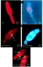
Fig. 1. Development of acidic compartments in cercariae of Schistosoma mansoni after treatment with poly-L-lysine. (A) Fresh cercariae were incubated for 1 h at 37 °C and then labelled with LysoTracker Red and Hoechst 33258 for 30 min at 37 °C. (B) Fresh cercariae were treated first with poly-L-lysine 24·0 kDa for 1 h at 37 °C, washed and then labelled with LysoTracker Red and Hoechst 33258. ac, Acidic compartments. (C) 2-h-old schistosomula incubated with LysoTracker for 30 min at 37 °C. ac, Acidic compartments; g, gut.

Fig. 2. Effect of poly-L-lysine treatment on the development of acidic compartments in cercariae. In the control group cercariae were labelled with LysoTracker Red. In cercariae treated with poly-L-lysine acidic compartments were quantified using the OpenLab. Histograms represent mean±S.E.M. (n=30).

Fig. 3. Two-h-old schistosomula were labelled with LysoTracker Red and quantified at 1 min, 5 min, 10 min, 15 min, 20 min, 30 min, 1 h and 2 h after incubation. Histograms represent mean fluorescence/schistosomulum. Error bars are standard errors (n=30).

Fig. 4. Two-h-old schistosomula were labelled with LysoTracker Red for 30 min at 4 °C and 37 °C. Histograms represent mean fluorescence/schistosomulum. Error bars are standard errors (n=30).
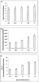
Fig. 5. Effect of age of schistosomula on the development of acidic compartments. Schistosomula prepared by mechanical transformation were labelled with LysoTracker Red at 0 min, 30 min, 1 h, 1·5 h and 2 h after transformation. Measurements were made with the OpenLab. Histograms represent mean±S.E.M. (n=30).
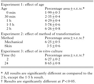
Table 1. The percentage of the total area of the schistosomulum that stains for acidic compartments
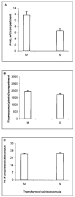
Fig. 6. Effect of mechanical (M) method or skin (S) method of transformation, on the area (μm2)/compartment, the quantity of fluorescence (pixels)/compartment and the total number of compartments/schistosomulum. Schistosomula were labelled with LyosTracker Red. Measurements were made with the OpenLab. Histograms represent mean±S.E.M. (n=30).
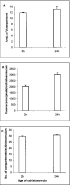
Fig. 7. Effect of in vitro culture on the acidic compartments of schistosomula. Schistosomula were transformed by the mechanical method and labelled with LysoTracker Red 2 h after transformation. The second group of schistosomula was cultured for 24 h and then labelled with LysoTracker Red. Measurements were made with the OpenLab. Histograms represent mean±S.E.M. (n=30).
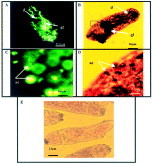
Fig. 8. Acid phosphatase activity in schistosomula of Schistosoma mansoni. (A) Schistosomula labelled with the fluorescence-labelled phosphatase substrate ELF 97. gl, Glands; d, duct. (B) Schistosomula treated with Na β-glycerophosphate substrate in Tris–maleate buffer using the Gomori method. gl, Glands; d, ducts. (C and D) Higher magnifications of the boxed areas in (A) and (B) respectively. Arrows point to labelled acidic compartments (ac). (E) Control schistosomula treated as described in (B) but in the absence of substrate.
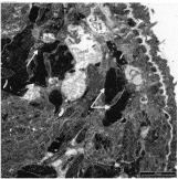
Fig. 9. Section through the body of 2-h-old schistosomula prepared by mechanical transformation and stained by the Gomori method to detect acid phosphatase activity. Tegument (white arrow heads); muscles and parenchyma (white arrows).
 You have
Access
- 7
- Cited by