Introduction
Nicotinic acetylcholine receptors (nAChRs) are neurotransmitter-gated ion channels that are widely distributed throughout the nervous system, where they have been attributed an important role in processes such as learning and memory. Their deficit has been related to diseases, such as Alzheimer’s and Parkinson’s as well as to schizophrenia (Lindstrom, Reference Lindstrom1997; Newhouse et al., Reference Newhouse, Potter and Levin1997). nAChRs are formed by the homomeric or heteromeric combination of five protein subunits. At least 12 different subunits have been described in the mammalian nervous system (α2–α10 and β2–β4), which can coassemble to form functional pentamers with different properties depending on their composition (Millar, Reference Millar2003; Gotti & Clementi, Reference Gotti and Clementi2004).
It is now fully recognized that nAChRs also play important roles in nonexcitable cells. Nonneuronal cholinergic systems are expressed in different cells and tissues and are involved in the regulation of their functions. Moreover, it has been shown that cholinergic dysfunction is related to the pathophysiology of diseases even though a systematic analysis of all the components of the nonneuronal cholinergic system in different diseases has not yet been performed (Kawashima & Fujii, Reference Kawashima and Fujii2008; Wessler & Kirkpatrick, Reference Wessler and Kirkpatrick2008). Thus, expression of nAChRs has been reported in several types of cells, including the epithelial cells of bronchial epithelium (Maus et al., Reference Maus, Pereira, Karachunski, Horton, Navaneetham, Macklin, Cortes, Albuquerque and Conti-Fine1998; Wang et al., Reference Wang, Pereira, Maus, Ostlie, Navaneetham, Lei, Albuquerque and Conti-Fine2001), keratinocytes (Zia et al., Reference Zia, Ndoye, Lee, Webber and Grando2000), nasal epithelium (Blank et al., Reference Blank, Ruckes, Clauss and Weber1997), vascular endothelial cells (Macklin et al., Reference Macklin, Maus, Pereira, Albuquerque and Conti-Fine1998; Wang et al., Reference Wang, Pereira, Maus, Ostlie, Navaneetham, Lei, Albuquerque and Conti-Fine2001), or lymphocytes and peripheral blood cells (Toyabe et al., Reference Toyabe, Iiai, Fukuda, Kawamura, Suzuki, Uchiyama and Abo1997; Sato et al., Reference Sato, Fujii, Watanabe, Yamada, Ando, Kazuko and Kawashima1999; Benhammou et al., Reference Benhammou, Lee, Strook, Sullivan, Logel, Raschen, Gotti and Leonard2000). The nAChRs have also been involved in different cell processes (reviewed by Wessler & Kirkpatrick, Reference Wessler and Kirkpatrick2008). In keratinocytes, α7 nicotinic receptors have been proposed as mediators in maturation, differentiation, migration, and apoptosis (Zia et al., Reference Zia, Ndoye, Lee, Webber and Grando2000; Nguyen et al., Reference Nguyen, Ndoye, Hall, Zia, Arredondo, Chernyavsky, Kist, Zelickson, Lawry and Grando2001; Chernyavsky et al., Reference Chernyavsky, Arredondo, Marubio and Grando2004). It has also been stated that α3 and α7 receptors control the migratory function of these cells through different effector proteins (kinases and GTPases).
Nicotinic receptors are modulated by different conditions and agents. In this sense, hypoxia appears to modulate α7 nAChRs in cholinergic and noncholinergic systems, both increasing and decreasing expression of nicotinic receptors, depending on the conditions tested. For instance, Heeschen et al. (Reference Heeschen, Weis, Aicher, Dimmeler and Cooke2002) showed that a hypoxic stimulus of 4 h increases expression of α7 nAChRs in human umbilical vein endothelial cells. Otherwise, it has been reported that long-term hypoxia decreases the expression of nicotinic α7 receptors in the adrenomedullar gland (Ducsay et al., Reference Ducsay, Hyatt, Mlynarczyk, Root, Kaushal and Myers2007).
In vivo nonneuronal activity of nAChRs is also modulated by some members of the Ly-6/neurotoxin gene family, such as SLURP-1, SLURP-2, or lynx1, among others (Miwa et al., Reference Miwa, Ibanez-Tallon, Crabtree, Sánchez, Sali, Role and Heintz1999; Grando, Reference Grando2008; Kawashima & Fujii, Reference Kawashima and Fujii2008). Lynx1, homologous to α-bungarotoxin, colocalizes with α7 and β2 nicotinic subunits and acts as an allosteric modulator of nAChRs in vivo, balancing neuronal activity. It has been proposed that lynx1 maintains both a low ACh sensitivity and a safety margin against agonist overstimulation of nAChRs (Miwa et al., Reference Miwa, Stevens, King, Caldarone, Ibanez-Tallon, Xiao, Fitzsimonds, Pavlides, Lester, Picciotto and Heintz2006). Expression of these endogenous modulators of nAChRs such as lynx1, SLURP-1, or SLURP-2 has not yet been described in the retina, although some works have reported the expression of SLURP1 in whole eye preparation (Mastrangeli et al., Reference Mastrangeli, Donini, Kelton, He, Bressan, Milazzo, Ciolli, Borrelli, Martelli, Biffoni, Serlupi-Crescenzi, Serani, Micangeli, El Tayar, Vaccaro, Renda, Lisciani, Rossi and Papoian2003) or tears (Favre et al., Reference Favre, Plantard, Aeschbach, Brakch, Christen-Zaech, de Viragh, Sergeant, Huber and Hohl2007).
There is some evidence suggesting that the retinal pigment epithelium (RPE) can express nAChRs. RPE is an epithelial monolayer of highly specialized nonexcitable cells, which is located between the neural retina and the vascular choroid. This layer plays a key role in the mechanical and metabolic support of photoreceptors. Moreover, evidence suggests that RPE is also involved in functions, such as eye growth control (Stone et al., Reference Stone, Sugimoto, Gill, Liu, Capehart and Lindstrom2001). This process may involve cholinergic mechanisms that act through muscarinic receptors. In this sense, a number of studies have shown that the muscarinic antagonist atropine delays the development of myopia in several animal models (Raviola & Wiesel, Reference Raviola and Wiesel1985; Stone et al., Reference Stone, Lin and Laties1991; Tigges et al., Reference Tigges, Iuvone, Fernandes, Sugrue, Mallorga, Laties and Stone1999). Stone et al. (Reference Stone, Sugimoto, Gill, Liu, Capehart and Lindstrom2001) suggest a role for nAChRs on RPE function. They reported that chlorisondamine, which is a substance that accumulates in neurons with nicotinic receptors, induces RPE layer degeneration, while other classical nicotinic antagonists cause no damage to the epithelial layer. This fact led the authors to suggest the possibility that RPE could also express nAChRs.
The aim of this work was to characterize the presence and functionality of α7 nAChRs in RPE and test their modulation by stimulus such as hypoxia as well as to explore the expression of endogenous modulators of nAChRs in the retina that could influence activity of nAChRs in RPE.
Materials and methods
Cell culture
RPE from cow eyes obtained from the municipal slaughterhouse in Orihuela (Spain) was isolated using standard techniques for cellular dissociation and isolation. Briefly, after the retinas were removed, the eyecups were incubated with 0.5% trypsin–EDTA for 45 min. Dissociated cells were concentrated by centrifugation and resuspended in dulbecco’s modified eagle medium containing antibiotics (50 μg/ml gentamicin and 2.5 μg/ml amphotericin B) and bovine fetal serum (FCS, 10%). The cells were grown on plastic flasks at 37°C in 5% CO2. The culture medium was changed every 2 days. Primary cultures aged between 4 and 6 weeks were subcultured following treatment with trypsin/EDTA. In order to avoid opacities caused by the melanin granules present on the RPE explants and first passages, unless otherwise indicated, cells cultures were used after four passages, where pigmented granules were not present. The confluent cultures appeared to be monolayered, with RPE cells showing a flattened appearance.
Reverse transcriptase–PCR protocol
Total RNA was extracted using the GenElute Mammalian Total RNA Miniprep Kit (Sigma, St. Louis, MO), according to the manufacturer’s instructions. Contaminating DNA was removed using DNAse I (1 U/μg total RNA; Fermentas, Glen Burnie, MD). Reverse transcription was performed with 1 μg RNA in the presence of 1.25 μM random nonamers (Sigma), 1 mM dNTPs (Fermentas), 20 U of ribonuclease inhibitor (Fermentas), 50 mM Tris-HCl (pH 8.3), 50 mM KCl, 4 mM MgCl2, 10 mM dithiothreitol, and 200 U of reverse transcriptase (RT, RevertAid M-MuLV Reverse Transcriptase; Fermentas) at 42°C for 60 min. Negative controls without RT were added. For the PCR reaction, we used specific primers for α7 nicotinic receptor subunits, based on Criado et al. (Reference Criado, Dominguez, Carrasco-Serrano, Smillie, Juiz, Viniegra and Ballesta1997). As control amplifications, we used the glyceraldehyde-3-phosphate dehydrogenase (GAPDH) primers described by Yuan et al. (Reference Yuan, Van Soom, Coopman, Mintiens, Boerjan, Van Zeveren, de Kruif and Peelman2003). The following CRALBP- and RPE65-specific oligonucleotides were used for RPE characterization: CRALBP forward: GAAGAGGAACAGGAGCTCCG, CRALBP reverse: CTTTGAACCGAGCTGGGAAGG, RPE65 forward (described by Vogel et al., Reference Vogel, Bullen, Teygong and Howard2007): TGGAGATTAACATCCCCGTCACC, and RPE65 reverse: TATCAGCTGTAGAGGACTTTGGCT.
To analyze endogenous modulator expression, the primers used were the following: lynx1 bovine forward: CCACATCCCTGCTCTCACTC, reverse: GTGGCCAGGAGTATAAGGGC; SLURP1 rat forward: CCACGGCCATTAACTCATGC, reverse: CACGGAAGCAACAGAAGACAG; RGD1308195 rat forward (based on Moriwaki et al., Reference Moriwaki, Yoshikawa, Fukuda, Fujii, Misawa and Kawashima2007): ATGAGGCTTCCCTTCTGGTTC, reverse (based on Moriwaki et al., Reference Moriwaki, Yoshikawa, Fukuda, Fujii, Misawa and Kawashima2007): TCAGTCCCTGTTGCAGAGATT; and lynx1 rat forward: CATGACCCACTTGCTGACAG, reverse: GGATGAAGGTGAGGAAGTCC.
Complementary DNA (cDNA) was PCR amplified under standardized conditions as follows: denaturation at 94°C for 2 min, followed by 35 cycles of 30 s each at 94°C, 1 min at 57°C (CRALBP and RPE65) or 55°C (α7) or 30 s at 59.5°C (bovine lynx1, rat SLURP1, and RGD1308195) or 58°C (rat lynx1), 1 min at 72°C, and a final elongation step at 72°C for 15 min. Negative control without cDNA was present in all amplifications. PCR products were separated by electrophoresis in 1.5% agarose gels. Identity of the amplified sequences was confirmed by RT-PCR.
Western blotting
Western blot was performed using standard techniques. After washing with sodium phosphate-buffered saline (PBS), the dissociated cells were lysed in the presence of a protease inhibitor cocktail from Sigma. Protein concentrations were determined by the Bradford method, using the Bio-Rad Protein Assay Dye Reagent (Bio-Rad, Herculez, CA). Denatured proteins were run on 10% sodium dodecyl sulfate-polyacrylamide gels. Proteins were blotted onto polyvinylidene difluoride membranes. Bands were identified using Lumi-light Western Blotting Substrate (Roche Applied Science, Indianapolis, IN) according to the manufacturer’s instructions. Molecular weight standard markers were used (Pageruler Unstained Protein Ladder and Prestained Protein Molecular Weight Marker; Fermentas). For α7 nicotinic subunit, primary antibody anti-α7 monoclonal antibody mAb319 (Sigma) was used at 2.5 μg/ml. Secondary antibody: 40 ng/ml goat anti-rat IgG conjugated with horseradish peroxidase (HRP) (Jackson Immunoresearch Laboratories, West Grove, PA). For lynx-1 detection, primary antibody T-15 (Santa Cruz Inc, Santa Cruz, CA) was used at 2 μg/ml. Secondary antibody: donkey anti-goat IgG–HRP conjugated (Santa Cruz Inc) at 0.2 μg/ml. The anti-β-actin monoclonal antibody (clone AC-15) was used as loading control.
Immunocytochemistry for cultured cells
Using standard techniques, RPE cells were grown on glass coverslips until confluence, washed with PBS, and fixed for 20 min with 4% paraformaldehyde in PBS at 4°C. Cells were permeabilized with 0.2% Triton X-100 in PBS for 20 min. Nonspecific binding was blocked by incubating with 1% BSA in PBS for 45 min at room temperature. Cells were incubated with primary antibody in PBS plus 1% bovine serum albumin (BSA) overnight at 4°C in a wet chamber. Secondary antibody was applied in PBS for 1 h at room temperature. Cells were observed with Leica confocal microscopy (Leica TCS SP2; Leica Microsystems, Wetzlar, Germany). Negative controls lacking primary and secondary antibodies were added. The thin morphology of the cultured monolayers limited the use of confocal Z-sections and discriminations about cell polarization and labeling distribution.
Immunohistochemistry for tissue sections
Three Sprague Dawley were anesthetized with a lethal dose of ketamine (0.5 g/kg i.p.) and eyes were enucleated. The eyecups were fixed in 4% paraformaldehyde in 0.1 m PBS at pH 7.4 for 2 h and then washed in 0.1 m PBS before being cryoprotected in 10% sucrose for 0.5 h, 20% sucrose for 1 h, and 30% sucrose overnight at 4°C. The next day, they were embedded in optimal cutting temperature compound and cut into 14-μm thick radial sections on a cryostat and mounted on glass slides. Sections were incubated in 10% normal donkey serum (Jackson Immunoresearch Laboratories) to avoid nonspecific staining. Without washing, they were then immunostained for rat anti-α7 nicotinic receptor subunit antibody diluted in PBS containing 0.5% Triton X-100. Sections were incubated overnight at room temperature. The next day, sections were washed in 0.1 m PBS. To visualize antibody binding patterns, they were transferred to donkey anti-rat IgG coupled to fluorescein isothiocyanate (FITC) in 0.1 m PBS 0.5% Triton X-100 for 1 h. Finally, the sections were washed in 0.1 m PBS, mounted in watermount (Vector Laboratories, Burlingame, CA), and coverslipped for viewing by confocal microscopy. To control for nonspecific staining, some sections were stained omitting the primary antibody.
Primary antibodies: 5 μg/ml mAb319 (Sigma) for α7 nicotinic subunit. For RPE characterization, 4 μg/ml of goat polyclonal anti-CRALBP (L-19) antibody were used (Santa Cruz Inc). Secondary antibodies: Goat anti-rat Alexa 546 (10 μg/ml; Molecular Probes, Eugene, OR), donkey anti-rat Alexa 488 (10 μg/ml; Molecular Probes), and donkey anti-goat 4 μg/ml (Santa Cruz Inc). Negative controls without primary or/and secondary antibodies were included.
Fluorescence measurements
Fluorescence determinations were made using a conventional fluorescence microscopy system. Prior to the recordings, cells were incubated (37°C) with the fluorescent Ca2+ indicator fura-2 acetoxy methyl ester (fura-2 AM, 4 μM; Molecular Probes) for 45 min in culture medium. After extensive washing with external solution, the cells were tested with brief applications of 200 μM nicotine or 200 μM acetylcholine plus 0.5 μM atropine sulfate to block muscarinic responses. In order to prevent nAChR desensitization, successive nicotine and acethylcholine applications were separated by at least 60-s intervals. In some experiments, the external medium was supplemented with 10 mM Ca2+ (12 mM final concentration) in order to amplify the fluorescence response. The level of intracellular free Ca2+ concentration was taken as the ratio between the digital images obtained with 340 and 380-nm excitation wavelengths. Emission was monitored at 510 nm. During recordings, cells were continuously superfused using a gravity-driven perfusion system with separate tubes for normal and drug-containing external solutions. The tube ends were positioned 50–100 μm away from the cells and connected on the other side to a fast exchanger system (RSC-200; Biologic, Claix, France).
Hypoxia induction
Flasks containing approximately 7 × 10⁶ bovine RPE and glass coverslips containing RPE confluent cultures were incubated without FCS in a sealed incubator at 3% O2 for 72 h. Control cells were maintained in normoxic conditions. After the stimulus, flask cells were collected and total proteins were immunoblotted. Cells in coverslips were fixed and used for immunofluorescence. Both were probed against mAB319 antibody for α7 nAChR.
Solutions and chemicals
The external solution had the following composition (in millimolar): NaCl, 140; KCl, 5.6; MgCl2, 2; CaCl2, 2; Glucose, 10; and HEPES–NaOH, 10; pH 7.4. Culture media were purchased from GIBCO (Grand Island, NY). Unless otherwise indicated, all the other chemicals used came from Sigma.
Results
RPE characterization
Using specific oligonucleotides for the specific RPE markers CRALBP and RPE65, messenger RNA (mRNA) expression was shown by RT-PCR from RPE cultured cell RNA. Sequence was confirmed by DNA sequencing. Moreover, 100% of cultured monolayers were stained with a goat polyclonal anti-CRALBP antibody (Fig. 1A–1C).
Bovine RPE cells characterization and expression of α7 nAChRs. (A) Representative 10× image of a four-passages culture. Cells were cultured for up to 7 days till they were grown to confluence. (B) Representative RPE cell culture stained with a goat polyclonal antibody against the RPE-specific marker CRALBP. (C) RT-PCR amplification with specific primers for CRALBP (C) and RPE-65 (R). (D) RT-PCR amplification from bovine RPE mRNA. 1 μg of total RNA purified from cultured RPE (E) and whole retina (R) was reverse transcribed and amplified with specific oligonucleotides for α7 nicotinic subunit. Negative PCR control without cDNA (C) and RNA standards (St) are also shown. A total volume of 12.5 μl was loaded in each lane, onto ethidium bromide–stained 1.5% agarose gel. (E) Western blot from freshly isolated RPE (F), total retina (R), and cultured RPE (E) total proteins expected immunoreactive bands for anti-α7 is observed at 57 kDa. Monoclonal antibody mAb319 HRP conjugated was used. Each lane contained 20 μg of total protein. (F) Immunocytochemistry confocal images of representative RPE cells in culture labeled with mAb319 antibody (against α7 nAChRs). Note the immunoreactive prolongations of cultured cells marked by arrows. (G) Representative negative control image without primary antibody.

α7 nAChRs mRNA expression
In order to test for the presence of nAChRs in RPE, the expression of α7 nicotinic receptor subunits was analyzed by RT-PCR in RPE cultured cells. As shown in Fig. 1D, the oligonucleotide primers for nicotinic α7 subunit (see Materials and methods) amplified a 280 bp fragment. This same fragment was also amplified when RNA from total retina was used (positive control). Negative controls did not amplify any fragment. The GAPDH primers described by Yuan et al. (Reference Yuan, Van Soom, Coopman, Mintiens, Boerjan, Van Zeveren, de Kruif and Peelman2003) (see Materials and methods) amplified a 238 bp band corresponding to an mRNA template. No 458 bp band from contaminating DNA was observed (not shown).
Protein expression
To determine whether transcripts encoding the α7 nicotinic subunits detected in RPE by RT-PCR were translated, Western blotting was performed with cultured RPE cells. Total retina protein extract was used as positive control. Nicotinic α7 subunit expression was also tested in freshly isolated RPE cells. Samples were probed with anti-α7 monoclonal antibody mAb319. As shown in Fig. 1E, mAb319 detected a ~57 kDa band corresponding, which was also present in all three protein extracts.
Immunofluorescence staining using antibodies against α7 subunit was performed in cultured RPE cells to confirm the expression of nAChRs on these cells. As can be seen in Fig. 1F, fluorescence activity was observed under the microscope in RPE cells labeled with mAb319, indicating the expression of α7 nicotinic subunits. Fluorescence was mostly distributed around the cell nucleus. No evident labeling was observed when no primary antibody was added (Fig. 1G), or when the secondary antibody was absent (not shown).
Effect of hypoxia on expression of α7 nAChRs
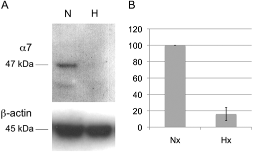
In order to study the possible modulator effect of hypoxia on expression of α7 nAChRs in RPE cells, we applied a long-term hypoxia stimulus of 72 h to the RPE cell culture. After this time, protein extracts were probed with mAb319 antibody. As Fig. 2 shows, α7 expression decreased significantly after long-term hypoxia compared with normal conditions.
Hypoxia modulation of expression of α7 nAChRs. (A) Western blot showing total proteins extract from RPE cells maintained in hypoxia for 72 h (H) or in control normoxic conditions (N), probed with mAb319 antibody. Bands for β-actin expression are shown as control. (B) Histogram showing expression of α7 nAChRs normalized to β-actin under control (normoxia) and hypoxia conditions. The values represent the means ± s.d. from triplicates.

Functional nAChRs detection
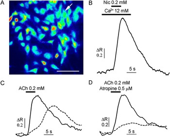
In order to test for the presence of functional nAChRs in RPE in culture, we examined the Ca2+ transients elicited by both nicotine and acetylcholine (200 μM) in fura-2–loaded cells. As shown in Fig. 3A and 3B, a small subset of fura-2–loaded RPE cells displayed an apparent elevation of intracellular free calcium concentration when stimulated with nicotine pulses (four responsive cells from a total of 70 optic fields viewed, representing 0.18% of total cells). Similar Ca2+ transients were observed when 200 μM acetylcholine was applied (29 responsive cells in 50 microscopic fields, representing 2% of the cells, Fig. 3C). However, only a subset of acetylcholine-sensitive RPE cells (approximately 10%) responded to 200 μM acetylcholine when coapplied with 0.5 μM atropine sulfate, a selective inhibitor of muscarinic acetylcholine receptors, resulting in a responsiveness of 0.21% (Fig. 3D).
Nicotine- and acetylcholine-evoked responses in cultured RPE cells. (A) Typical RPE cells in culture seen under fluorescence microscopy. Cells were incubated with the fluorescent Ca2+ indicator fura-2 acetoxy methyl ester for 45 min. Bar 100 μm. (B) Intracellular Ca2+ transient elicited by nicotine (Nic) application in the cultured cell pointed at in (A) by the arrow. In order to obtain measurable responses, external medium was supplemented with 10 mM Ca2+ prior, during, and after the nicotine pulse. Horizontal black bars indicate the time point and duration of drug applications. (C) Intracellular Ca2+ response evoked by acetylcholine (ACh) on another two single RPE cells. (D) Intracellular Ca2+ response recorded in the same cells than in (C) when acetylcholine was coapplied with atropine sulfate.

Tissue sections
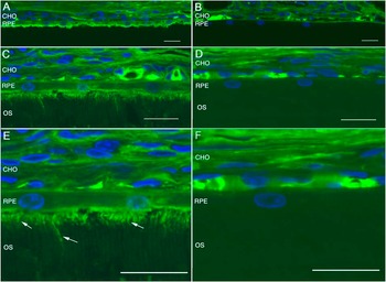
To further assess expression of α7 nAChRs in RPE, sections of albino rat retina were immunostained with antibodies against α7 nAChRs. Fig. 4A shows pigment epithelial cells immunoreactivity for α7 antibodies. An increase of immunoreactivity was found in the apical surface of these cells. No immunostaining was found in control sections (Fig. 4B, 4D, and 4F). In the retinal sections where the photoreceptors’ outer segment was not detached, the immunostained apical processes of the pigment epithelium were identified, located between outer segments of the photoreceptors (Fig. 4C and 4E, arrows).
Immunohistochemistry confocal images of expression of α7 nAChRs in rat RPE. (A, C) Vertical section of rat retina showing pigment epithelium cells labeled with antibodies against α7 nAChRs. (B, D) Control retinas. (E) High magnification of (C) showing α7 nAChRs immunoreactivity processes of pigment epithelium cells located between photoreceptor outer segments, marked by arrows. (F) High magnification of (D). Nuclei stained with TOPRO-3 in blue. Bar 20 μm. CHO, Choroid; OS, Photoreceptor outer segments.

Expression of endogenous modulators of nAChRs
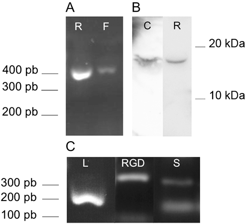
In order to test the expression of endogenous modulators of nAChRs in the retina and RPE, we tested the expression of lynx1 (a member of Ly-6/neurotoxin gene family) reported as α7 modulator. By using total mRNA from bovine retina and RPE and with specific primers, RT-PCR amplified a band of 371 pb corresponding to the lynx1 gene in both bovine retina and freshly isolated RPE (Fig. 5A). By Western blot, we detected a single band of 17 kDa, which was higher than the expected 9-kDa band. The same fragment size was obtained for the rat brain sample used as positive control. Although further investigation is needed to clarify this aspect, it could be due to protein phosphorylation (Fig. 5B). The same positive results for RT-PCR amplification and Western blot analysis were obtained for rat retina (not shown). We also obtained positive RT-PCR amplification for lynx1, RGD1308195, and SLURP-1 expression in rat retina (Fig. 5C).
Retinal expression of nAChRs’ endogenous modulators. (A) RT-PCR amplification with lynx1 specific oligonucleotides from bovine retina (R) and freshly isolated RPE (F). (B) Western blot showing immunoreactive bands obtained with lynx1 antibody in total bovine retinal extract proteins (R). Band obtained from rat brain is shown as positive control (C). (C) RT-PCR amplification from rat retinal total RNA using specific lynx1 (L), RGD1308195 (RGD), and SLURP-1 (S) oligonucleotides.

Discussion
The present work provides evidence that α7 nAChRs are present in RPE cells. Stone et al. (Reference Stone, Sugimoto, Gill, Liu, Capehart and Lindstrom2001) had already reported that chlorisondamine, which is a substance that accumulates in neurons with nicotinic receptors, induced RPE layer degeneration, thus suggesting the possibility of expression of nAChRs in RPE cells. However, to our knowledge, this is the first direct evidence that nAChRs are expressed in RPE. These nicotinic receptors can be part of the nonneuronal cholinergic system in the eye.
The nAChRs are widely expressed in the eye. The retina is one of the tissues that expresses the greatest variety of nAChRs. Nicotinic receptors have a specific distribution among retinal layers, and their expression is regulated during development and by visual experience (Vailati et al., Reference Vailati, Moretti, Longhi, Rovati, Clementi and Gotti2003; Moretti et al., Reference Moretti, Vailati, Zoli, Lippi, Riganti, Longhi, Viegi, Clementi and Gotti2004; Marritt et al., Reference Marritt, Cox, Yasuda, McIntosh, Xiao, Wolfe and Kellar2005). Acting through nAChRs, acetylcholine seems to play an important role in retinal and central visual pathways. It is possible that the increased expression of certain receptor subtypes is dependent on activity and only appears after the first use of the visual system (Stone et al., Reference Stone, Sugimoto, Gill, Liu, Capehart and Lindstrom2001; Vailati et al., Reference Vailati, Moretti, Longhi, Rovati, Clementi and Gotti2003; Moretti et al., Reference Moretti, Vailati, Zoli, Lippi, Riganti, Longhi, Viegi, Clementi and Gotti2004; Marritt et al., Reference Marritt, Cox, Yasuda, McIntosh, Xiao, Wolfe and Kellar2005). It has also been demonstrated that visual deprivation triggers changes in expression of nAChRs (Moretti et al., Reference Moretti, Vailati, Zoli, Lippi, Riganti, Longhi, Viegi, Clementi and Gotti2004).
Understanding of the nonneuronal cholinergic system is just beginning to emerge. ACh is present in the vast majority of human cells, including epithelial, endothelial, or immune cells, and it can play different roles in cell biology and homeostasis with biological and clinical significance (Grando, Reference Grando2008; Kawashima & Fujii, Reference Kawashima and Fujii2008; Wessler & Kirkpatrick, Reference Wessler and Kirkpatrick2008). In the eye, the role of nonneuronal cholinergic system has not yet been widely studied. However, if we take into account the relevant roles of this system in most other tissues, it would not be surprising to find similar roles in retina and RPE layers. In this sense, one could expect the presence of nonneuronal nAChRs in RPE given that they are expressed in numerous epithelial cells from different tissues, such as airway surface epithelium, alveolar type 2 cells, skin (e.g., keratinocytes and melanocytes), intestine (e.g., surface epithelium or colonic epithelial cell line), ovary, or urothelium (Wessler & Kirkpatrick, Reference Wessler and Kirkpatrick2008).
In this work, nicotinic subunit α7 transcripts were detected in bovine RPE by RT-PCR, and protein expression was confirmed by Western blot and immunocytochemistry. Protein expression was detected in both freshly isolated and cultured bovine RPE after four passages. Immunofluorescence was mostly distributed around the cell nucleus and may be associated with the endoplasmic reticulum, as described for α7 nAChRs in rat hippocampus by Fabian-Fine et al. (Reference Fabian-Fine, Skehel, Errington, Davies, Sher, Stewart and Fine2001) and on glutamatergic axon terminals in the rat ventral tegmental area by Jones and Wonnacott (Reference Jones and Wonnacott2004).
Immunohistochemistry in sections of rat retina showed fluorescence in RPE layer, indicating that nAChRs are expressed in the RPE layer under normal conditions. As supporting data of RPE expression of nAChRs, we also detected the expression of α3 nicotinic receptors in RPE, at least at the mRNA level (not shown).
Our data indicate that cultured RPE cells, at least at early culture passages, seem to be a good model for studying nicotinic receptor composition and function in RPE. They are also useful in studying the relationship of nAChRs and the retina and the role in the visual system. In order to characterize our cell cultures and to discard other cell type contamination, expression of the RPE-specific markers CRALBP and RPE65 was tested and confirmed by RT-PCR. Moreover, CRALBP expression was detected by immunocytochemistry, with 100% of the cells being positively stained. To our knowledge, nicotinic receptors have been described in the eye in bipolar, amacrine, and ganglion cells (Dmitrieva et al., Reference Dmitrieva, Strang and Keyser2007). However, any probability of neuronal survival was eliminated after four culture passages (about 2 months of culture). Under better conditions, retinal ganglion cells can survive for about 4 weeks without successive passages (Romano & Hicks, Reference Romano and Hicks2007). Furthermore, while the medium used is suitable for RPE cells, it lacks enrichment factors to promote neuronal survival. Cell contamination could also come from nonneuronal cells, such as fibroblasts, but this possibility has also been discarded, since the presence of these cells would be evident in the first two culture passages, since RPE cells grow slowly and their melanin granules give them a characteristic morphology (Lu et al., Reference Lu, Yan, Zhou, Hu and Qu2007).
Since evidence exists showing that nAChRs are modulated by hypoxia, we have carried out studies to determine if RPE nAChRs are also modulated by oxidative stress. Expression of α7 nAChRs appeared to be modulated by long-term hypoxia, decreasing its expression after 72 h of hypoxic stimulus. These data are in accordance with data of Ducsay et al. (Reference Ducsay, Hyatt, Mlynarczyk, Root, Kaushal and Myers2007). The biological or clinical relevance of this result should be clarified.
Calcium imaging analysis was used to test for the presence of functional nAChRs in cultured RPE cells. It is well known that adequate stimulation of nAChRs induces Ca2+ influx through ion pores (Fucile et al., Reference Fucile, Renzi, Lauro, Limatola, Ciotti and Eusebi2004), which can be detected by monitoring intracellular calcium levels (Lax, Reference Lax2008). The calcium transients measured will vary depending on both current amplitude and fractional Ca2+ permeability. In cultured cerebellar granule neurons (CGN), cells with detectable Ca2+ transients displayed nicotine-evoked currents more than three times larger than those from randomly recorded cells (Fucile et al., Reference Fucile, Renzi, Lauro, Limatola, Ciotti and Eusebi2004). In our experiments, the percentage of cells with detectable Ca2+ transients were 0.18% for 200 μM nicotine and 0.21% for 200 μM acethylcholine plus 0.5 μM atropine. The low number of cells with measurable responses contrast with the immunostaining in Fig. 1F. This difference could be due to (a) the low sensibility of the calcium imaging analysis, in part because of the thinness of RPE cells; (b) cytoplasmic location of nAChRs and/or; (c) low functionality of nAChRs in RPE cultured cells, for example, because of endogenous modulation.
These results per se can neither confirm nor reject the role of nAChRs as traditional neurotransmitter-gated ion channels in RPE. On one hand, they are very similar to those reported in previous experiments in which Ca2+ transients were measured in CGN (Fucile et al., Reference Fucile, Renzi, Lauro, Limatola, Ciotti and Eusebi2004; Lax, Reference Lax2008); however, on the other hand, the small number of cells responding to nicotinic agonist stimulus could better fit with a role in the nonneuronal cholinergic system in the RPE.
Since there is increasing evidence of the relevant role of nonneuronal nAChRs ligands in nicotinic function and modulation (Miwa et al., Reference Miwa, Ibanez-Tallon, Crabtree, Sánchez, Sali, Role and Heintz1999; Grando, Reference Grando2008; Kawashima & Fujii, Reference Kawashima and Fujii2008), we tested and confirmed the expression of lynx1, the endogenous modulator of α7 receptor, in the bovine retina and RPE. We also showed the expression of other members of the Ly-6/neurotoxin gene family in rat retina: lynx1, RGD1308195, and SLURP-1. These data support the idea that nicotinic receptors in RPE may have a significant role in aspects similar to those observed in other epithelial cells (Grando, Reference Grando2008; Kawashima & Fujii, Reference Kawashima and Fujii2008).
We propose that nAChRs in RPE could be involved in cell protection mechanisms. Available data show that nicotine and some nicotinic agonists exert a protective role against amyloid beta–induced toxicity through nAChRs (Kihara et al., Reference Kihara, Shimohama and Akaike1999; Shimohama & Kihara, Reference Shimohama and Kihara2001; Martin et al., Reference Martin, De Fiebre and de Fiebre2004; de Fiebre & de Fiebre, Reference de Fiebre and de Fiebre2005). In this regard, it is known that amyloid beta depositions (a major component of senile plaques found in Alzheimer’s patients) are present in RPE drusen of patients with age-related macular degeneration (AMD), and accumulation of this peptide induces RPE atrophy (Yoshida et al., Reference Yoshida, Ohno-Matsui, Ichinose, Sato, Iwata, Saido, Hisatomi, Mochizuki and Morita2005). AMD is one of the leading causes of irreversible vision loss and blindness in industrialized countries, occurring in about 10–25% of people over 65 years of age (Klein et al., Reference Klein, Klein and Linton1992; Zarbin, Reference Zarbin1998; Klaver et al., Reference Klaver, Assink, van Leeuwen, Wolfs, Vingerling, Stijnen, Hofman and de Jong2001; Chakravarthy et al., Reference Chakravarthy, Evans and Rosenfeld2010). On the other hand, in PC12 cells, it has been demonstrated that during early stages of damage by oxidative stress, there is a reduction in α-bungarotoxin binding sites and expression of nAChRs selectively decreases (Guan et al., Reference Guan, Zhang, Mousavi, Tian, Unger and Nordberg2001).
In the mechanisms of cell protection involving nAChRs, endogenous modulators such as SLURP1, SLURP2, or lynx1 can play a relevant role. In this sense, it has been reported that endogenous ligand SLURP1 binds to α7 nAChRs and facilitates apoptosis, while SLURP2 binds to α3 nAChRs and prevents apoptosis (Arredondo et al., Reference Arredondo, Chernyavsky, Webber and Grando2005, Reference Arredondo, Chernyavsky, Jolkovsky, Webber and Grando2006, Reference Arredondo, Chernyavsky and Grando2007; Moriwaki et al., Reference Moriwaki, Yoshikawa, Fukuda, Fujii, Misawa and Kawashima2007).
Future studies should be conducted in order to clarify the subunit composition of nAChRs in RPE cells and to understand nAChRs function under normal and pathological conditions. We can conclude that RPE cells express α7 nAChRs as well as at least one endogenous nicotinic modulator, lynx1. These receptors may be involved in the pathological progression of ocular degenerative diseases, such as AMD, and their study could provide new therapeutic approaches for achieving successful treatments.
Acknowledgments
We are grateful to both Dr. Jonathan Rojo for his assistance in optimizing Western blot conditions and his additional help and to Laura Campello for kindly providing rat retinal mRNA. We also would like to thank Miguel Ángel Company for his technical assistance. This work was supported by Spanish Ministerio de Educación y Ciencia (grants BFU 2007-60658/BFI and BFU2009-07793/BFI) and Ministerio de Sanidad y Consumo (grant RETICS RD07/0062/0012).