INTRODUCTION
RNA interference (RNAi) can be defined as a gene silencing process that is triggered by double-stranded RNA (dsRNA). By delivering gene-specific dsRNA to a competent cell, the RNAi pathway is engaged leading to suppression of target gene expression. As a molecular tool for analyzing gene function, RNAi has permeated all fields of eukaryote biology. This is particularly so in organisms such as schistosomes that are not amenable to classical genetic approaches. The technology has already provided valuable insight into diverse areas of schistosome biology including digestive pathways (Correnti et al. Reference Correnti, Brindley and Pearce2005; Delcroix et al. Reference Delcroix, Sajid, Caffrey, Lim, Dvorak, Hsieh, Bahgat, Dissous and Mckerrow2006; Krautz-Peterson and Skelly, Reference Krautz-Peterson and Skelly2008b; Morales et al. Reference Morales, Rinaldi, Gobert, Kines, Tort and Brindley2008), water movement (Faghiri and Skelly, Reference Faghiri and Skelly2009) and development (Dinguirard and Yoshino, Reference Dinguirard and Yoshino2006; Freitas et al. Reference Freitas, Jung and Pearce2007; Pereira et al. Reference Pereira, Pascoal, Marchesini, Maia, Magalhaes, Zanotti-Magalhaes and Lopes-Cendes2008; Rinaldi et al. Reference Rinaldi, Morales, Alrefaei, Cancela, Castillo, Dalton, Tort and Brindley2009). Given the abundance of schistosome transcriptome and genome sequences now available (Hoffmann and Dunne, Reference Hoffmann and Dunne2003; Oliveira, Reference Oliveira2007; van Hellemond et al. Reference Van Hellemond, Van Balkom and Tielens2007; Verjovski-Almeida et al. Reference Verjovski-Almeida, Demarco, Martins, Guimaraes, Ojopi, Paquola, Piazza, Nishiyama, Kitajima, Adamson, Ashton, Bonaldo, Coulson, Dillon, Farias, Gregorio, Ho, Leite, Malaquias, Marques, Miyasato, Nascimento, Ohlweiler, Reis, Ribeiro, Sa, Stukart, Soares, Gargioni, Kawano, Rodrigues, Madeira, Wilson, Menck, Setubal, Leite and Dias-Neto2003; Wilson et al. Reference Wilson, Ashton, Braschi, Dillon, Berriman and Ivens2007; Berriman et al. Reference Berriman, Haas, Loverde, Wilson, Dillon, Cerqueira, Mashiyama, Al-Lazikani, Andrade, Ashton, Aslett, Bartholomeu, Blandin, Caffrey, Coghlan, Coulson, Day, Delcher, Demarco, Djikeng, Eyre, Gamble, Ghedin, Gu, Hertz-Fowler, Hirai, Hirai, Houston, Ivens, Johnston, Lacerda, Macedo, Mcveigh, Ning, Oliveira, Overington, Parkhill, Pertea, Pierce, Protasio, Quail, Rajandream, Rogers, Sajid, Salzberg, Stanke, Tivey, White, Williams, Wortman, Wu, Zamanian, Zerlotini, Fraser-Liggett, Barrell and El-Sayed2009; Liu et al. Reference Liu, Zhou, Wang, Lu, Zheng, Brindley, Mcmanus, Blair, Zhang, Zhong, Wang, Han and Chen2009) RNAi technology has the potential to revolutionize investigation of the roles and importance of the genes of this globally significant parasite.
In addition to acting as a key tool for molecular studies, RNAi plays an important role in regulating normal gene expression. Endogenous short non-coding RNA substrates such as microRNAs (miRNAs) engage the RNAi pathway leading to gene silencing either through mRNA destruction or the suppression of protein synthesis (Chua et al. Reference Chua, Armugam and Jeyaseelan2009; Liu et al. Reference Liu, Fortin and Mourelatos2008). Five miRNAs have been identified in S. japonicum (Xue et al. Reference Xue, Sun, Zhang, Wang, Huang and Pan2008).
A simple model for RNAi is based on two steps, each centering on a ribonuclease enzyme. First, the trigger RNA (either dsRNA or miRNA primary transcript) is processed into a short inhibitory RNA (siRNA) by RNaseIII enzymes called Dicer and Drosha, with dsRNA binding domain (dsRBD) proteins acting as cofactors. In the second step, siRNAs are loaded into the effector protein complex called the RNA-induced silencing complex (RISC) (Pratt and Macrae, Reference Pratt and Macrae2009). The siRNA is unwound in a strand-specific manner during RISC assembly. This single-stranded siRNA locates its cognate mRNA target by Watson–Crick base pairing. Gene silencing results from the nucleolytic degradation of the targeted mRNA by the RNaseH enzyme Argonaute (also called Slicer) (Pratt and Macrae, Reference Pratt and Macrae2009). If the siRNA/mRNA duplex contains mismatches at the scissile site, as is often the case for miRNAs, the mRNA is not cleaved and gene silencing results from translational inhibition (Siomi and Siomi, Reference Siomi and Siomi2009). In addition, small RNAs targeted to gene promoters can control gene expression not only through transcriptional suppression but also, in some cases, by activation (Janowski et al. Reference Janowski, Younger, Hardy, Ram, Huffman and Corey2007; Morris et al. Reference Morris, Santoso, Turner, Pastori and Hawkins2008).
From gene suppression work, it is clear that schistosomes possess the molecular machinery that mediates RNAi. Furthermore, a number of potential protein participants in the RNAi pathway in schistosomes have been identified from analysis of transcriptome and genomic sequences. For instance, a single Dicer homologue (SmDicer) has been characterized from S. mansoni (Krautz-Peterson and Skelly, Reference Krautz-Peterson and Skelly2008a). At an estimated 2641 amino acids in size, it is the largest Dicer protein yet described. SmDicer contains all domains that are characteristic of metazoan dicers including an amino terminal helicase domain, DUF283, a PAZ domain, two RNAse III domains and an RNA binding domain (Krautz-Peterson and Skelly, Reference Krautz-Peterson and Skelly2008a). A Drosha gene has been identified in the S. mansoni genome which, through alternative splicing, is predicted to generate 2 protein homologues (Gomes et al. Reference Gomes, Cabral, Jannotti-Passos, Carvalho, Rodrigues, Baba and Sa2009). These proteins, SmDrosha1 & 2, comprise 1531 and 1577 amino acids, respectively, and possess conserved endonuclease domains and double-stranded RNA binding motifs (Gomes et al. Reference Gomes, Cabral, Jannotti-Passos, Carvalho, Rodrigues, Baba and Sa2009). Four Argonaute homologues have been identified (SmAgo1–4) with SmAgo 3 and 4 being splice variants (Gomes et al. Reference Gomes, Cabral, Jannotti-Passos, Carvalho, Rodrigues, Baba and Sa2009). Each SmAgo is ∼900 amino acids in size and all possess conserved PAZ and Piwi domains (Gomes et al. Reference Gomes, Cabral, Jannotti-Passos, Carvalho, Rodrigues, Baba and Sa2009). Finally, analysis of the schistosome transcriptome has revealed several expressed sequence tags with homology to components of the RNAi pathway of other organisms (Verjovski-Almeida et al. Reference Verjovski-Almeida, Demarco, Martins, Guimaraes, Ojopi, Paquola, Piazza, Nishiyama, Kitajima, Adamson, Ashton, Bonaldo, Coulson, Dillon, Farias, Gregorio, Ho, Leite, Malaquias, Marques, Miyasato, Nascimento, Ohlweiler, Reis, Ribeiro, Sa, Stukart, Soares, Gargioni, Kawano, Rodrigues, Madeira, Wilson, Menck, Setubal, Leite and Dias-Neto2003). These analyses permit us to derive here a draft of potential RNAi pathways in schistosomes.
Beyond the basic pathway leading to mRNA cleavage or translational inhibition, additional molecular components bear upon how efficient the process is in different organisms or tissues. Foremost among these is the ability of the RNAi effect to spread from the point of first engagement with dsRNA. In the nematode Caenorhabditis elegans RNAi is systemic (Whangbo and Hunter, Reference Whangbo and Hunter2008). The effect can spread from a site of RNA introduction to suppress a targeted gene at a different site or throughout the animal and, in some cases, in the animal's progeny (Whangbo and Hunter, Reference Whangbo and Hunter2008). Furthermore, RNAi can take effect when worms are simply soaked in solutions of dsRNA or if they are fed bacteria that express dsRNA (Conte and Mello, Reference Conte and Mello2003). It is clear therefore that dsRNA can enter C. elegans from the environment and spread internally. In C. elegans, mutants that were defective for spreading of the RNAi effect were identified (and designated systemic RNAi defective (Sid)) (Winston et al. Reference Winston, Molodowitch and Hunter2002). This led to the identification of the sid-1 gene that encodes a multiple membrane-spanning protein (Winston et al. Reference Winston, Molodowitch and Hunter2002). This protein has been shown to be required for uptake of dsRNA in C. elegans and is expressed in all cells sensitive to systemic RNAi (Winston et al. Reference Winston, Molodowitch and Hunter2002; Feinberg and Hunter, Reference Feinberg and Hunter2003). Furthermore, expression of SID-1 in Drosophila cells results in marked potentiation of dsRNA soaking-induced RNAi by facilitating dsRNA uptake from the growth medium (Shih et al. Reference Shih, Fitzgerald, Sutherlin and Hunter2009). Current data suggest that SID-1 functions as an energy-independent channel capable of importing dsRNA into cells (Shih et al. Reference Shih, Fitzgerald, Sutherlin and Hunter2009). RNAi triggered by environmental exposure to dsRNA has also been documented in several other invertebrates including schistosomes (Skelly et al. Reference Skelly, Da'dara and Harn2003; Skelly, Reference Skelly, Maule and Marks2006; Ndegwa et al. Reference Ndegwa, Krautz-Peterson and Skelly2007). This suggests that these parasites also possess mechanisms to transport dsRNA into and between cells. An examination of the S. mansoni sequence database revealed a clear SID-1 homologue which we describe in this work.
To promote the use of RNAi in schistosome research, we also present recent work on the methodology and efficiency of RNAi in these parasites including data on the variation observed in the level of gene suppression for different target genes in schistosomes and for the same target gene in different individual worms.
MATERIALS AND METHODS
Parasites
The Puerto Rican strain of Schistosoma mansoni was maintained at the Biomedical Research Institute, Rockville, MD, and obtained from Dr. Fred Lewis. Cercariae were obtained from infected Biomphalaria glabrata and isolated parasite bodies were prepared as described previously (Skelly et al. Reference Skelly, Da'dara and Harn2003). Parasites were cultured in complete RPMI culture medium which is RPMI medium supplemented with 10 mm Hepes, 2 mm glutamate, 5% foetal calf serum and antibiotics (100 U/ml penicillin and 100 μg/ml streptomycin) at 37°C, in an atmosphere of 5% CO2. Adult male and female parasites were recovered by perfusion from BALB/c mice that were infected with 125 cercariae, 7 weeks previously. Parasites were subjected to RNAi after 6–8 days in culture.
Preparation and delivery of dsRNA
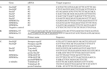
Small inhibitory RNAs (siRNAs) targeting specific schistosome genes were synthesized commercially by Integrated DNA Technologies (IDT, Coralville, IA) with the help of the on-line IDT RNAi Design Tool (https://www.idtdna.com/Scitools/Applications/RNAi/RNAi.aspx). Table 1A lists the siRNAs used in this work. The primers shown in Table 1B were used in a PCR with S. mansoni adult cDNA to generate a 943 bp fragment of the SPRM1hc coding DNA with flanking T7 and T3 polymerase promoter sites (underlined). This purified PCR fragment was then used to generate RNA by in vitro transcription with T7 and T3 RNA polymerase (Megascript kits, Ambion). Equimolar amounts of complimentary, single stranded RNAs were mixed and heated to 68°C for 15 min for annealing. A negative control, long dsRNA was generated in a similar manner using a 990 bp PCR fragment derived from a yeast expression vector, as previously described (Skelly et al. Reference Skelly, Da'dara and Harn2003).
List of siRNAs, primers and probes. A. siRNA designations and sequences. SmAQP, S. mansoni aquaporin; SmAP, S. mansoni alkaline phosphatase; SPRM1hc, S. mansoni permease heavy chain. B. Oligonucleotide sequences of primers used to generate an SPRM1hc long double stranded RNA in vitro. The underlined sequences are promoters for T7 (top) and T3 (bottom) RNA polymerase. C. Gene specific primer sets and reporter probes labeled with 6-carboxyfluorescein (FAM) used in quantitative real time PCR TaqMan assays

To deliver the siRNAs, parasites (1000 schistosomula or 8 adults/group), in 100 μl electroporation buffer (Ambion, TX), or RPMI medium (as indicated), containing 2·5 μg siRNA (unless otherwise specified), or 20 μg long dsRNA, were electroporated in a 4 mm cuvette by applying a square wave with a single 20 ms impulse, at 125 V and at room temperature, as described (Correnti et al. Reference Correnti, Brindley and Pearce2005). Parasites were then transferred to 1 ml complete RPMI. After overnight culture, medium was replaced with 2 ml of fresh complete RPMI culture medium. Suppression was monitored by quantitative real-time PCR (qRT-PCR) 2 days after siRNA treatment.
Gene expression analysis
Gene expression was monitored by qRT-PCR. RNA was first extracted from the parasites using the Trizol method (Invitrogen, CA) following the manufacturer's instructions. Any residual DNA remaining in the RNA preparations was removed by DNase digestion using a TurboDNAse kit (Ambion, TX). cDNA was synthesized using 1 μg RNA and an oligo (dT)20 primer and Superscript reverse transcriptase (Invitrogen, CA). Quantitative real-time PCR was performed using custom TaqMan Assays with primer sets and reporter probes labeled with 6-carboxyfluorescein (FAM), obtained from Applied Biosystems (Foster City, CA). Table 1C lists the primers & probes used in this work.
Each real-time TaqMan PCR was performed using cDNA equivalent to 10 ng total parasite RNA according to the manufacturer's universal conditions PCR protocol, in a final volume of 25 μl. All samples were run in triplicate and underwent 45 amplification cycles on a 7500 ABI PRISM® Sequence Detection System Instrument. For relative quantification, the ΔΔCt method was employed, using alpha tubulin as the endogenous standard for each sample. Results obtained from parasites treated with irrelevant dsRNA were used for calibration (Livak and Schmittgen, Reference Livak and Schmittgen2001). For graphical representation, the ΔΔCt values were normalized to controls and expressed as percent difference (Livak and Schmittgen, Reference Livak and Schmittgen2001).
RESULTS
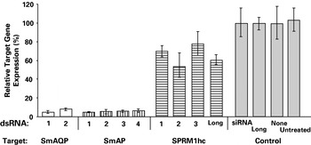
Fig. 1 shows the expression level of 3 different target genes (encoding an S. mansoni aquaporin (SmAQP), alkaline phosphatase (SmAP) and an amino acid permease heavy chain (SPRM1hc)) following RNAi treatment of schistosomula using electroporation (Faghiri and Skelly, Reference Faghiri and Skelly2009; Krautz-Peterson et al. Reference Krautz-Peterson, Camargo, Huggel, Verrey, Shoemaker and Skelly2007a; Ndegwa et al. Reference Ndegwa, Krautz-Peterson and Skelly2007). SmAQP was targeted with 2 distinct siRNAs, SmAP was targeted with 4 distinct siRNAs and SPRM1hc was targeted with 3 distinct siRNAs (Fig. 1). Sequences of each siRNA are given in Table 1. The levels of suppression achieved with each siRNA for its specific target are comparable. Both siRNAs targeting SmAQP suppress the expression of this gene >90%. Likewise all 4 of the siRNAs targeting SmAP yield >90% gene suppression (Fig. 1). However, none of the 3 different siRNAs targeting SPRM1hc (here used at a four fold higher concentration than the siRNAs targeting SmAQP and SmAP) gives rise to more than 50% suppression of this target gene expression. Delivering long dsRNA (at the relatively high dose of 20 μg) targeting SPRM1hc, does not improve on the level of suppression achieved using siRNAs. The controls (Fig. 1, grey) include parasites treated with an irrelevant siRNA (10 μg, set at 100% in Fig. 1), the irrelevant long dsRNA (20 μg) or no dsRNA. The final control (Fig. 1, far right) represents parasites that were not exposed to dsRNA and were not electroporated. These data demonstrate that not all schistosome genes can be suppressed to the same extent, using the methods employed here.
Relative expression of 3 S. mansoni target genes, SmAQP (aquaporin), SmAP (alkaline phosphatase) and SPRM1hc (amino acid permease, heavy chain), following RNAi treatment of schistosomula using electroporation, as determined by quantitative real-time PCR. Numbers refer to different siRNAs targeting each gene, delivered at 2·5 μg (for SmAQP and SmAP) and at 10 μg (for SPRM1hc). ‘Long’ refers to long dsRNA, delivered at 20 μg. The controls (grey bars) include parasites treated with an irrelevant siRNA (10 μg, set at 100%), an irrelevant long dsRNA or no dsRNA. The final control (far right) represents parasites that were not exposed to dsRNA and were not electroporated. Sequences of all siRNAs are shown in Table 1.

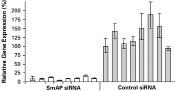
Fig. 2 examines the variation in the level of suppression achieved in individual adult female parasites following RNAi treatment using SmAP siRNA #1 by electroporation. The level of expression of the SmAP gene in 8 individual adult female worms treated with control siRNA (Fig. 2, grey bars) is compared with SmAP gene expression in 8 parasites treated with SmAP siRNA (Fig. 2, white bars). It is clear that there is a large variation in relative SmAP gene expression (as much as two fold) between different individual control worms. While there are likewise differences in the levels of gene suppression achieved in individual worms treated with SmAP siRNA, it is clear that all SmAP siRNA treated worms exhibit robust (>80%) gene suppression.
Relative expression of the SmAP gene in 8 individual adult female parasites treated via electroporation with SmAP siRNA (white) versus 8 individual parasites treated with an irrelevant control siRNA (grey). The value achieved by control individual 1 is arbitrarily set at 100%.

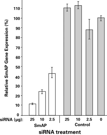
Fig. 3 shows the level of suppression of the SmAP gene that is achieved after adult female parasites are soaked in siRNA. Greater suppression is achieved following exposure to higher levels of siRNA and a clear dose response is recorded. However the level of suppression achieved is lower than that seen when siRNAs are delivered by electroporation; soaking parasites in 2·5 μg siRNA leads to ∼60% SmAP gene suppression (Fig. 3), whereas electroporating the parasites with this amount routinely yields >80% suppression (Fig. 2).
Relative expression of the SmAP gene in parasites treated with different concentrations of SmAP siRNA by soaking (white) versus controls (grey).

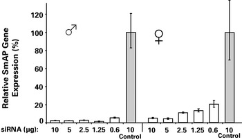
Male/female parasite pairs were electroporated with increasing amounts of siRNA while the parasites were in copula. Fig. 4 shows the level of SmAP gene suppression achieved in separated male and female parasites 8 days later. It is clear that robust suppression (>80%) is seen in all cases. However, at lower doses of siRNA (e.g. 1·25 and 0·6 μg) the level of gene suppression, while still substantial, declines in females.
Relative expression of the SmAP gene in male (left) and female (right) parasites treated while in copula with different concentrations of SmAP siRNA by electroporation (white) versus controls (grey).

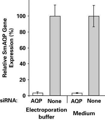
Fig. 5 compares the level of SmAQP gene suppression achieved following schistosomula electroporation with siRNA either in electroporation buffer (Fig. 5, left) or in RPMI medium (Fig. 5, right). Comparable levels of suppression (both ∼95%) are seen using both approaches.
Relative expression of the SmAQP gene in untreated control parasites (grey) and parasites treated with SmAQP siRNA (white) by electroporation in commercial electroporation buffer (left) versus in RPMI medium.

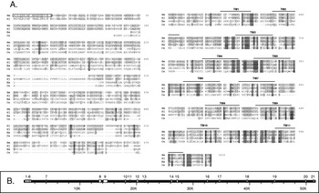
An examination of the predicted S. mansoni protein database at S. mansoni genedb (http://www.genedb.org/genedb/smansoni/) for homologues of the C. elegans RNA channel SID-1 revealed a match coded on sequence Smp_152020. This sequence potentially encodes a 1018 amino acid protein, here designated SmSID-1, with a predicted size of 115 556 and pI of 8·3. The SmSID-1 sequence is shown in Fig. 6A along with an alignment of this protein with diverse members of this protein family generated using ClustalW. Conserved motifs are shaded. SmSID-1 exhibits high sequence similarity (81%) with its counterpart from S. japonicum (Genbank accession number BAH22347), moderate similarity (40%) with a human homologue (NP_060169) and lower similarity (19%) with C. elegans SID-1 (AF478687) (Fig. 6A). Amino acids 1–21 are predicted to constitute a signal peptide (1MIRLLFAIAFAVSCVIFPTYS21) at http://www.cbs.dtu.dk/services/SignalP-3.0/ and this is boxed in Fig. 6A. In common with the other members of this protein family, SmSID-1 is predicted to contain 11 transmembrane domains (TM1-11, Fig. 6A) and to possess a large (here >600 amino acid) extracellular N-terminal domain.
Alignment of SmSID-1 with other members of the SID protein family. Sm, Schistosoma mansoni; Sj, Schistosoma japonicum (GenBank accession number BAH22347); Hs, Homo sapiens (NP_060169); Ce, C. elegans SID-1 (AF478687). Predicted transmembrane domains are indicated, TM1-11, and a predicted signal sequence is boxed. Shading highlights similarity between family members. B. The S. mansoni SID-1 (SmSID-1) gene. Exons 1 to 21 are indicated by white boxes ‘K’ indicates kilobase pairs.

The entire SmSID-1 gene was identified on contig Smp_scaff000150 at S. mansoni genedb. As illustrated in Fig. 6B, the gene is >50 kb and consists of 21 exons that range in size from 690 bp (exon 9) to 44 bp (exon 13). Introns range in size from 9284 bp (intron 7) to 43 bp (intron 3). All introns possess canonical GT:AG splice donor and acceptor sites.
DISCUSSION
In this work, we show that all genes are not susceptible to RNAi to the same degree in S. mansoni. Techniques that lead to the efficient suppression of some target genes (here SmAQP and SmAP) lead to very modest suppression of a third target – the SPRM1hc gene. The inability to suppress efficiently the SPRM1hc gene is not likely due to its expression in cell types that are restrictive for RNAi; SPRM1hc is widely expressed in intravascular schistosomes and this is the case for the highly suppressible SmAP too (Dusanic, Reference Dusanic1959; Halton, Reference Halton1967; Krautz-Peterson et al. Reference Krautz-Peterson, Camargo, Huggel, Verrey, Shoemaker and Skelly2007a). The lower susceptibility of SPRM1hc to RNAi may be due to the secondary structure of its mRNA. Perhaps this mRNA is less accessible to the RISC than are other targets. To ensure that the 3 siRNAs originally designed to target SPRM1hc did not derive from particularly inaccessible regions, a 943 bp long dsRNA was generated and parasites were electroporated at a high dose (20 μg) with this RNA. This long dsRNA should be processed by SmDicer to generate an array of siRNAs. The expectation was that some of these siRNAs would be able to reach the SPRM1hc mRNA to effect suppression but this was not observed. The result with SPRM1hc is unusual. In published and unpublished work, it is the only gene target that is recalcitrant to robust RNAi, of 12 targets tested so far in this laboratory (Faghiri and Skelly, Reference Faghiri and Skelly2009; Krautz-Peterson and Skelly, Reference Krautz-Peterson and Skelly2008b; Ndegwa et al. Reference Ndegwa, Krautz-Peterson and Skelly2007). Note that very high levels of gene suppression may not be essential for a phenotype to emerge. Depending on the importance of each gene product, a modest diminution in expression of an important gene may be detrimental for the parasites and may be informative. For instance, suppressing the gene that encodes the schistosome TGF-β homologue SmInAct leads to a modest ∼40% suppression at the RNA level, nevertheless eggs produced by SmInAct dsRNA–treated female parasites fail to develop (Freitas et al. Reference Freitas, Jung and Pearce2007).
Following exposure of a schistosomula population to dsRNA, the outcome for each parasite is not uniform and this is likely due to differences in dsRNA uptake and/or RNAi pathway activation and/or target gene expression in different individuals. For instance, following cathepsin B gene suppression in a population of schistosomula, the level of cathepsin B protein present in individuals within the population was assessed after 6 days by immunostaining using anti-cathepsin B antibodies. While most parasites (63%) lacked detectable protein, a sizable percentage (37%) stained clearly and some of these (5%) very brightly (Skelly et al. Reference Skelly, Da'dara and Harn2003). To monitor individual variability directly in gene suppression achieved following dsRNA exposure, the level of SmAP gene expression was assessed in each of eight adult worms that had been subjected, as a group, to RNAi. This work reveals some variability (up to 2 fold) in normal SmAP gene expression in control parasites as well as a remarkably robust suppression of SmAP gene expression (by 80–95%) in each worm tested. This high level of gene suppression in each individual is encouraging and may lead to a more uniform and informative phenotypic manifestation following gene suppression in adult parasites.
In an effort to simplify the RNAi protocol and minimize excessive handling of the parasites, which can damage them, parasite pairs were not separated before treatment. Instead, worms in copula were subjected to RNAi by electroporation. Eight days later the level of gene suppression achieved was assessed in now separated males and females. It is clear that both genders are highly susceptible to RNAi following this protocol and that separation of females from their male partners is not essential for efficient gene suppression. However, at the lower doses of siRNA used, females do display some diminution in the effect suggesting that at these levels their residence within the gynaecophoric canal of their male partners lessens their exposure to sufficient siRNA for maximal gene suppression.
It has been reported that dsRNA can be delivered to schistosomes by electroporation in RPMI culture medium (Morales et al. Reference Morales, Rinaldi, Gobert, Kines, Tort and Brindley2008). In work described here, the efficiency of gene suppression was compared when siRNA was delivered by electroporation to parasites in RPMI medium versus in commercial electroporation buffer (from Ambion Inc). Very high and comparable levels of suppression of the SmAQP target gene were observed in both cases. The use of medium makes each experiment more economical given its considerably lower cost versus electroporation buffer. It remains possible that differences in efficiency, following the use of medium versus electroporation buffer, may be revealed at lower concentrations of dsRNA or for different gene targets.
The preferred protocol for RNAi in schistosomes that is followed in this laboratory involves treating parasites with relatively low doses (2·5 μg) of gene-specific siRNA by electroporation. This routinely leads to robust target gene suppression. In work described here, the level of suppression achieved by soaking parasites in siRNA was assessed. In a Drosophila cell line cultured in medium containing dsRNAs, it was reported that longer dsRNAs are preferentially taken up versus siRNAs (Saleh et al. Reference Saleh, Van Rij, Hekele, Gillis, Foley, O'farrell and Andino2006). Here we show that schistosomes can take up siRNAs in a dose-dependent manner. However, the level of suppression achieved is substantially lower than that seen using electroporation. Approximately 55% suppression of SmAP gene expression is achieved after soaking parasites in 2·5 μg siRNA versus >80% following electroporation. Electroporation likely delivers dsRNA more efficiently into the body of the parasite. Since schistosomes can take up siRNA by soaking this suggests that the parasites in the blood stream will be amenable to RNAi if they are exposed to a sufficiently high dsRNA dose. It has previously been shown that gene expression in schistosomes can be efficiently suppressed by soaking the parasites in long dsRNA (Ndegwa et al. Reference Ndegwa, Krautz-Peterson and Skelly2007; Skelly et al. Reference Skelly, Da'dara and Harn2003). Long dsRNA cannot be used in vivo to suppress gene expression in intravascular parasites since nucleases in the blood are known to rapidly degrade any introduced long dsRNAs (Aagaard and Rossi, Reference Aagaard and Rossi2007). Additionally, long dsRNAs are rapid inducers of a systemic/nonspecific interferon response when delivered in vivo, resulting in cell apoptosis (Aigner, Reference Aigner2006).This implies that siRNAs will need to be used for use with blood-dwelling schistosomes and it was recently reported that injecting siRNAs into schistosome-infected mice could suppress gene expression in parasites in the blood (Pereira et al. Reference Pereira, Pascoal, Marchesini, Maia, Magalhaes, Zanotti-Magalhaes and Lopes-Cendes2008). Three siRNAs targeting the parasite hypoxanthine–guanine phosphoribosyltransferase (HGPRTase) gene were injected in phosphate buffered saline intravenously into schistosome-infected mice and 6 days later, parasites were recovered. The total number of parasites was reduced by ∼27% after treatment and the level of HGPRTase gene expression was significantly reduced in the HGPRTase siRNA-treated group versus controls (Pereira et al. Reference Pereira, Pascoal, Marchesini, Maia, Magalhaes, Zanotti-Magalhaes and Lopes-Cendes2008).
By scanning the available schistosome DNA sequence databases, a number of groups have identified homologues of proteins in schistosomes that are known to be involved in the RNAi pathway in other organisms (Gomes et al. Reference Gomes, Cabral, Jannotti-Passos, Carvalho, Rodrigues, Baba and Sa2009; Lendner et al. Reference Lendner, Doligalska, Lucius and Hartmann2008). This, coupled with our own work, has permitted the generation of a first draft of RNAi pathways in schistosomes that are represented in Fig. 7. The pathway engaged following the uptake of exogenous dsRNA is shown in Fig. 7A while that engaged by endogenous dsRNA is shown in Fig. 7B.
Proposed RNAi pathways in S. mansoni. A: Double stranded RNA (dsRNA) taken in via SmSID-1 (i) engages SmDicer/Partner Dicer (ii) and is cleaved into short inhibitory RNAs (siRNAs). These engage the RNA induced silencing complex (RISC) containing an Argonaute (SmAgo) and other proteins (Tudor-SN & Fmr-1) (iii). A cognate mRNA is identified and cleaved to effect gene silencing (iv). B: Pri-miRNAs are processed in the nucleus by SmDrosha/Partner Drosha (i) and the resultant pre-miRNAs are exported to the cytoplasm via Exportin-5. Here they are acted upon by Dicer/Partner Dicer (ii) to generate miRNAs which enter the RISC (iii). This interaction generally results in translational repression (iv).

Exogenous dsRNA that is taken up (Fig. 7A (i)) engages SmDicer and an RNA-binding protein (Partner Dicer) (Fig. 7A (ii)). This interaction leads to dsRNA cleavage and siRNA generation. The siRNA duplex is unwound and one strand is preferentially loaded into the short interfering RNA induced silencing complex (siRISC) (Fig. 7A (iii)). The minimal RISC, sufficient for target RNA recognition and cleavage is simply an Argonaute protein bound to a small RNA (Rivas et al. Reference Rivas, Tolia, Song, Aragon, Liu, Hannon and Joshua-Tor2005). However Argonaute proteins can have several associated binding partners (Pratt and Macrae, Reference Pratt and Macrae2009). Here we postulate that in a schistosome RISC, Argonaute (SmAgo) associates with homologues of the RNA binding protein Fmr1 and the nuclease Tudor-SN (Fig. 7A (iii)); the S. mansoni genome is reported to contain homologues of these proteins and they exist in other invertebrate RISCs (Gomes et al. Reference Gomes, Cabral, Jannotti-Passos, Carvalho, Rodrigues, Baba and Sa2009; Lendner et al. Reference Lendner, Doligalska, Lucius and Hartmann2008). The RISC, now loaded with a single stranded RNA (called the guide RNA), searches the transcriptome to find potential target mRNAs. Targets are engaged by Watson-Crick base pairing which directs the Argonaute endonuclease (SmAgo) to cleave them (Fig. 7A (iv)) (Siomi and Siomi, Reference Siomi and Siomi2009). Many rounds of mRNA cleavage result in substantial suppression of target gene expression and effectively results in gene silencing.
In the case of the microRNA pathway (Fig. 7B), first, primary precursor miRNA transcripts (pri-miRNAs), generated in the nucleus, are cleaved by the RNaseIII SmDrosha (in association with the RNA binding protein Partner Drosha) (Fig. 7B (i)). These pri-miRNAs, containing stem-loop structures depicted in Fig. 7B, harbour the mature miRNAs in the 5′ or 3′ half of the stem. The Drosha-cleaved product is generally 60–70 nucleotides and is called a precursor miRNA (pre-miRNA). This pre-miRNA is then exported, via the carrier protein Exportin 5, to the cytoplasm. Here the SmDicer/Partner Dicer complex may engage it and cleave it to generate a mature miRNA (Fig. 7B (ii)). Next, one strand of the miRNA is loaded into the microRNA induced silencing complex (miRISC) whose composition may be similar, as depicted in Fig. 7B (iii), or different from the postulated siRISC. In other systems different proteins, including distinct Argonautes, comprise different RISCs (Okamura et al. Reference Okamura, Ishizuka, Siomi and Siomi2004) and this may be the case for schistosomes too since miRNA engagement often results in gene suppression via translational repression (Fig. 7B (iv)) as opposed to target mRNA cleavage. In the S. mansoni genome 4 Argonaute homologues have been identified (Gomes et al. Reference Gomes, Cabral, Jannotti-Passos, Carvalho, Rodrigues, Baba and Sa2009) and it is possible that different forms occupy distinct RISCs, or perform other functions within the cell. It is noteworthy that of the 4 Argonaute proteins in humans only Ago2 is a catalytically active slicer (Liu et al. Reference Liu, Carmell, Rivas, Marsden, Thomson, Song, Hammond, Joshua-Tor and Hannon2004).
One factor determining the efficiency of RNAi is the ability of a cell to directly take up dsRNA. Recalcitrance to RNAi can perhaps be overcome by electroporation. In the case of schistosomes, it is clear that electroporation with gene-specific dsRNA leads to efficient target gene suppression in most cases. It has been demonstrated here, and previously, that simply soaking schistosomes in dsRNA can also lead to efficient gene suppression (Krautz-Peterson et al. Reference Krautz-Peterson, Radwanska, Ndegwa, Shoemaker and Skelly2007b; Skelly et al. Reference Skelly, Da'dara and Harn2003). How dsRNA enters the soaked parasites remains unclear. In C. elegans the SID-1 (systemic RNAi defective-1) protein has been shown to be required for uptake of dsRNA (Feinberg and Hunter, Reference Feinberg and Hunter2003; Winston et al. Reference Winston, Molodowitch and Hunter2002). In this work we describe the identification in silico of a schistosome homologue of this C. elegans protein. The schistosome protein, SmSID-1, has a close homologue in S. japonicum and both predicted schistosome proteins are substantially larger when compared to the SID-1 proteins of other organisms. All members of this family have 11 predicted transmembrane domains toward the carboxyl end. The distance from the initiator methionine to the first of these transmembrane domains is >600 amino acids in the case of the schistosomes. This amounts to >200 amino acids more than for the remaining homologues. Since both the C. elegans and human SID-1 proteins have been shown to be capable of effecting dsRNA uptake (Duxbury et al. Reference Duxbury, Ashley and Whang2005; Shih et al. Reference Shih, Fitzgerald, Sutherlin and Hunter2009), it is reasonable to assume that SmSID-1 likewise acts as a channel to import dsRNA into schistosomes and is depicted as such in Fig 7A (i). It is noteworthy that many schistosome tissues are syncytial (Morris, Reference Morris1968; Silk et al. Reference Silk, Spence and Gear1969; Spence and Silk, Reference Spence and Silk1970, Reference Spence and Silk1971) so that, once dsRNA has entered a tissue, it may be able to traverse relatively large distances internally without the need to cross additional plasma membranes.
ACKNOWLEDGEMENTS
Schistosome-infected snails were provided by the Biomedical Research Institute through NIH-NIAID Contract N01-AI-30026.
FINANCIAL SUPPORT
This work was supported by grant AI-056273 from the National Institutes of Health-National Institute of Allergy & Infectious Diseases.