Introduction
Many plants and plant products used as food in tropical countries contain cyanogenic glycosides (Oke, Reference Oke1979; Osuntokun, Reference Osuntokun1981; Jones, Reference Jones1998). On hydrolysis, these glycosides yield cyanide, a sugar and a ketone or aldehyde. Cyanide is a potent cytotoxic agent that kills the cell by inhibiting cytochrome c oxidase in the fourth complex of the mitochondrial electron transport chain.
Cassava is a known cyanogenic plant containing the cyanoglucosides linamarin (α-hydroxyisobutyronitrile-β-d-glucose) in large amounts and lostaustralin (α-hydroxyl-α-methylbutyronitrile-β-d-glucose) in a smaller amount in all parts of the plant (De Bruijn, Reference De Bruijn, Nestel and MacIntyre1973). Linamarin is degraded in cassava by the action of the enzymes linamarase (linamarin β-glucosidase glucohydrolase, EC 3.2.1.21) and hydroxynitrile lyase (EC 4.1.2.37) that hydrolyse linamarin to cyanohydrins and then to cyanide in a process known as cyanogenesis (Butler et al., Reference Butler, Barley and Kennedy1965; Nartey, Reference Nartey1968; Cooke, Reference Cooke1978). Chewing insects induce cyanogenesis during feeding and are thereby exposed to the toxic effects of the released cyanide (Conn, Reference Conn, Rosenthal and Janzen1979; Lindroth, Reference Lindroth and Bernays1991). When ingested, cyanide activates the body's own mechanisms of detoxification, resulting in the transformation of cyanide into a less toxic compound, thiocyanate (Ballantyne, Reference Ballantyne, Ballantyne and Marrs1987). Detoxification of cyanide so produced in both plants and chewing insects has been attributed to the presence of a group of enzymes known as sulphur transferases (Roy and Trudinger, Reference Roy and Trudinger1970; Westley, Reference Westley1973; Anosike and Ugochukwu, Reference Anosike and Ugochukwu1981), which are a group of enzymes widely distributed in plants, bacteria and animals that catalyses the transfer of a sulphur atom from a donor molecule to a thiophilic acceptor substrate (Westley, Reference Westley1973, Reference Westley and Jacoby1980). Examples of sulphur transferases are rhodanese, 3-mercaptopyruvate synthase and thiosulphate thiol transferase (Roy and Trudinger, Reference Roy and Trudinger1970; Westley, Reference Westley1973). However, sulphur transferases have other primary functions (Nagahara et al., Reference Nagahara, Ito and Minami1999; Al-Qarawi et al., Reference Al-Qarawi, Mousa and Ali2001). Some of the metabolic functions carried out by these enzymes are transfer of sulphur from thiosulphate to a nucleophilic acceptor such as reduced lipoate (Volini et al., Reference Volini, DeToma and Westley1967; Tomati et al., Reference Tomati, Giovanni, Anni, Dupres and Cannella1976), reactivation of ferredoxin from apoferridoxin (Tomati et al., Reference Tomati, Giovanni, Anni, Dupres and Cannella1976) and supply of sulphide centre for the electron transport chain in mammalian tissues (Tanka and Gatai, Reference Tanka and Gatai1983).
The variegated grasshopper Zonocerus variegatus (L.) (Orthoptera: Pyrgomorphidae) is a brightly coloured grasshopper found from the West Coast of Africa to the north-east and south-west of the continent (Youdeowei, Reference Youdeowei1974). They feed mainly on leaves and stems of cassava, Manihot spp. (Euphorbiaceae), for their survival and development. Grasshoppers also attack a wide range of crops mainly in the seedling stage. The survival of this insect on cassava plants is hinged on the possession of an enzymatic detoxification process based on sulphur transferase. It has been shown in our laboratory that cyanoalanine synthase is present in the gut of the variegated grasshopper (Ogunlabi and Agboola, Reference Ogunlabi and Agboola2007). In this study, we report the presence of rhodanese and its properties in the gut of Z. variegatus.
Materials and methods
Materials
Potassium cyanide, sodium thiosulphate, boric acid, sodium borate, formaldehyde, glycine, ferric nitrite, nitric acid, citric acid, sodium citrate, ammonium sulphate (enzyme grade), disodium hydrogen phosphate, monosodium hydrogen phosphate, potassium chloride, sodium chloride and ɛ-amino-n-caproic acid were obtained from BDH Chemicals Ltd (Poole, UK). Glycerol, sodium acetate, ethylenediaminetetraacetic acid, Coomassie Brilliant Blue, Blue Dextran and bovine serum albumin (BSA) were obtained from Sigma (St Louis, USA). CM-Sephadex C-25, DEAE-Sephadex A-50 and Sephadex G-75 were obtained from Pharmacia Fine Chemical (Uppsala, Sweden). All other analytical reagents were obtained from either Sigma-Aldrich or BDH Chemicals Ltd.
Methods
Enzyme and protein assays
Rhodanese activity was measured routinely according to the method of Lee et al. (Reference Lee, Hwang, Lee and Cho1995). The reaction mixture contained 50 mm borate buffer (pH 9), 0.25 mm KCN and 0.25 mm Na2S2O3 in a 1 ml cuvette. Sorbo reagent was used as the colour developer according to the method of Sorbo (Reference Sorbo1951, Reference Sorbo1953a, Reference Sorbob). Absorbance was then read at 460 nm against a reagent blank containing no enzyme sample. One rhodanese unit (RU) was taken to be the amount of enzyme per min that under the given conditions produced an optical density reading of 1.08 at 460 nm (Sorbo, Reference Sorbo1951, Reference Sorbo1953a, b). Protein concentration was determined using the Bradford (1976) method using BSA as the standard.
Enzyme isolation
All experiments, including enzyme assay, were carried out at room temperature and all buffers used contained 10 mm sodium thiosulphate to stabilize the enzyme. The grasshoppers were handpicked from Babajakan Village, Orile Owu Road, Gbongan, Osun State, Nigeria in November 2008, and brought to the laboratory and kept frozen until required. The grasshoppers were dissected and the guts removed. Guts with a wet weight of 102 g (obtained from 500 grasshoppers) were homogenized in three volumes of 0.1 m acetate glycine buffer (pH 7.8) containing 10 mm sodium thiosulphate and 10 mm ɛ-amino-n-caproic acid (buffer A) for about 5 min in a Waring blender. The homogenate was then centrifuged at 15,000 rpm at 4 °C for 30 min using an IEC DPR 600 refrigerated centrifuge.
Ammonium sulphate fractionation
The supernatant obtained was brought to 70% ammonium sulphate saturation (472 g/l) by the addition of solid ammonium sulphate. This solution was kept over a period of 1 h with occasional stirring and then allowed to stand for 12 h before centrifugation at 15,000 rpm at 4 °C for 30 min. The precipitate was collected and stored in a minimum amount of the homogenization buffer.
CM-Sephadex ion-exchange chromatography
A CM-Sephadex C-25 cation exchanger was soaked and washed with 50 mm citrate buffer, pH 5.0 (buffer B). The precipitated enzyme was dialysed in an acetylated dialysis bag and layered on the column (2.5 × 40 cm). The column was then washed with 240 ml of buffer B to remove the unbound protein, followed by elution with a 200 ml linear gradient of 0–1 m KC1 in the same buffer. Fractions (5 ml) were collected from the column at a rate of 30 ml/h. Protein was monitored spectrophotometrically at 280 nm. The fractions were assayed for rhodanese activity. The active fractions were pooled and immediately dialysed against 50% glycerol in the elution buffer to store the enzyme.
DEAE-Sephadex ion-exchange chromatography
A DEAE-Sephadex A-50 anion exchanger was prepared by swelling 50 g in distilled water for 3 days. This was equilibrated in buffer B followed by the packing of the resin into a 2.5 × 40 cm column. Then, 20 ml of the previous sample were applied on the column. The column was then washed with 240 ml of buffer B to remove the unbound protein, followed by elution with a 200 ml linear gradient of 0–1.0 m KC1 in buffer B. Fractions (5 ml) were collected from the column at a rate of 32 ml/h. Protein was monitored at 280 nm. The fractions were also assayed for rhodanese activity. The active fractions were pooled and immediately dialysed against 50% glycerol to store the enzyme.
Gel filtration on Sephadex G-75
Sephadex G-75 resin was swollen in boiling distilled water for 2 h and then equilibrated with 10 mm sodium phosphate, pH 7.2 (buffer C). The sample obtained was layered on the column (2.5 × 70 cm). The column was then eluted with 380 ml of buffer C to remove the unbound protein. Fractions (5 ml) were collected from the column at a rate of 10 ml/h. The activities of enzyme and protein were routinely determined.
Agarose-Blue affinity chromatography
Agarose Blue resin was treated by adding 40 ml of pre-treated resin in buffer B. The resin was then packed into a 1.5 × 20 cm column and equilibrated with buffer B. Then, 10 ml of the previous sample were applied on the column. The column was then washed with 240 ml of buffer B to remove the unbound protein, followed by elution with a 200 ml linear gradient of 0–1 m KC1 in buffer B. Fractions (5 ml) were collected from the column at a rate of 42 ml/h. Protein was monitored at 280 nm and the fractions assayed for rhodanese activity. The active fractions were pooled and immediately dialysed against 50% glycerol (in buffer B) to store the enzyme.
Native and subunit molecular weight determinations
The native molecular weight of the enzyme was determined on a Sephadex G-75 column (2.5 × 90 cm). The standard proteins were 3 ml each of creatinine phosphatase (88,000; 3 mg/ml), BSA (66,000; 3 mg/ml), ovalbumin (45,000; 3 mg/ml) and chymotrypsinogen (25,000; 3 mg/ml). The column was eluted with 10 mm phosphate buffer (pH 7.2) at a flow rate of 8 ml/h. The void volume (V o) of the column was determined by the elution volume of Blue Dextran (2 mg/ml). The molecular weight of the enzyme was intrapolated from the standard calibration curve according to the method of Andrews (Reference Andrews1964). Sodium dodecyl sulphate polyacrylamide gel electrophoresis (SDS–PAGE) was carried out to determine the subunit molecular weight of the enzyme on a 10% acrylamide gel as described by Weber and Osborn (Reference Weber, Osborn, Neurath and Hill1975). Standard proteins were BSA (66,000 Da), ovalbumin (45,000 Da), pepsinogen (34,700 Da), trypsinogen (24,000 Da) and β-lactoglobulin (20,100 Da).
Determination of kinetic parameters
The kinetic parameters (K m and V max) of the enzyme were determined by varying the concentration of KCN between 10 and 100 mm at a fixed concentration of 50 mm Na2S2O3. In addition, the concentration of Na2S2O3 was varied between 20 and 200 mm at a fixed concentration of 200 mm KCN.
Effect of temperature
Rhodanese activity was assayed at temperatures between 0 and 70 °C to determine the optimum temperature of the enzyme. The assay mixture was incubated at the indicated temperature for 10 min prior to initiating a reaction by the addition of an aliquot of the enzyme that had been equilibrated at the same temperature. Residual activity was then assayed.
Effect of pH
The effect of pH on the activity of rhodanese from the gut of the variegated grasshopper was carried out according to the method of Agboola and Okonji (Reference Agboola and Okonji2004). The enzyme was assayed using the following buffers at the indicated pH values: 50 mm citrate buffer (pH 4.0–6.0), 10 mm phosphate buffer (pH 6.0–8.0) and 50 mm borate buffer (pH 9.0–9.5).
Effect of salts
The salts used were NH4C1, MgCl2, CoCl2, CaCl2, MnCl2, NiCl2, SnCl2, BaCl2 and ZnCl2 at final concentrations of 0.05 and 0.1 mm in the enzyme assay.
Results
Purification and homogeneity
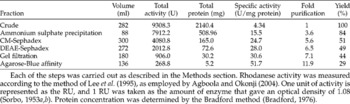
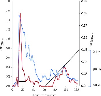
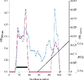
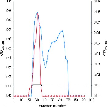
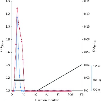
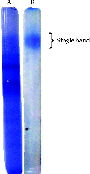
The summary of a typical purification procedure for rhodanese from the gut of the variegated grasshopper is shown in Table 1. The elution profile of the enzyme using ion-exchange chromatography on both CM-Sephadex C-25 and DEAE-Sephadex A-50 is shown in Figs 1 and 2, respectively. The elution profile of the enzyme using Sephadex G-75 and Agarose-Blue affinity chromatography is shown in Figs 3 and 4. The results of PAGE in the absence of SDS using the continuous phosphate buffer system are shown in Fig. 5.
Purification of rhodanese from the gut of the grasshopper Zonocerus variegatus

U = RU/min under the assay condition.
Each of the steps was carried out as described in the Methods section. Rhodanese activity was measured according to the method of Lee et al. (Reference Lee, Hwang, Lee and Cho1995), as employed by Agboola and Okonji (Reference Agboola and Okonji2004). One unit of activity is represented as the RU, and 1 RU was taken as the amount of enzyme that gave an optical density of 1.08 (Sorbo, Reference Sorbo1953a, b). Protein concentration was determined by the Bradford method (Bradford, 1976).
(colour online) Elution profile of rhodanese from the gut of the variegated grasshopper using ion-exchange chromatography on a CM-Sephadex C-25 column (2.5 × 40 cm). The column was first washed with 300 ml of buffer B and then eluted with another 300 ml linear salt gradient of 0–1 m KCl. Elution was carried out at a flow rate of 30 ml/h and 5 ml fractions were collected. One active activity peak was obtained.  , OD280nm;
 , OD460nm; ■, pooled fraction;
 , KCl gradient.

(colour online) Elution profile of rhodanese from the gut of the variegated grasshopper using ion-exchange chromatography on a DEAE-Sephadex A-50 column (2.5 × 40 cm). The column was first washed with 300 ml of buffer B and then eluted with another 300 ml linear salt gradient of 0–1 m KCl. Elution was carried out at a flow rate of 30 ml/h and 5 ml fractions were collected. One active activity peak was obtained. , OD280nm; , OD460nm; ■, pooled fraction; , KCl gradient.

(colour online) Elution profile of rhodanese from the gut of the variegated grasshopper using gel filtration chromatography on a Sephadex G-75 column (2.5 × 70 cm). The column was first washed with 460 ml of buffer C. Elution was carried out at a flow rate of 10 ml/h and 5 ml fractions were collected. One active activity peak was obtained. , OD280nm; , OD460nm; , pooled fraction.

(colour online) Elution profile of rhodanese from the gut of the variegated grasshopper using affinity chromatography on an Agarose Blue resin column (1.5 × 40 cm). The column was first washed with 300 ml of buffer B and then eluted with another 300 ml linear salt gradient of 0–1 m KCl. Elution was carried out at a flow rate of 60 ml/h and 5 ml fractions were collected. One active activity peak was obtained. , OD280nm; △, OD460nm; □, pooled fraction; , KCl gradient.

(colour online) PAGE of rhodanese from the gut of the variegated grasshopper. Electrophoresis was carried out in the absence of SDS on a 7.5% acrylamide gel, where A represents the crude enzyme and B is the purified enzyme.

Molecular weight determination
The native molecular weight of rhodanese from the gut of the variegated grasshopper was estimated to be 35,400 ± 482 Da using gel filtration on Sephadex G-75. The subunit molecular weight of the enzyme was found to be 33,000 ± 212 Da using SDS–PAGE on a 10% acrylamide gel.
Kinetic parameters of rhodanese
The K m value of KCN was found to be 22.63 ± 02.87, while that of Na2S2O3 was 29.41 ± 03.04 as estimated from the double-reciprocal plot by varying KCN concentrations at a fixed Na2S2O3 concentration and that of varying concentrations of Na2S2O3 at a fixed KCN concentration.
Effect of pH and temperature on rhodanese
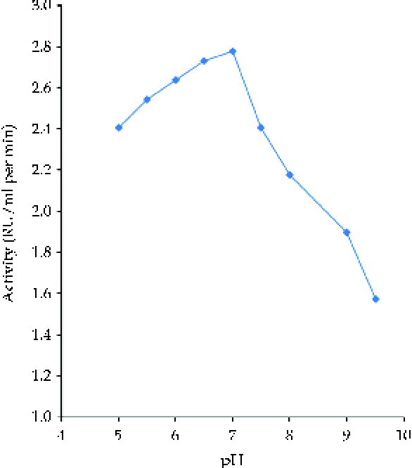
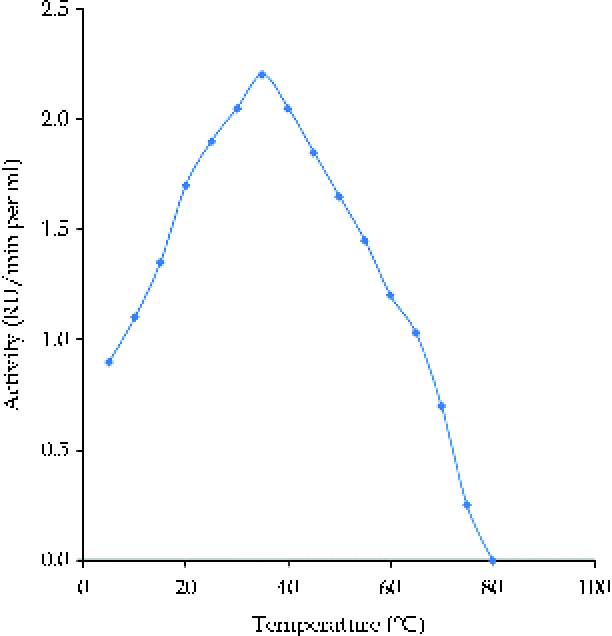
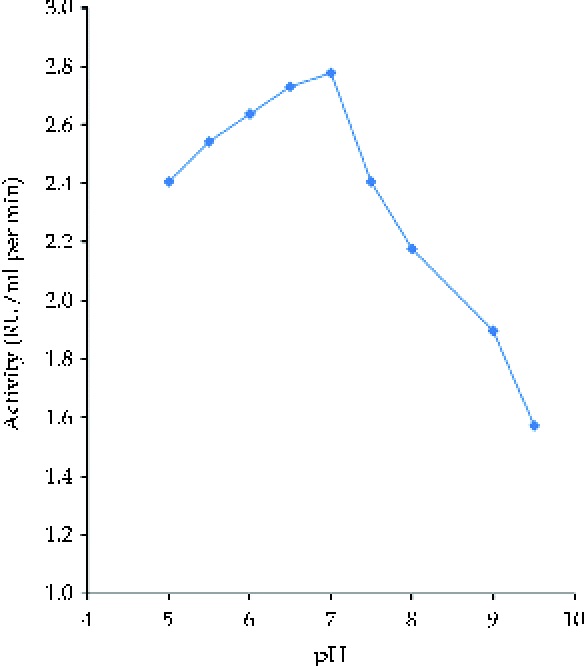
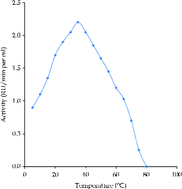
The influence of pH on the rate of rhodanese activity is shown in Fig. 6. The optimum pH of 7.0 was obtained for rhodanese. The effect of temperature on the activity of rhodanese from the gut of the variegated grasshopper is shown in Fig. 7. The optimum temperature of the enzyme was found to be 35°C.
(colour online) Effect of pH on rhodanese from the gut of the variegated grasshopper. The optimum pH was determined using 50 mm citrate buffer (pH 5.0–6.0), 50 mm phosphate buffer (pH 6.0–8.0) and 50 mm borate buffer (pH 9.0–9.5).
(colour online) Activity–temperature profile showing the optimum temperature of rhodanese from the gut of the variegated grasshopper. For the experimental procedures, see the Methods section. Percentage of residual activity is compared with the activity of the native enzyme at 0 °C.
Effect of salts on rhodanese
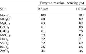
The results of the effect of various salts on the activity of rhodanese from the gut of the variegated grasshopper (Table 2) showed that CaCl2, CoCl2, MgCl2, MnCl2, NH4Cl, NiCl2 and SnCl2 had little or no effect on enzyme activity. However, BaCl2 and ZnCl2 inhibited the activity of the enzyme at concentrations of both 0.5 and 1.0 mm.
Effect of cations on the activity of rhodanese from the gut of the variegated grasshopper
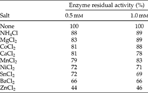
For the experimental procedures, see the Methods section. Percentage of residual activity is compared with the activity of the enzyme in the absence of metal ions.
Discussion
Several animals and insects are capable of feeding on or parasitize cyanogenic plants due to mechanisms involving detoxification of cyanide occurring in the organisms. Cyanide is a naturally occurring chemical present in many food items such as almonds, maize, apple seeds, millets, bamboo, mustard, beans, peas, elderberry, sorghum, cassava root, sugarcane, lemon, sweet potato, lima beans, wild cherry, lime and yam (Claus et al., Reference Claus, Tyler and Brady1970). The organs that are most susceptible to cyanide are the central nervous system and the heart. Cyanide is a potent cytotoxic agent that kills the cell by inhibiting cytochrome c oxidase in the mitochondrial electron transport chain (Warburg, Reference Warburg1911; Keilin, Reference Keilin1929). Hazards of cyanide to fish, wildlife, livestock and humans have been well documented. Rhodanese, or 3-mercaptopyruvate sulphur transferase, represents the chief detoxifying enzyme of cyanide (Lang, Reference Lang1933; Himwich and Saunders, Reference Himwich and Saunders1948; Sorbo, Reference Sorbo1951, Reference Sorbo1953a, b; Meister, Reference Meister1953; Westley, Reference Westley and Jacoby1980; Nagahara et al., Reference Nagahara, Ito and Minami1999). Other detoxifying enzymes of cyanide in the system include cyanoalanine synthase, thiosulphate reductase, cystathionase and albumin (enzyme-like behaviour in cyanide detoxification) (Lieske et al., Reference Lieske, Clark, Zoeffel, von Tersch, Lowe, Smith, Broomfield, Baskin and Maxwell1996).
The variegated grasshopper attacks a wide range of crops (Youdeowei, Reference Youdeowei1974; Modder, Reference Modder1994). It feeds on the plants, leaves and stems of cassava and may cause defoliation and debarked stems (Nicol et al., Reference Nicol, Assadsolimani, Langewald and Schmutterer1995). Cassava contains cyanogenic glucosides, mainly linamarin (92–98%), which release hydrogen cyanide after hydrolysis by an endogenous linamarase (Butler et al., Reference Butler, Barley and Kennedy1965; Conn, Reference Conn, Rosenthal and Janzen1979). Despite the high level of cyanide in cassava, no harmful effects have been observed on grasshoppers upon ingestion of this poisonous compound from their food. Since Z. variegatus feeds mainly on cassava leaves, it is therefore reasonable to assume that this insect possesses an efficient enzymatic mechanism to detoxify the ingested cyanide.
This study shows the presence of rhodanese in the gut of the variegated grasshopper. To the best of our knowledge, no previous effort has been made to purify and study rhodanese from the grasshopper. Rhodanese from the gut of Z. variegatus was purified to homogeneity by a combination of chromatographic procedures that involved ion-exchange chromatography on CM-Sephadex and DEAE-Sephadex, gel filtration on Sephadex G-75 and Agarose-Blue affinity chromatography. The pattern employed in the purification of the enzyme rhodanese from the gut of Z. variegatus is different from that followed by previous studies on the enzyme. Lee et al. (Reference Lee, Hwang, Lee and Cho1995) purified rhodanese from the mouse liver to homogeneity by ammonium sulphate precipitation, CM-Sephadex ion-exchange chromatography, hydroxyapatite chromatography and Sephacryl S-200 gel filtration chromatography. Rhodanese was isolated and purified from the cytosolic fraction of the liver tissue homogenate of the straw-coloured fruit bat Eidolon helvum Kerr (Pteropodidae), by using ammonium sulphate precipitation and CM-Sephadex ion-exchange chromatography (Agboola and Okonji, Reference Agboola and Okonji2004). Two forms of rhodanese were purified from the liver of the African sharptooth catfish Clarias gariepinus (Burchell) (Clariidae) by ion-exchange chromatography on a CM-Sepharose CL-6B column and gel filtration on a Sephadex G-75 column (Akinsiku et al., Reference Akinsiku, Agboola, Kuku and Afolayan2010). The inclusion of affinity gel seemed to be a novel approach, which was an effective method in eluting impurities bound to the column at a very high salt concentration (Fig. 4). The preparation was adjudged pure by the presence of a single protein band after electrophoresis in the presence and absence of SDS (Fig. 5).
The specific activity of the enzyme was found to be 51.7 RU/mg protein. Sorbo (Reference Sorbo1953a, b) and Himwich and Saunders (Reference Himwich and Saunders1948) obtained a specific activity of 256 and 131 RU/mg protein, respectively, for rhodanese from the bovine liver. The specific activity of purified rhodanese from the mouse liver was estimated to be 1070 RU/mg protein (Lee et al., Reference Lee, Hwang, Lee and Cho1995). Agboola and Okonji (Reference Agboola and Okonji2004) obtained a specific activity of 136.6 RU/mg protein for rhodanese from the fruit bat liver, while Akinsiku et al. (Reference Akinsiku, Agboola, Kuku and Afolayan2010) obtained a specific activity of 73 and 72 RU/mg protein for two isozymes, cRHD I and cRHD II, respectively, of rhodanese from the catfish liver.
The apparent molecular weight of 35,400 ± 482 Da was obtained for rhodanese, which agrees with reported molecular weight values estimated to be between 33,000 and 37,000 Da (Wang and Volini, Reference Wang and Volini1968; Blumenthal and Heinrikson, Reference Blumenthal and Heinrikson1971; Nagahara et al., Reference Nagahara, Ito and Minami1999). The following molecular weights have been obtained for rhodanese from various sources: bovine liver (37,000 Da; Sorbo, Reference Sorbo1953a, b, Reference Sorbo1955), human liver (37,000 Da; Jarabak and Westley, Reference Jarabak and Westley1978), mouse liver (34,800 Da; Lee et al., Reference Lee, Hwang, Lee and Cho1995) and rat liver (34,000 Da; Nagahara et al., Reference Nagahara, Okazaki and Nishino1995). The molecular weight obtained in this study is very close to that obtained by Lee et al. (Reference Lee, Hwang, Lee and Cho1995) and Agboola and Okonji (Reference Agboola and Okonji2004), which were 34,000 and 35,700 Da for rhodanese from the liver of mice and fruit bats, respectively. Akinsiku et al. (Reference Akinsiku, Agboola, Kuku and Afolayan2010) found an apparent molecular weight of 33,200 and 36,800 Da for cRHD I and cRHD II, respectively, in the catfish liver. The subunit molecular weight of 33,000 ± 212 Da is very close to that of 34,000 Da obtained by Lee et al. (Reference Lee, Hwang, Lee and Cho1995) for rhodanese from the mouse liver. This result suggests that the enzyme is a monomeric protein. Jarabak and Westley (Reference Jarabak and Westley1978) and Lee et al. (Reference Lee, Hwang, Lee and Cho1995) have shown that purified rhodanese from the liver of mice and humans are monomeric proteins. The molecular weight and the number of polypeptides in rhodanese have been subjects of controversy. Since Sorbo (Reference Sorbo1953a, b) initially reported a molecular weight of 37,000 Da for rhodanese, numerous investigators have suggested that the molecular weight of this enzyme was approximately 33,000–37,000 Da and a dimer composed of two identical subunits, M r 18,000–19,000 Da (Blumenthal and Heinrikson, Reference Blumenthal and Heinrikson1971; Volini et al., 1978). The dimer theory was later found to be incompatible with the results of three-dimensional structure analysis, and the well-characterized rhodanese from bovine liver mitochondria has been found to possess a single polypeptide chain of 32,000 Da composed of 293 amino acids (Russell et al., 1978; Ploegman et al., 1978). Since both forms of the enzyme from catfish were found to have the same molecular weight of about 35,000 Da, the probability that the two forms are monomeric and aggregate forms of one of the other does not arise (Blumenthal and Heinrikson, Reference Blumenthal and Heinrikson1971; Akinsiku et al., Reference Akinsiku, Agboola, Kuku and Afolayan2010).
The kinetic parameters of rhodanese from the gut of Z. variegatus when compared with the ones obtained from other mammalian sources (Akinsiku et al., Reference Akinsiku, Agboola, Kuku and Afolayan2010) were found to be close to those of rhodanese obtained from the liver of fruit bats and catfish (Agboola and Okonji, Reference Agboola and Okonji2004; Akinsiku et al., Reference Akinsiku, Agboola, Kuku and Afolayan2010). However, it was noted that the K m value of KCN from the gut of the variegated grasshopper is higher than that of KCN from the liver of the fruit bat. The K m value of Na2S2O3 from the gut of Z. variegatus is close to that of the enzyme from the liver of catfish and fruit bat.
Various optimum temperatures have been reported for rhodanese from different organisms. Himwich and Saunders (Reference Himwich and Saunders1948) obtained an optimum temperature between 38 and 40 °C for rhodanese from the bovine liver. A value as high as 50–59 °C was obtained by Sorbo (Reference Sorbo1953a, b) and Chew and Boey (Reference Chew and Boey1972) for rhodanese from the bovine liver and tapioca leaf, respectively. Ezzi et al. (Reference Ezzi, Pascual, Gould and Lynch2003) obtained a wide range of temperature optima of 35–55 °C for rhodanese from the fungus Trichoderma (Hypocreaceae). Agboola and Okonji (Reference Agboola and Okonji2004) reported an optimum temperature of 35 °C for rhodanese in the cytosolic fraction of the fruit bat liver. Furthermore, Akinsiku et al. (Reference Akinsiku, Agboola, Kuku and Afolayan2010) reported an optimum temperature of 40 °C for rhodanese from the liver of catfish from Lake Asejire in Nigeria. The optimum temperature obtained for rhodanese from Z. variegatus was 35 °C, which could be a result of temperature adaptation in their habitat. This possibility needs further investigation.
An optimum pH of 7.0 was obtained for rhodanese from the gut of Z. variegatus. Lang (Reference Lang1933) reported a pH of 8.3 for the enzyme from the rabbit liver. The optimum pH is in good agreement with that from the bovine liver, which is between 8.0 and 9.0 (Sorbo, Reference Sorbo1953a, b). Chew and Boey (Reference Chew and Boey1972) reported pH values between 10.2 and 11.0 for the enzyme from tapioca leaves, while Lee et al. (Reference Lee, Hwang, Lee and Cho1995) obtained a pH of 9.4 for the enzyme from the mouse liver. Agboola and Okonji (Reference Agboola and Okonji2004) reported an optimum pH of 9.0 for rhodanese from the fruit bat liver. Akinsiku et al. (Reference Akinsiku, Agboola, Kuku and Afolayan2010) reported an optimum pH of 6.5 for rhodanese from catfish.
It has been reported that metal ions such as Cu2+ and Fe2+ inhibit the activity of rhodanese significantly (Lang, Reference Lang1933). Metal ions showing enzyme inhibition are those that have a strong affinity for ligands such as phosphate, cysteinyl and histidyl side chains of protein (Stokinger, 1981). The results of enzyme inhibition with a number of chloride salts showed that Ca2+, Co2+, Mg2+, Mn2+,  $$NH _{4}^{ + } $$, Ni2+ and Sn2+ had little or no effect on the activity of the enzyme. However, both Ba2+ and Zn2+ inhibited the activity of the enzyme considerably. This result could be compared with that of rhodanese from the fruit bat liver, reporting the inhibition of enzyme activity by Ba2+ and Hg2+ (Agboola and Okonji, Reference Agboola and Okonji2004). It has been reported that the enzyme rhodanese from the liver of catfish was inhibited by Zn2+ and Ni2+ (Akinsiku et al., Reference Akinsiku, Agboola, Kuku and Afolayan2010).
The properties of rhodanese from the gut of Z. variegatus are similar to those of the enzyme from other animal sources. It is plausible to assume that the enzyme is involved in detoxification when feeding on cassava, a highly cyanogenic plant. Biophysical and kinetic properties of this enzyme, which will reveal how this ability can possibly be compromised by enzyme inhibition, may lead, in the long term, to the potential use of this enzyme for pest control or to the development of an antidote in case of cyanide poisoning.