Introduction
Diversity of bacteria and role of communities
The diversity and prevalence of bacteria is astonishing. Bacteria rapidly adapt to their extracellular conditions, which has made it possible for them to establish themselves in nearly all habitats in the biosphere, including humans. To survive in diverse and fluctuating environmental conditions, cells have evolved mechanisms of attaching to surfaces and forming communities, including biofilms.Reference Hall-Stoodley, Costerton and Stoodley1 Surface-associated communities protect bacteria from predators and the immune system, support the division of labor, provide a physical and structural barrier against mechanical and physical stimuli, and promote the conservation of the genotype.Reference Flemming and Wingender2 These communities may be persistent and difficult to remove once formed, and efforts to understand their mechanism of growth and homeostasis have broad applications that include biomedicine, dentistry, ecology, agriculture, and industrial processing.Reference Dunne3
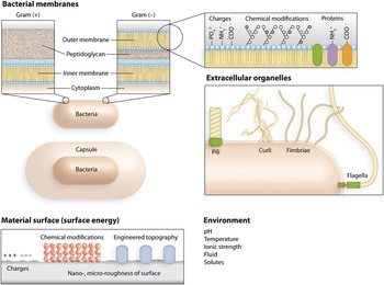
Some bacterial biofilms are beneficial to human health. The existence of some strains of bacteria that evolved to form biofilms that persist in specific human niches is important for establishing the diverse group of symbiotic bacteria that are referred to as the human microbiome. These bacteria shape human behavior, physiology, and development.Reference Neufeld and Foster4, Reference Turnbaugh, Ley, Hamady, Fraser-Liggett, Knight and Gordon5 Bacterial biofilms may also be detrimental to human health. The attachment of bacteria on biomedical devices that are in contact with, or within, the human body provides a starting point for the onset of clinical infections. Bacterial biofouling of surgical implants, catheters, and contact lenses interferes with the function of these systems and provides a mechanism for introducing pathogenic bacteria into the human body, which may lead to infections and disease.Reference Bryers6 The “race for the surface” was a phrase coined to describe a model for mammalian cells and bacteria competing to adhere to the surface of implantable biomedical devices.Reference Gristina7 The model describes the interplay between the substrate, species of bacteria, and fluid on the surface attachment of bacteria and their growth and development into a biofilm (Figure 1).
Parameters that influence the interactions between bacteria and surfaces. The cell wall of Gram-positive bacteria consists of an inner lipid membrane surrounded by a layer of cross-linked polysaccharide referred to as the peptidoglycan. The cell wall of Gram-negative bacteria consists of an inner lipid membrane surrounded by a layer of peptidoglycan, which is surrounded by an outer lipid membrane. Outer membrane proteins and lipopolysaccharide provide surface charge. Some bacteria have a capsule that extends beyond the cell wall and consists of a thick layer of alginate and other complex polysaccharides. Extracellular organelles for attachment and motility include pili, curli, fimbriae, and flagella. The surface of substrates has intrinsic charge from functional groups that are solvent exposed. The composition of the surrounding environment also influences the interactions during bacterial cell attachment.

The process of biofilm formation is characterized by five stages. (1) Cells attach to surfaces reversibly. In this step, bacteria use a variety of extracellular organelles and proteins for sensing and attaching to surfaces, including flagella, pili, fimbriae, curli fibers, and outer membrane proteinsReference Bullitt and Makowski8, Reference Thomas, Nilsson, Forero, Sokurenko and Vogel9 (Figure 1). Cells attach to substrates that are immersed in, or are in contact with, fluids containing electrolytes and macromolecules (e.g., DNA, proteins, and humic acids, which are formed by the degradation of biomolecules). These soluble components adsorb on surfaces and screen the intrinsic physical and chemical properties of materials. There are similarities between bacteria adhering to these “preconditioned” surfaces and the attachment and spreading of mammalian cells on substrates that are remodeled by the adsorption of matrix proteins secreted by cells.Reference Loeb and Neihof10, Reference Lutolf and Hubbell11 (2) Cells attach to surfaces irreversibly. The secretion of an extracellular polymeric substance (EPS) that consists of DNA, proteins, lipids, and lipopolysaccharides facilitates adhesion between cells and surfaces.Reference Flemming and Wingender2 (3) Cells adsorbed on surfaces replicate and grow into microcolonies, which are named for their physical dimensions of tens or hundreds of microns in diameter. These bacteria secrete EPS and become encapsulated in a layer of the hydrogel, which forms a physical barrier between the community and the extracellular environment. The composition of EPS varies between species and growth conditions, and chemical communication between cells in the community stimulates its formation and secretion.Reference Borlee, Goldman, Murakami, Samudrala, Wozniak and Parsek12 Quorum sensing (QS) is the best-characterized example of chemical communication in bacteria. QS is a central process in biofilm formation and a mechanism that cells use to query their extracellular environment (see the Shrout et al. article in this issue). QS modulates a variety of cellular functions, including pathogenesis, nutrient acquisition, conjugation, motility, and secondary metabolite production.Reference Harmsen, Yang, Pamp and Tolker-Nielsen13 (4) The community grows into a three-dimensional structure and matures into a biofilm as cells replicate and the EPS accumulates. Cells in an established biofilm are “glued” together by the EPS, which resists mechanical stresses and detachment of the community from the surface of the substrate. (5) Some cells detach from regions of the biofilm and disperse into the bulk fluid, where they may adsorb on surfaces and form biofilms in new environmental niches.Reference Hall-Stoodley, Costerton and Stoodley1, Reference Costerton, Lewandowski, Caldwell, Korber and Lappin-Scott14 This step is important for propagation and self-renewal of the community.
Many physical, chemical, and biological interactions facilitate the attachment of bacteria to surfaces. Specific (e.g., receptor ligand binding) and non-specific interactions (e.g., hydrophobicity) participate in cell attachment. Dissecting the molecular, mechanical, and topographical factors that contribute to attachment and adhesion is complicated, as these factors may vary with bacterial strains and extracellular conditions, including the immediate environment around the substrate and conditions for cell growth (e.g., temperature, carbon source, fluid flow, and the composition of nutrient media and growth factors). The manipulation of individual environmental factors to prevent biofilm formation has been met with limited success. Control over surface chemistry has been used to reduce cell attachment, including the development of “dynamic” surfaces that degrade or reorganize in response to temperature and other environmental conditions and shed adsorbed bacteria into the bulk fluid.Reference Ista, Mendez, Balamurugan Sreelatha, Balamurugan, Rama Rao Venkata and Lopez Gabriel15 Surface structuring has also been explored by engineering high-aspect ratio topographical features that decrease substrate wettability and render surfaces superhydrophobic. This structural characteristic is conceptually reminiscent of a lotus leaf.Reference Bhushan, Jung and Koch16, Reference Guo, Liu and Su17 Chemically modified polymer coatings also reduce cell adsorption.Reference Bruellhoff, Fiedler, Moller, Groll and Brenner18, Reference Lewis and Klibanov19 However, these strategies do not eliminate the attachment of bacteria or prevent the formation of biofilms. Surfaces that can successfully prevent bacterial adsorption and biofilm formation over time scales longer than several days are just beginning to emerge after many years of research.Reference Zhang, Lalani and Liu20 These efforts support the view of biofilms as ineluctable structures.
How do chemical and physical cues affect cell attachment and biofilm formation? Insight into these mechanisms will provide clues for creating successful antifouling surfaces. To facilitate the design of new materials, we review the role of the physical, chemical, and structural properties of surfaces on biofilm formation. We discuss how the physicochemical properties of substrates affect the adhesion of cells to surfaces and influence biofilm growth and development. The complex milieu in which bacteria are suspended influences the properties of surfaces and transforms bacterial resistant surfaces into substrates for attachment, growth, and biofilm formation. The development of biofilm-resistant materials will likely require integrated approaches combining chemical, mechanical, and topographical elements into the design of surfaces and interfaces. This challenge is ideally suited to the expertise of material scientists and engineers. Multidisciplinary research on surface design and engineering may have a deep impact on both fundamental and applied microbiological science and technology.
Physical properties of surfaces
Physical interactions
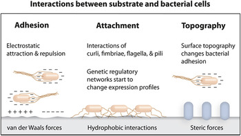
This section discusses electrostatic interactions and surface energy on bacterial adhesion to surfaces. The interactions between the bacterial cell wall and surfaces (including other cell walls) are primarily influenced by interfacial electrostatic (e.g., repulsion, attraction) and van der Waals forcesReference McClaine and Ford21, Reference Vigeant, Ford, Wagner and Tamm22 (Figure 2). However, many different non-specific interactions and interfacial forces influence cell attachment, including hydration forces, hydrophobic interactions, and steric forces.Reference Israelachvili23 Hydrophobic (e.g., low surface energy) and electrostatic interactions (e.g., charge) are among the best studied of these phenomena. Properties of substrates that influence adsorption, adhesion, and diffusion regulate the physiology of bacteria and their growth into biofilms include stiffness, mechanical stability, elasticity, and topography. In response to surface properties, cells secrete DNA, proteins, lipids, and lipopolysaccharide that accumulate and form the EPS, influence the stiffness and elasticity of biofilms, and pose a challenge to eradicating these communities.Reference Flemming and Wingender2 Many studies have concluded that individual physical properties of a surface, such as those mentioned previously, may have a dramatic impact on bacterial attachment. For example, the adhesion of Staphylococcus epidermidis is correlated with the stiffness of the polymer substrate.Reference Derjaguin and Landau24 However, a detailed understanding of the mechanisms underlying cell/surface interactions is not known and makes it difficult to gauge the relative importance of each physical property on cell attachment. This limitation is, in part, a consequence of the techniques and capabilities that are available for studying these interactions. The state of the art for studying bacterial adhesion still relies on the theoretical framework developed for studying colloidal systems, such as DLVO theory (named after Derjaguin, Landau, Verwey, and Overbeek).Reference Lichter, Thompson, Delgadillo, Nishikawa, Rubner and Van Vliet25, Reference Verwey and Overbeek26 This model was developed to study “hard” particles that are non-deformable, but its application to studying bacteria has limitations. Recent modifications to DLVO theory, including the mathematical treatment of bacteria as “soft” particles, improves the accuracy of simulated interactions between cells and interfaces.Reference Ohshima27
The initial attachment of bacteria to substrate surfaces is characterized by electrostatic repulsion or attraction. Once this obstacle is overcome, hydrophobic interactions influence the attachment of bacteria to surfaces. This binding event initiates the genetic regulation and expression and secretion of chemical factors such as quorum sensing molecules to induce biofilm formation and expression of extracellular polymeric substance. The design of specific substrate topography can influence the initial attachment of bacterial cells and regulate biofilm formation.

Electrostatic interactions
Electrostatic forces are among the earliest interactions that influence the attachment of bacterial cells to surfaces (Figure 2). Most bacterial genera have a net negative charge as determined by zeta-potential measurements.Reference Soni, Balasubramanian, Beskok and Pillai28, Reference Katsikogianni and Missirlis29 Bacteria attach rapidly and tightly to positively charged surfaces, and electrostatic repulsion destabilizes cell contact with negatively charged surfaces. Destabilizing interactions between cells and anionic surfaces during the initial stages of attachment can be overcome by extracellular organelles that promote adhesion, including fimbriae, flagella, curli, and pili (Figure 1).Reference Bullitt and Makowski8 The charge discrimination of surfaces disappears in high ionic strength liquids. The layer of the bacteria cell wall that is in contact with the extracellular environment is complex and exposes many different functional groups that may interact with substrates (Figure 1). These functional groups include carboxylate, hydroxyl, phosphate, and amine moieties.Reference Hong and Brown30 In their native environments, bacterial cells are not in contact with “naked” surfaces. Diffusion and mass transport influence the adsorption of small molecules, ions, and proteins on surfaces and alter surface chemistry and charge. The layer of adsorbed molecules screens the intrinsic surface charge and promotes the adsorption of bacteria and their growth into biofilms.
Low-energy surfaces
After overcoming electrostatic repulsions, the preferential alignment of hydrophobic functional groups on surfaces and hydrophobic moieties on the bacteria cell wall, and extracellular organelles, stabilizes interfacial interactions (Figure 2). The preference of different aquatic bacteria attaching to hydrophobic, low-energy surfaces demonstrates this phenomenon.Reference Pringle and Fletcher31 The authors studied bacterial attachment in nutrient-free media to avoid nutrients remodeling the surface and concluded that physical interactions between cell surfaces and substrate were responsible for attachment. This research uncovered a recurring theme in the description of cell-surface interactions: the physical interactions between hydrophobic surfaces and flagella, fimbriae, and pili facilitate the attachment of bacteria to non-polar, low-energy substrates.Reference Thomas, Nilsson, Forero, Sokurenko and Vogel9, Reference Pringle and Fletcher31 During the initial approach and attachment, bacteria experience short-range repulsions in close proximity to negatively charged surfaces (Figure 2). The displacement of water molecules near surfaces enhances hydrophobic interactions and promotes close contact between cells and surfaces.Reference Norde32
High-energy surfaces
Thermodynamic predictions of surface energies can explain the behavior of bacteria during adhesion.Reference Absolom, Lamberti, Policova, Zingg, van Oss and Neumann33 Adhesion of bacteria to hydrophilic surfaces is enhanced if the surface tension of the bacterial cell wall is higher than the surface tension of the surrounding liquid.Reference Absolom, Lamberti, Policova, Zingg, van Oss and Neumann33 Fluorinated materials exhibit large contact angles that are characteristic of low energy surfaces. The oxidation of fluorinated surfaces revealed that the initial hydrophilic properties of a substrate reduces the initial attachment of bacteria onto surfaces.Reference Davidson and Lowe34 Unfortunately, a complication of engineered surfaces in real-life applications is that materials are exposed to environments that present solutes that adsorb at the interface. Consequently, the preliminary effects of surface energy on attachment disappear. The initial hydrophilic property therefore does not guarantee resistance to bacterial attachment. As already mentioned, many bacteria attach preferentially to hydrophobic surfaces, but others, including the human-associated bacterium Staphylococcus epidermidis, prefer polar, hydrophilic substrates.Reference Ista, Fan, Baca and Lopez35
The influence of surface energy on bacterial attachment is still not completely understood, and its extrapolation into a general principle and design rule for engineering and preventing adhesion is unrealized. A recent study concluded that hydrophobic interactions may not be responsible for the attachment of bacteria to surfaces and the formation of biofilms.Reference Heistad, Scott, Skaarer, Seidu, Hanssen and Stenstrom36 The initial adhesion of bacteria to hydrophobic surfaces and the maturation in biofilms is reduced compared to other commonly used surfaces in industrial applications (e.g., steel, glass, polypropylene).Reference Hyde, Alberg and Smith37 The ability of bacteria to adhere to both hydrophilic and hydrophobic substrates suggests a strategy for biofilm formation (and survival) in diverse environmental conditions.
Topographic properties of surfaces
The interaction of bacteria with surface topography requires an understanding of their physical dimensions. Cells of most strains of bacteria are typically 1 micrometer in diameter; by comparison, mammalian cells are typically larger than ten micrometers. Surface roughness has an effect on bacterial attachment. Nano- and microscale surface roughness enhances the adhesion of bacteria to substrates during the initial steps of colonization as it provides more surface area for cell attachment. Surface roughness reduces the shear force on bacterial cells and communities positioned in flowing liquids. This characteristic is particularly relevant to biofilms, as these structures frequently form in environments in which fluids are flowing, often at high flow rates (e.g., water pipes in industrial plants).
Engineering surface roughness and topography
We refer to roughness as an intrinsic property of surfaces and topography as a user-defined characteristic of a surface (Figure 1). There are many techniques available for creating nanopatterned topography, including photolithography, electron beam lithography, soft lithography, dip pen nanolithography, wet chemical etching, self-assembly, and Langmuir-Blodgett deposition. Techniques in this area have been reviewed recently.Reference Anselme, Davidson, Popa, Giazzon, Liley and Ploux38 Of these techniques, soft lithography has become one of the most widely used methods for creating defined surface topography because the techniques are straightforward and inexpensive.Reference Xia and Whitesides39
Surface roughness
Titanium is a commonly used biomaterial. A study on the effect of titanium surfaces on the attachment of bacteria demonstrated that roughness on the nanometer scale—and not micrometer scale—increases the attachment of bacteria.Reference Truong, Rundell, Lapovok, Estrin, Wang, Berndt, Barnes, Fluke, Crawford and Ivanova40 The authors compared all of the physical and chemical variables of their measurements (e.g., cell surface charge, surface energy, and surface zeta potential) and concluded that topography is the most influential factor on bacterial adhesion, and other interfacial parameters had little or no influence in their study. Nanoscale topography can change the physicochemical properties of materials, including the surface energy. The chemical etching of poly(vinyl chloride) (PVC) to introduce nanoscale roughness changed the surface energy of the polymer and reduced the initial attachment of bacterial strains.Reference Machado, Cheng, Tarquinio and Webster41 It has been suggested that there may be an optimal feature size—on the microscale—that decreases the attachment of bacteria to surfaces.Reference Anselme, Davidson, Popa, Giazzon, Liley and Ploux38 However, it is unlikely that there is a “one-size-fits-all” relationship between roughness and attachment as bacterial strains—even within the same species—can vary significantly in size and shape.Reference Young42 To complicate matters, many bacteria sense and respond to surfaces using mechanisms that remain uncharacterized. Some bacteria become morphologically differentiated in contact with surfaces. For example, Escherichia coli and Proteus mirabilis elongate into filaments, increase the surface density of flagella, and increase their flexibility and adhesion potential on rough surfaces.Reference Copeland and Weibel43
Topography
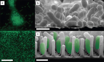
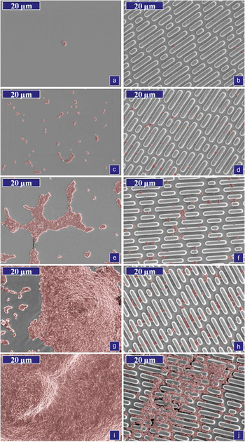
Surface roughness and topography influence the adhesion of mammalian cells.Reference Hoffman-Kim, Mitchel and Bellamkonda44 This effect involves the spreading of cells into the features on the surface. Bacteria are stiffer than mammalian cells and do not typically deform to accommodate the topographical constraints of surfaces. The observation that biofilm forming cells are stiffer than their planktonic counterpart supports the hypothesis that the mechanism by which topography affects bacterial attachment and growth into biofilms is different from mammalian cells.Reference Volle, Ferguson, Aidala, Spain and Nunez45 Engineering surface topography is a bona-fide strategy for influencing the adhesion of bacterial cells. A recent paper demonstrates that the pattern of adhered bacteria is affected by surface topography.Reference Hochbaum and Aizenberg46 The study found that bacterial cells became aligned normal to an epoxy surface etched with high aspect ratio structures that formed a nanopillar array (Figure 3). The spacing between the polymer posts was 1.2–1.5 μm, and flagella and pili had no influence on the pattern of adhesion. The authors concluded that maximizing surface contact influenced the alignment of cells. One promising approach is the creation of the micropatterned surface Sharklet AF in the silicone elastomer, poly(dimethylsiloxane) (PDMS) that reduces the biofilm colonization of the human pathogen Staphylococcus aureus compared to smooth PDMS surfaces (Figure 4).Reference Chung, Schumacher, Sampson, Burne, Antonelli and Brennan47 The same group later reported the patterning of surfaces with nanoforce gradients that enabled the regulation of the attachment of the zoospores of Ulva linza.Reference Schumacher, Long, Callow, Finlay, Callow and Brennan48 Future research will demonstrate whether the concept of nanoforce gradients can also be applied to reduce or enhance the attachment of bacteria and the formation of biofilms.
Comparison of P. aeruginosa adhesion on topographically patterned and non-patterned surfaces. (a) The image shows the adhesion of cells to a flat region of an epoxy substrate (top left) and to a topographically patterned epoxy substrate (bottom left). (b–c) Cross-sectional scanning electron microscopy images of cells cultured on flat and topographically patterned epoxy surfaces, respectively, showing the difference in attachment morphology. Scale bars are 10 μm in (a) and 1 μm in (b) and (c). Reproduced with permission from Reference 46. ©2010, American Chemical Society.

Representative scanning electron microscopy images of Staphylococcus aureus on polydimethylsiloxane (PDMS) surfaces over the course of 21 days (areas of bacteria highlighted with color to enhance contrast). On the left are smooth PDMS surfaces, and the right column shows Sharklet AF PDMS surfaces. (a) and (b) Day 0, (c) and (d) Day 2, (e) and (f) Day 7, (g) and (h) Day 14, and (i) and (j) Day 21. The patterned surface decreases the number of attached cells significantly. Reproduced with permission from Reference 47. ©2007, AVS Science & Technology of Materials, Interfaces, and Processing.

A combination of chemical and topographical surface modifications may reduce the surface attachment of cells significantly and the formation of biofilms. The fine-tuning of topological constraints and chemical characteristics of nanopatterned surfaces can be combined in a way that may lead to the development of non-fouling surfaces.Reference Anselme, Davidson, Popa, Giazzon, Liley and Ploux38, Reference Campoccia, Montanaro, Agheli, Sutherland, Pirini, Donati and Arciola49
Chemical properties of surfaces
The chemical modification of surfaces presents an important strategy for regulating the attachment of bacteria on substrates and their growth into biofilms. A general approach for controlling cell attachment entails controlling the surface chemistry of the substrate. General strategies for the design of substrate surface chemistry include covalent modification, non-covalent modification, controlled release of small molecules, and degradation of polymeric surfaces (also see the Khoo et al. article in this issue).Reference Qiu, Zhang, An and Wen50 These strategies have been successfully used to control bacterial attachment; several examples are discussed later.
An example of the influence of surface chemistry on the attachment of bacteria explored the polymer poly(N-isopropylacrylamide) (PNIPAAm).Reference Ista, Mendez, Balamurugan Sreelatha, Balamurugan, Rama Rao Venkata and Lopez Gabriel15, Reference Ista, Perez-Luna and Lopez51, Reference Ista, Mendez and Lopez52 The temperature-responsive switching of PNIPAAm changes the surface energy of the polymer and thus modulates the adsorption of cells and the attachment of biofilms. Cycling the polymer through different temperatures makes it possible to shed EPS and cells accumulating on the surface. Grafting polymer coatings on surfaces can reduce attachment and affect biofilm organization. Several examples of polymer coatings that have been used to modify the interfacial interaction of bacteria with surfaces include dextrans,Reference Bosker, Patzsch, Stuart and Norde53 poly(ethylene oxide) (PEO),Reference Patel, Ebert, Ward and Anderson54 poly(ethylenimine) (PEI),Reference Haldar, An, Alvarez de Cienfuegos, Chen and Klibanov55, Reference Wong, Li, Veselinovic, Kim, Klibanov and Hammond56 and poly(sulfobetaine methacrylate)(pSBMA).Reference Cheng, Xue, Zhang, Chen and Jiang57
Several research groups have tested the influence of the chemical composition of defined substrate surfaces on bacterial attachment and biofilm formation using a range of techniques spanning from grafted polymers to self-assembled monolayers (SAMs) displaying a diverse selection of functional groups.Reference Ista, Fan, Baca and Lopez35, Reference Wong, Li, Veselinovic, Kim, Klibanov and Hammond56, Reference Burton, Simon, Hou, Ren and Luk58 Modified surface chemistry influences the initial attachment of bacteria to substrates but may not completely inhibit cell adsorption and biofilm formation. Bacteria adsorbed on surfaces secrete EPS, which triggers the cohesion between cells and the adhesion of biofilms to surfaces. The affinity of the secreted EPS to the surface determines the maturation of the biofilm.Reference Flemming and Wingender2, Reference Rodrigues and Elimelech59 The surface properties may further influence general transport processes (adsorption/desorption rates), such as diffusion between bacteria and within a biofilm community.Reference Carnes, Lopez, Donegan, Cheung, Gresham, Timmins and Brinker60, Reference Hornemann, Lysova, Codd, Seymour, Busse, Stewart and Brown61 For more discussion on the topic of diffusion and its effect on community formation, see the last section in the review.
Self-assembled monolayers as a model system for biofilm research
SAMs are a particularly useful class of materials for fabricating surfaces with homogeneous or heterogenous chemical properties and studying interfacial interactions with bacteria.Reference Love, Estroff, Kriebel, Nuzzo and Whitesides62 SAMs make it possible to control the functional groups presented to cells and the surface density of ligands. SAMs can be prepared reproducibly and are a class of materials that have been characterized in detail.Reference Ulman63 This approach provides control over surface energy and charge density. The attachment of bacteria to SAMs presenting gradients of hydrophilic (hydroxyl-terminated) and hydrophobic (methyl-terminated) groups has been used to study bacterial attachment.Reference Burton, Simon, Hou, Ren and Luk58 The authors found that these SAMs were excellent substrates for bacterial attachment. In contrast, cell attachment and biofilm formation on SAMs terminated with monosaccharides, and PEO was reduced;Reference Hou, Burton, Wu, Luk and Ren64 the effects of these functional groups have also been demonstrated through their incorporation into polymeric materials.Reference Ista, Fan, Baca and Lopez35, Reference Buck, Breitbach, Belgrade, Blackwell and Lynn65 The mechanism underlying the biophysical properties of these surfaces is unknown and likely to involve the regulation of the structure of solvent molecules at the interfaces.Reference Israelachvili23, Reference de Gennes66 One setback of using SAMs is the timescale over which they are stable, which limits experiments to several weeks before thiol desorption occurs and surface defects form.Reference Jiang, Bruzewicz, Thant and Whitesides67 In real-life applications, surface stability would ideally be months or years.
Antimicrobial and bactericidal surfaces
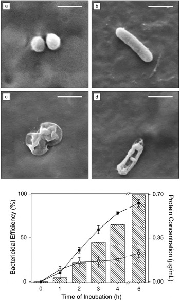
Hydrophilic, PEO coatings inhibit protein adsorption and repel bacterial adhesion. These properties are attributed to the steric repulsion of proteins and cells at interfaces where water molecules are coordinated to the PEO chains.Reference Currie, Norde and Cohen Stuart68 Grafting PEO coatings on surfaces provides a route to prevent the irreversible attachment of bacteria, which sets the stage for biofilm formation.Reference Adout, Kang, Asatekin, Mayes and Elimelech69 Another mechanism that has been applied to disrupting biofilm formation is to covalently attach bactericidal molecules to the surface of a substrate. The display of quaternary ammonium groups formed from N,N’-disubstituted PEI polymers or N-substitute polyvinylpyridine polymers have been described as antimicrobial coatings for surfacesReference Lewis and Klibanov19, Reference Haldar, An, Alvarez de Cienfuegos, Chen and Klibanov55, Reference Wong, Li, Veselinovic, Kim, Klibanov and Hammond56, Reference Hsu, Ouyang, Wong, Hammond and Klibanov70 (Figure 5). The flexibility of the chains makes it possible for the positively charged polymer to interact with the bacterial membrane and outer cell wall, which is lethal to E. coli and S. aureus. Cells that attach to substrates presenting these polymers are lysed. Although the quaternary ammonium groups have been described as penetrating into the cell, it seems more reasonable that these functional groups reorganize the membrane/cell wall in a manner similar to antimicrobial peptides.Reference Yang, Gordon, Trinkle, Schmidt, Davis, DeVries, Som, Cronan, Tew and Wong71, Reference Yang, Gordon, Mishra, Som, Purdy, Davis, Tew and Wong72
(a–d) Scanning electron microscopy (SEM) images of S. aureus and E. coli K12 in contact with bare silicon wafers (a and b, respectively) and on silicon wafers coated with N,N’-dodecyl-methyl-PEI (c and d, respectively); PEI, poly(ethylenimine). The scale bars are 1 μm. (e) A plot depicting the effect of the N,N’-dodecyl-methyl-PEI coating on the viability of E. coli K12 and on the concentration of intracellular proteins released into solution via cell lysis. The shaded bars represent bactericidal efficiencies; error bars were omitted for clarity. Total protein in solution after incubation with plain (empty square) and N,N’-dodecyl-methyl-PEI-coated (filled square) polypropylene tubes are shown with lines. Reproduced with permission from Reference 70. ©2011, Springer.

This class of surface functional groups is generally toxic to cells—including eukaryotic cells—and limits its application in biomedical devices and implants. The use of quaternary ammonium presenting surfaces in areas in which there is a high risk of bacterial contamination (e.g., surfaces in hospitals) has advantages over conventional methods of sterilization. However, the activity of these surfaces over time is not well understood. It is unclear how the effectiveness of these surfaces changes as cells lyse and their intracellular components adsorb on the surface of the substrate and shield the cationic interface. One solution is to combine a bactericidal surface with a stimuli responsive polymer that can be tuned to respond to external stimuli such as pH or temperature. Surface coating based on pSBMA combine antimicrobial and antifouling characteristics.Reference Cheng, Xue, Zhang, Chen and Jiang57 pSBMA coatings are bactericidal and kill cells upon contact—the polymer hydrolyzes slowly into a non-fouling, zwitterionic form that has excellent resistance to bacterial adhesion. 4-nitro-pyridine-N-oxide is also used to prevent cell attachment. This compound is an inhibitor of QS in Pseudomonas aeruginosa and adsorbs to substrate and cell surfaces and reduces attractive electrostatic forces by decreasing the surface potential.Reference Vanoyan, Walker, Gillor and Herzberg73
Other chemical factors
The initial attachment of microorganisms to surfaces strongly depends on the “conditioning layer.”Reference Loeb and Neihof10 As described earlier, this material consists of an adsorbed layer of molecules (e.g., proteins, sugars, fatty acids, lipids, and nucleic acids) that has a structure that varies with the composition of the nutrients and growth factors, and the environmental conditions (e.g., temperature, pH). For example, the layer adsorbed on an implanted medical device primarily contains proteins, nucleic acids, and salts.Reference Bryers6 In contrast, the stringent environmental conditions of desalination plants produce preconditioning layers that are devoid of proteins. Humic acids are a primary component of the conditioning layer in nutrient-poor environments.Reference Siboni, Lidor, Kramarsky-Winter and Kushmaro74 Divalent cationic ions (e.g., Mg2+, Ca2+) may enhance the attachment of bacteria to surface by reducing electrostatic repulsion and stabilizing interactions between the negatively charged surface of bacteria and anionic substrates.
The design and fabrication of materials for the controlled release of antimicrobial small molecules and secondary metabolites provides another route to controlling cell attachment and biofilm formation. The general principles behind the design of these materials can capitalize on the extensive studies of the controlled drug release field and can be incorporated into biomaterials and the surface chemistry of implanted devices.Reference Hetrick and Schoenfisch75, Reference Breitbach, Broderick, Jewell, Gunasekaran, Lin, Lynn and Blackwell76
Diffusion/signaling
Cells adsorbed on surfaces secrete EPS, QS molecules, and secondary metabolites that regulate cell physiology and behavior, including growth, motility, biofilm formation, and pathogenicity.Reference Flemming and Wingender2, Reference Bassler and Losick77 The implementation of materials-based techniques for confining and manipulating the diffusion of secondary metabolites has been used to study chemical communication, engineer syntrophic communities of bacteria, and to design and construct bacterial communities, including biofilms, with new functions.Reference Carnes, Lopez, Donegan, Cheung, Gresham, Timmins and Brinker60, Reference Kim, Du and Ismagilov78–Reference Flickinger, Copeland, Downes, Braasch, Tuson, Eun and Weibel81 Control over the spatial confinement of cells makes it possible to understand how the diffusion of nutrients, metabolic waste, and soluble small molecules that regulate chemical communication participate in biofilm formation, growth, homeostasis, and replication.Reference Boedicker, Vincent and Ismagilov82, Reference Vincent, Liu, Haney and Ismagilov83 This area at the interface of microbiology and materials science and engineering is fascinating and is developing rapidly.Reference Weibel, Diluzio and Whitesides84
Conclusion
In this review, we have summarized the physicochemical factors that govern the initial attachment and adhesion of bacteria to surfaces, which is the first step in biofilm formation. The general rule-of-thumb is that bacteria will preferentially colonize surfaces that are hydrophobic, have surface roughness on the nano- and microscale, and are exposed to a conditioning layer in contrast to smooth, hydrophilic surfaces. This trend is not absolute for all bacteria; however, it provides a general design principle for developing bacteria-resistant surfaces. A key challenge in this area is the prevention of the formation of a conditioning layer that passivates the exposed surface chemistry and provides a site of attachment for bacteria. Thus, a critical parameter to consider in surface design is the composition of solutes in the liquid in contact with surfaces.
Bacteria adapt to environmental changes using extracellular organelles that improve their chances of survival. As mentioned earlier, cells use these structures to sense their extracellular environment. A model organism in this area of research has been Vibrio parahaemolyticus.Reference McCarter and Silverman85, Reference Pruss, Besemann, Denton and Wolfe86 As most bacteria have an outer cell wall organization that is different from V. parahaemolyticus, it is likely that these organisms use different mechanisms for extracellular sensing. The characterization of these mechanisms and the stimuli that they respond to may guide the development of new materials for controlling bacterial attachment and biofouling.
The design of materials for studying bacteria at interfaces may uncover the mechanisms that regulate biofilm formation and provide insight into fundamental areas of microbiology. These studies will almost certainly guide and advance the design of surfaces for controlling attachment and biofilm formation. There are several unmet challenges in this area. One limitation is that substrates designed to control bacterial attachment may have synergistic and antagonistic effects on one strain of bacteria that may not work for other strains. Considering the remarkable diversity of bacteria in the biosphere, it is difficult to imagine a universal set of guidelines for designing materials. Another limitation is that for studies on biofilms in which these communities are reproduced with their native structure and in their habitat, it will be necessary to create multispecies biofilms. However, the vast majority of bacteria in the biosphere have not yet been cultivated in the lab, presumably because the physicochemical requirements for their growth are unknown. Materials science may have an important impact on this area of microbiology. Another limitation is that the design of substrate surfaces has to accommodate the variety of shapes and sizes of bacteria, which will respond differently to the physical characteristics of surfaces.Reference Young42 Finally, the tallest hurdle may be how to engineer a surface and keep it “clean.” The milieu of solutes in liquids and the significant biomass secreted and shed by bacteria pose a unique challenge to preserving the properties of surfaces designed to regulate attachment and biofilm formation. Engineering surface properties for studying and controlling bacterial biofilms may be difficult. However, the fundamental science and applied technology that emerges in this area of materials science and engineering certainly will be exciting and will open new doors in microbiology.
Acknowledgments
Our research in this area of microbiology is supported by DARPA, USDA (WIS00974), DuPont, 3M Searle Scholars Award (DBW), Deutsche Forschungsgemeinschaft (LDR), and the National Science Foundation under Grant No. DMR-0520527.