Introduction
In this review, we aim to describe the pathophysiology of biofilm communities and their role in otorhinolaryngological disease, with reference to the published literature.
We performed a review of the relevant literature, using Medline and the search term ‘biofilms’ combined with ‘otorhinolaryngology’, and also various related keywords such as ‘tonsil’ and ‘adenoid’.
Biofilms
Biofilms are microbial communities which attach to biological or non-biological surfaces and produce their own matrix. Most bacteria have the ability to form biofilms and to exist in this state (i.e. sessile) rather than in a free-floating state (i.e. planktonic).Reference Dunne1 Since the 1970s, it has been recognised that biofilms are the most prevalent microbial form in biological environments.Reference Costerton, Geesey and Cheng2 Their importance in human disease was first described during research into dental plaque, largely because teeth are accessible and have a non-shedding surface.Reference Wilson3
Scanning confocal laser microscopy has revealed that, within biofilm communities, there are cell towers with pores and water channels for nutrient and waste exchange.Reference Dunne1 The shape of the colony is dictated by the shear forces it experiences, with colonies varying from mushroom shaped to tadpole shaped.Reference Donlan and Costerton4 Colony cells have different roles; their cell specialisation is achieved by differential gene expression which alters the phenotype of individual microbes.Reference Wimpenny, Manz and Szewzyk5 Phenotypic changes occur in response to intercellular signalling or physicochemical gradients within the biofilm. The microbes secrete a matrix of polysaccharides, nucleic acids and extracellular polymeric substances, termed a glycocalyx;Reference Post, Stoodley, Hall-Stoodley and Erlich6 this has a slimy consistency and is predominantly water.Reference Wilson3 The organisms have varying growth rates, and a proportion will be in a dormant phase at any particular time.
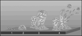
Four phases of biofilm development are recognised (Figure 1).
Phases of biofilm development.

In phase one, cells attach to a mucosal surface and become sessile. The surface may be reached randomly or in response to chemotactic, aerotactic or phototactic stimuli.Reference Wilson3 This phase is reversible;Reference Dunne1 attachment only occurs if the balance between attracting and repulsing forces is correct. These include hydrophobic, electrostatic, stearic and environmental forces (an example of the latter being flow across surfaces). In general, adhesion to abiotic surfaces is mediated by hydrophobic forces, whereas attachment to biological surfaces is controlled through specific molecular ligands such as lectin or adhesions.Reference Dunne1
In phase two, following adherence, the microbe phenotype is altered, colonisation occurs and extracellular matrix is produced.Reference Wilson3 Ongoing adherence is not limited to a single species (the initialising species).Reference Dunne1
In phase three, cells multiply and aggregate into communities and mature into complex, three dimensional, mushroom-shaped colonies. Eventually, colonies reach an equilibrium at which size limits their growth and they become static.Reference Dunne1 Within biofilms, cells are subject to varying conditions and different oxygen tensions; consequently, anaerobic and aerobic microbes can thrive within the same biofilm.Reference Post, Stoodley, Hall-Stoodley and Erlich6
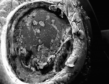
In phase four the final stage is reached, with detachment and dispersal of microbes, either singly or in aggregates (i.e. planktonic form); these then attempt to establish satellite colonies (Figure 2).Reference Post, Stoodley, Hall-Stoodley and Erlich6
Scanning electron microscope view of a biofilm on a voice prosthesis valve (×10).

Forming such biofilm communities confers survival advantages for microbes by increasing their resistance to environmental stresses such as extremes of light, moisture and temperature.Reference Post, Stoodley, Hall-Stoodley and Erlich6 There is increased resistance to phagocytosis because of the biofilm's size and anti-phagocytic properties. The glycocalyx may block complement activation.
Quorum-sensing is an important phenomenon among microbes which allows them to communicate. Quorum-sensing signals are specifically released, diffusible substances. When sufficient cells are present, signal intensity reaches a threshold which causes changes in gene transcription within the cells.Reference Withers, Swift and Williams7 This influences adhesion, virulence, metabolite production and biofilm formation.Reference Withers, Swift and Williams7 It is now recognised that these signals occur even when there is a low cell density, and that their function is to enable communication between cells.Reference Withers, Swift and Williams7 Gram-negative bacteria often use N-acylhomoserine lactone molecules as quorum-sensing signals, whereas most Gram-positive bacteria use secreted peptides.Reference Withers, Swift and Williams7
Quorum-sensing signals in pseudomonas species have been extensively studied and described. In these microbes, the effects of quorum-sensing signals include transcriptional activation of proteins (such as proteases, catalase and superoxide dismutase) which protect the bacteria from host defences.Reference Prince8 A balance exists between host defence functionality and microbe activity. For example, individuals with normal lung function can clear inhaled microbes, via lactoferrin and effective mucociliary clearance, before sufficient bacteria and quorum-sensing signals accumulate to activate transcription of more pathogenic proteins.Reference Prince8 This explains why Pseudomonas aeruginosa infection is rare except in immunocompromised hosts.
Mutant pseudomonas strains have been developed which help indicate the roles of certain genes in the stages of colony development. For example, pseudomonas with defective type IV pili development is unable to form microcolonies from a microlayer. Another mutant pseudomonas type with a defective flagellum cannot properly adhere to surfaces and therefore cannot even form a microlayer.Reference Palmer9 In the chinchilla model of non-typeable Haemophilis influenzae otitis media, lipo-oligosaccharides on bacterial surfaces have been studied to determine whether they assist evasion of host defences, thus promoting the establishment of biofilm colonies. Studies of deficient, mutant bacteria show that they produce less dense biofilms which are eliminated by host defences within 14 days, compared with wild-type bacteria.Reference Hong, Mason, Jurcisek, Novotny, Bakaletz and Swords10
Bacterial biofilms have greater antibiotic resistance than their planktonic counterparts. Ceri et al. studied the differences in antibiotic susceptibility between planktonic and biofilm forms of Escherichia coli, Staphylococcus aureus and P aeruginosa.Reference Ceri, Olson, Stremick, Read, Morck and Buret11 The biofilm form of each bacterial type was less sensitive, but the difference was more pronounced with certain antibiotics.
Several mechanisms have been proposed to explain this phenomenon.
Firstly, environmental gradients within the biofilm may result in varying antibiotic concentrations reaching the individual target bacterium, thus protecting some cells within the colony.
Secondly, varying chemical and pH gradients may affect antimicrobial action.Reference Stewart and Costerton12
Thirdly, properties of the biofilm matrix may prevent antibiotic penetrance.Reference Stewart and Costerton12, Reference Parsek and Fuqua13 For example, in the case of pseudomonas, aminoglycosides are known to be bound by the alginate in the extra-cellular polymeric substances matrix.Reference Gordon, Hodges and Marriott14
Fourthly, within the colony, non-dividing or metabolically quiescent cells (termed ‘persistors’) can regenerate the biofilm.Reference Parsek and Fuqua13 Studies showing decreased antibiotic efficacy on metabolically quiescent cells support this theory.Reference Eng, Padberg, Smith, Tan and Cherubin15 Matrix components may bind the antibiotic and inactivate it or prevent its penetration.Reference Gordon, Hodges and Marriott14
Fifthly, there may be altered gene expression within cells of the biofilm, with some phenotypes being less sensitive to a particular antibiotic.Reference Wilson3Pseudomonas aeruginosa has a single gene locus that plays a role in the adherence and formation of biofilms, and also in the resistance to multiple antibiotics. When this pvrP gene is over-expressed, antibiotic-sensitive strains emerge that have a poor ability to form biofilms.Reference Drenkard and Ausubel16 The greater density of microbes in a biofilm makes the transfer of resistance genes easier.Reference Parsek and Singh17
The properties conferring antibiotic resistance to biofilms also allow them to act in a probiotic fashion to fend off colonisation by other pathogenic bacteria, on a biological surface where a microbial biofilm may be a commensal. Biofilms can also prevent pathogen colonisation by secreting acids, bacteriocins, hydrogen peroxide and biosurfactants.Reference Wilson3 This is exemplified in the vagina, where the presence of a lactobacillus biofilm can prevent anaerobic infections such as Gardnerella vaginalis.Reference Wilson3
It is unusual for biofilms to cause disease in humans unless there is a predisposing factor, such as immunocompromise or a medically implanted device. Common examples of biofilm disease include periodontal disease, endocarditis, renal calculi, and prosthetic joint or catheter infection.Reference Parsek and Singh17, Reference Stephens18 Most bacterial species are able to form biofilms; however, certain species have a predilection, and these are often the species found as natural flora at that anatomical site – e.g. staphylococci on implantable devices penetrating skin, Escherichia coli on urinary catheters, and lactobacilli in vaginal biofilms.Reference Wilson3Pseudomonas aeruginosa can form biofilms, but it is an opportunistic pathogen which tends to infect immunocompromised hosts, patients with burns or those with cystic fibrosis.Reference Wilson3
Strategies for preventing biofilm formation on medically implanted devices have been studied. These include: administration of prophylactic antibiotics; coating with or manufacturing the device from an antibiotic substance; and making the surface of the device less adherent. Newer techniques involve targeting the quorum-sensing signals.Reference Giacometti, Cirioni, Gov, Ghiselli, Del Prete and Mocchegiani19 RNAIII, is an RNA protein discovered in Staphylococcus aureus that regulates expression of many other proteins. It is one of the peptides involved in quorum signalling by S aureus, and RNAIII inhibiting peptide is an inhibitory peptide that blocks its action. One study coated a Dacron graft in RNAIII inhibiting peptide and then implanted the graft into a rat model. Biofilm growth was significantly decreased, especially when used in combination with antibiotics.Reference Giacometti, Cirioni, Gov, Ghiselli, Del Prete and Mocchegiani19 Theoretical strategies include using enzymes to dissolve the biofilm matrix and using chemicals that block matrix synthesis.Reference Stewart and Costerton12, Reference Elving, Van der Mei, Busscher, Van Weissenbruch and Albers20 A streptokinase-containing compound effectively reduced in vitro biofilm growth of S aureus. This compound and ofloxacin worked synergistically to effectively retard growth of the biofilm.Reference Elving, Van der Mei, Busscher, Van Weissenbruch and Albers20
Biofilm diseases in otorhinolaryngology
Biofilm-producing bacteria that cause infections in the head and neck include Streptococcus pneumoniae, S aureus, H influenzae and P aeruginosa. In addition, Candida albicans is well recognised for its biofilm colonisation of the oral cavity and silicone voice prostheses. Such biofilm infections can be divided into those which are associated with implants or protheses, and those associated with infective processes.
Biofilms in implant and prosthesis infection
Speech valve prostheses
Silicone voice prostheses commonly fail because of leakage through the valve, but also because of increased airflow resistance.Reference Elving, Van der Mei, Busscher, Van Weissenbruch and Albers20 A study of 845 valves in 158 patients found that the average valve lifespan was between three and five months,Reference Van Den Hoogen, Oudes, Hombergen, Nijdam and Manni21 with Groningen valves lasting on average three weeks longer than Provox valves. The valves were thought to fail because of biofilm colonisation with mixed colonies of yeasts and bacteria.Reference Elving, Van der Mei, Busscher, Van Weissenbruch and Albers20 Biofilm formation on silicone valve prostheses is retarded in the presence of saliva, suggesting that salivary proteins are involved in microbe adherence.Reference Holmes, van der Wielen, Cannon, Ruske and Dawes22
Bacterial growths from 692 Groningen valves have been analysed. Most of the organisms isolated were those found on the skin and in the oral cavity. A comparison of organisms found on valves lasting less than four months versus valves lasting more than 18 months found that Rothia dentacariosa, C albicans and Candida tropicalis were associated with early valve failure.Reference Elving, Van der Mei, Busscher, Van Weissenbruch and Albers23
Interestingly, it is not the absolute thickness of the biofilm which most affects airflow resistance but the combination of organisms in the biofilm. In a study of combined biofilm organisms, R dentacariosa, Streptococcus salivarius, S aureus, Staphylococcus epidermidis, C albicans and C tropicalis showed the greatest association with valve failure, when combined.Reference Elving, Van der Mei, Busscher, Van Weissenbruch and Albers20
Another study was performed to analyse the effect of synthetic salivary peptides, mucolytics and two antiseptics on the glycocalyx matrix, by assessing scanning electron microscopy pictures and by measuring microbial colony count and airflow resistance through valves as the study endpoint.Reference Oosterhof, Elving, Stokroos, Van Nieuw Amerongen, Van Der Mei and Busscher24 The mucolytic N-acetylcysteine significantly reduced airflow resistance by breaking down extra-cellular polymeric substances. The antiseptic triclosan had a similar effect through reducing bacteria and yeast numbers. The authors discussed the concept that long-term use of mucolytics might be preferable to long-term antimicrobial treatment, because of the possibility of microbes developing resistance and also the side-effect profile of the antimicrobials. The salivary peptides were not successful in reducing airflow resistance, implying that they did not have an effect on the biofilm matrix.
There is debate about the effect of antimicrobials on prosthetic valve colonisation. Anti-fungals such as amphotericin B lozenges, nystatin and fluconazole have been recommended in patients with short prosthesis lifespan. There are concerns over effectiveness and the development of antimicrobial resistance.Reference Elving, van der Mei, van Weissenbrunch, Albers and Busscher25 In a study evaluating anterograde placement of Provox 2 prostheses, analysis showed that anti-fungal medications did not prolong prosthesis lifespan.Reference Ackerstaff, Hilgers, Meeuwis, van der Velden, van der Hoogen and Marres26 In contrast, a double-blinded, randomised, controlled trial in 36 patients showed a significant decrease in colonisation of valve prostheses (as assessed by culture density) in patients taking oral miconazole.Reference Van Weissenbruch, Bouckaert, Remon, Nelis, Aerts and Albers27
The effect of prosthesis material upon biofilm adherence has been studied. Prostheses made from silicone and modified with perfluoro alkylsiloxane have been demonstrated to have less biofilm growth than those made from unmodified silicone material.Reference Everaert, Mahieu, van de Belt-Gritter, Peeters, Verkerke and van der Mei28
Tympanostomy tubes
Post-tympanostomy otorrhoea and tube occlusion have an estimated prevalence of 25 and 7 per cent, respectively in patients with tympanostomy tubes placed for otitis media.Reference Kay, Nelson and Rosenfeld29 Both may be influenced by biofilm formation. Biofilms have been demonstrated on tympanostomy tubes.Reference Saidi, Biedlingmaier and Whelan30, Reference Mehta, Lee, Stevens and Antonelli31 The role of ofloxacin otic solution in unplugging tubes has been examined.Reference Mehta, Lee, Stevens and Antonelli31 Eighteen tubes were incubated with P aeruginosa and S pneumoniae and verified as having biofilm growth; these were then compared with 18 controls. The ofloxacin otic solution was applied and the time taken to unblock the tubes was recorded. Fewer control tubes remained blocked at the end of the study, but this did not reach statistical significance. The tubes with biofilms took significantly longer to unblock.
Berry et al. examined the formation of S aureus and P aeruginosa biofilms on silver-coated, plain fluoroplastic and phosphorylcholine-coated fluoroplastic tympanostomy tubes.Reference Berry, Biedlingmaier and Whelan32 Both strains of bacteria grew biofilms on the silver-coated tubes, whereas only pseudomonas grew on the plain fluoroplastic tubes. Neither organism formed biofilms on the phosphorylcholine-coated fluoroplastic tubes. Another trial, comparing three tympanostomy tubes of various materials and growth of both P aeruginosa and S aureus, found that only the ionised fluoroplastic tube did not show growth.Reference Biedlingmaier, Samaranayake and Whelan33
Cochlear implants
Biofilms would be expected to form on cochlear implants, as they are long-term prostheses and are potentially exposed to middle-ear microbes. However, infection rates are relatively low, being quoted as 3.0 per cent for serious complications in a large case series.Reference Cunningham, Slattery and Luxford34 Two reports have directly visualised biofilms on cochlear implant devices removed because of chronic infection.Reference Pawlowski, Wawro and Roland35, Reference Antonelli, Lee and Burne36 One implant had a biofilm resembling S aureus; it was also noted that the biofilm was thicker in the depressed areas on the implant (created as part of its manufacture).Reference Pawlowski, Wawro and Roland35 The second study examined six implants: two that had been removed because of chronic infection, two that had been removed because of device failure, and two that had never been implanted. Biofilms were directly visualised on two of the implants, one each from the chronic infection and device failure groups. The other two implants from the chronic infection and device failure groups both showed some matrix, which may have been consistent with biofilm formation. No biofilms were observed on the non-implanted, control devices, only planktonic bacteria.Reference Antonelli, Lee and Burne36
Bone-anchored hearing aid implants
Biofilm formation has not been studied in bone-anchored hearing aid (BAHA) implants, but these implants have low rates of infection. One hypothesis is that biofilm infections are less common on BAHA implants than on cochlear implants because BAHAs are made of titanium and do not connect with the middle ear. Local infection rates are comparable to those for cochlear implants, with three out of 40 patients infected in one series, none requiring explantation.Reference Lustig, Arts, Brackmann, Francis, Molony and Megerian37
Biofilms in chronic infection
Sinusitis
Chronic rhinosinusitis, with its resistance to antibiotic therapy, is likely to be a biofilm-associated disease. Ramadan et al. analysed tissue samples from patients with chronic rhinosinusitis, using scanning electron microscopy.Reference Ramadan, Sanclement and Thomas38 All contained bacterial biofilms, present in the different phases of the growth cycle and resembling a staphylococcus species.
Sanderson et al. analysed sinus mucosa samples from 18 patients with chronic rhinosinusitis and five controls.Reference Sanderson, Leid and Hunsaker39 Fourteen of the 18 patients had biofilm formation, as well as two of the five controls. Fluorescent in situ hybridisation showed that the most common organisms were S pneumoniae, S aureus and H influenzae.
Palmer confirmed the presence of biofilm formation in sinusitis, using human peri-operative samples and also a rabbit model of sinusitis.Reference Palmer40 The same research group studied biofilm formation in rabbit sinus mucosa, using P aeruginosa and mutant pseudomonas strains incapable of producing biofilm colonies beyond a monolayer.Reference Palmer41 The study attempted to assess the effect of biofilm colonies on functional outcomes such as ciliary beat frequency. This activity was abolished in wild-type P aeruginosa, unaffected by a flagella mutant unable to form colonies, and doubled by the type IV pili mutant that forms only monolayer colonies.Reference Palmer41
The macrolides class of antibiotics are efficacious in treating sinusitis.Reference Wallwork, Coman, Mackay-Sim, Grieff and Cervin42 They have been shown to function as both antimicrobials and immunomodulators. This immunomodulatory function may be related to their specific effect on biofilm colony growth.Reference Wozniak and Keyser43 Macrolides have been shown to have a growth inhibitory effect on pseudomonas biofilms at sub-therapeutic levels.
Chronic tonsillitis
Biofilms have been detected in human tonsillar tissue.Reference Kania, Lamers, Vonk, Ba Huy, Hiemstra and Bloemberg44, Reference Chole and Faddis45 Kania et al. examined 24 tonsil samples removed from patients with chronic tonsillitis. Biofilms were detected in 17 of the 24 (70.8 per cent).Reference Abdul-Baqi, Shakhatreh and Khader46 Chole et al. studied 15 tonsil specimens removed because of chronic infection, and also four removed because of chronic hypertrophy, as controls.Reference Chole and Faddis45 Eleven of the 15 specimens showed biofilm formation, but there were also small clusters of colonies present in the hypertrophied tonsils. Because of the lack of suitable controls, the authors could not state conclusively that these biofilms were the cause of the tonsillitis.
Adenoid disease
Chronically infected adenoid tissue may seed infection to the sinuses or middle ear. Adenoidectomy has been shown to be effective for patients with otitis mediaReference Abdul-Baqi, Shakhatreh and Khader46 and chronic rhinosinusitis.Reference Ungkanont and Damrongsak47 Zuliani et al. observed the electron microscopy appearance of adenoids removed from seven children with chronic rhinosinusitis, compared with the adenoid appearance for nine children undergoing adenoidectomy primarily for obstructive sleep apnoea (OSA). Those adenoids removed for chronic rhinosinusitis had, on average, 95 per cent biofilm surface coverage, compared with 1.9 per cent coverage among adenoids removed for OSA.Reference Zuliani, Carron, Gurrola, Coleman, Haupert and Bert48, Reference Coticchia, Zuliani, Coleman, Carron, Gurrola and Haupert49
Middle-ear disease
Recently, two studies using a chinchilla model have shown the presence of bacterial biofilms in middle-ear effusions.Reference Post50, Reference Ehrlich, Veeh, Wang, Costerton, Hayes and Hu51 Also, confocal laser scanning microscopy has visualised biofilms on middle-ear mucosa biopsies from 46 out of 50 (92 per cent) symptomatic children.Reference Hall-Stoodley, Hu, Gieseke, Nistico, Nguyen and Hayes52 Post et al. proposed that the biofilm theory of otitis media with effusion would explain why tympanostomy tube insertion is an effective treatment for this condition, as the resultant increased oxygen tension, mechanical disruption and debulking help restore the middle-ear mucosa defences and clear the biofilm infection.Reference Post, Stoodley, Hall-Stoodley and Erlich6
Roland proposed that biofilms are the likely cause of chronic suppurative otitis media (CSOM), and this would explain the observed resistance to antibiotic therapy.Reference Roland53 Roland also noted that both P aeruginosa and S aureus are common pathogens in CSOM, and are both well known to form biofilms.Reference Roland53 Chronic suppurative otitis media has been induced in a primate model using P aeruginosa.Reference Dohar, Hebda, Veeh, Awad, Costerton and Hayes54
Cholesteatoma
Chole et al. have examined human and gerbil cholesteatomas for evidence of biofilms.Reference Chole and Faddis55 Most cholesteatomas showed evidence of biofilm formation by both Gram-positive and Gram-negative bacteria. Subsequently, Wang et al. analysed 12 P aeruginosa strains isolated from cholesteatomas, assessing their tendency to form biofilms.Reference Wang, Jung, Pashia, Nason, Scholnick and Chole56 Ten strains formed biofilms, showing strong adherence properties and expressing the Las and Rhl system quorum-sensing signals.
Conclusion
Biofilms play a central role in chronic infections and infections associated with implantable devices. Importantly, recent research has detected the presence of biofilms in infected tissue involved in otorhinolaryngological pathology. There is much exciting recent information on biofilms and their pathogenesis, signalling systems and genetics. This information needs to be used to design new ways of combating chronic infections and device-associated infections.
Acknowledgements
I would like to acknowledge Ms Liz Girvan, of the Otago University Electron Microscopy Unit, who provided the Scanning Electron Microscopy picture of a biofilm on a speech valve.