Screening of cardiac defects in the intrauterine period has increased the detection rate of congenital heart disease. In 1995, HoffmanReference Hoffman 1 showed that the incidence of prenatal congenital heart disease was around 1/100 live births. Hoffman’s data also indicated that the incidence of postnatal congenital heart disease ranged from 3/1000 to 12/1000 live births.Reference Hoffman 2 Currently, this incidence has increased owing to adequate screening during prenatal care.Reference Friedberg, Silverman and Moon-Grady 3 – Reference Clur, Van Brussel, Mathijssen, Pajkrt, Ottenkamp and Bilardo 8 The second trimester is considered the best time to perform echocardiography. 9 , Reference Lee, Allan and Carvalho 10 The foetal heart during this period is better to visualise because of the relatively large size of the foetus and because there are very few calcified structures that can block the image – acoustic shadowing. In this review, we intended to address the history of foetal echocardiography, the importance of early detection of congenital heart disease, and the impact of early detection on congenital heart disease prevalence in the postnatal period. We have discussed the common indications for foetal echocardiography, cardiac anatomy associated with normal echocardiographic findings, and notions about some heart defects. Finally, we performed a critical review of guidelines suggested by the International Society of Ultrasound in Obstetrics and Gynecology.
Materials and methods
We performed a comprehensive literature search of Medline and Scopus databases for English-language article publications released between 1980 and 2012. We used the following search terms: foetal echocardiography, foetal heart, congenital heart disease, prenatal diagnosis, cardiac screening examination, prenatal screening, three-dimensional foetal echocardiography, spatio-temporal image correlation and four-dimensional ultrasonography. Articles referenced within these sources were also selected if pertinent to this review.
A brief history of foetal echocardiography
The foetal echocardiogram was introduced almost 35 years ago.Reference Allan, Hornberger and Sharland 11 – Reference Kleinman, Donnerstein and DeVore 14 The technological developments expanded the extent of anatomical study of the foetal heart. Between 1950 and 1970, the first studies of foetal cardiac anatomy in animals were published.Reference Allan, Hornberger and Sharland 11 In the 1980s, the first papers on cardiac anatomy in human foetuses were published. Specialists in congenital heart disease correlated the heart structures with echocardiograms and schematic drawings for a better understanding of cardiac anatomy.Reference Allan, Tynan, Campbell, Wilkinson and Andersen 12 , Reference Sahn, Lange and Allen 13 Kleinman et alReference Kleinman, Donnerstein and DeVore 14 pointed out that foetal arrhythmia was one of the most common causes of non-immune hydrops. From that moment, there was a great progress in foetal echocardiography, with new researchers introducing pulsed, continuous and colour Doppler for haemodynamic heart evaluation.Reference Allan, Hornberger and Sharland 11
For several years, the diagnosis of congenital heart disease was conducted exclusively by using two-dimensional foetal echocardiography. The development of three-dimensional ultrasonography in the mid-1990s allowed evaluation of the heart in three planes – sagittal, axial and coronal – simultaneously.Reference Allan, Hornberger and Sharland 11 At the beginning of the past decade, a new software called Spatio-Temporal Image Correlation was released with the Voluson 730 Expert (General Electric Medical Systems, Zipf, Austria).Reference Viñals, Poblete and Giuliano 15 , Reference Gonçalves, Lee and Chaiworapongsa 16 Spatio-Temporal Image Correlation allows volumetric acquisition of the foetal heart with its vascular connections, and the images can be obtained and evaluated in the multiplanar mode. We can observe both static and moving – four-dimensional – images on through a cine loop sequence that simulates a complete cardiac cycle.Reference Viñals, Poblete and Giuliano 15 – Reference DeVore, Falkensammer, Sklansky and Platt 17
Importance of screening for congenital heart diseases
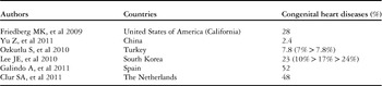
Heart defects are common congenital malformations of the foetus. The incidence of foetal heart defects is six times higher than that of chromosomal anomalies and four times greater than that of neural tube defects.Reference Viñals, Poblete and Giuliano 15 , Reference Allan 18 , Reference Carvalho, Mavrides, Shinebourne, Campbell and Thilaganathan 19 Second trimester screening has contributed to the increase in the detection rate of foetal congenital heart disease.Reference Friedberg, Silverman and Moon-Grady 3 – Reference Clur, Van Brussel, Mathijssen, Pajkrt, Ottenkamp and Bilardo 8 Since the introduction of the concept of heart screening, especially after standardisation of guidelines by International Society of Ultrasound in Obstetrics and Gynecology, the detection rate of heart defects has increased (Table 1).
Incidence of foetal heart defects.

Cardiac malformations are responsible for a high proportion of infant mortality and morbidity.Reference Ferencz, Rubin and McCarter 20 – Reference Cuneo, Curran, Davis and Elrad 22 Between 1950 and 1994, 42% of infant deaths reported to the World Health Organization were attributable to cardiac defects.Reference Rosano, Botto, Botting and Mastroiacovo 23 Prenatal diagnosis of congenital heart disease by foetal echocardiography realises three important purposes. First, it allows for family counselling about the nature, severity, clinical management and prognosis of the unborn child. Second, it allows practitioners to intervene in utero, when it is needed. Finally, it allows families and providers to appropriately plan for the delivery. 9 , Reference Lee, Allan and Carvalho 10 , Reference Carvalho, Mavrides, Shinebourne, Campbell and Thilaganathan 19
In the light of presented data, we can understand the advantages of early diagnosis and the reason for second trimester screening being so important to try to improve the rate of infant mortality and morbidity.
Indications of foetal echocardiography
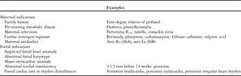
According to International Society of Ultrasound in Obstetrics and Gynecology guidelines, high-risk women should undergo more detailed foetal echocardiography, and those with lower risk should at least receive cardiac screening. Risk factors for cardiac anomaly are classified into maternal and foetal factors. Some indications are listed in Table 2. 9 , Reference Lee, Allan and Carvalho 10
Common indications for foetal echocardiography.

Recent research has found that compared with pregnant women in the high-risk group, those in the low-risk groups may be more likely to carry foetuses with heart disease. According to a retrospective study in 1395 foetuses in Turkey, 19% of the foetuses of women in low-risk groups were diagnosed with congenital heart disease, as opposed to 7% of the foetuses of women in high-risk groups.Reference Ozkutlu, Akça, Kafali and Beksaç 5 Therefore, all pregnant women should undergo cardiac screening.
A French group was the first to propose a greater role for obstetricians in evaluating the cardiac anatomy in order to improve cardiac prenatal screening.Reference Allan 18 Ideally, all pregnant women should undergo detailed foetal echocardiography, but the reality is that many countries do not follow this guideline. As the concept of cardiac screening gains strength, it may become possible to screen all pregnant women. It is our hope that women with suspected foetal anomalies and those at high risk will be referred for detailed foetal echocardiography. 9 , Reference Lee, Allan and Carvalho 10
Currently, our main challenge is to enable all our colleagues who perform prenatal examinations to also carry out cardiac screening for all foetuses. If there were a greater number of professionals performing this prenatal screening, diagnosis of congenital heart disease may be more effective. As a result, we would expect more positive outcomes, with better family planning, childbirth and treatment of the child.
How is the screening for congenital heart diseases performed in foetuses?
In 2006, the International Society of Ultrasound in Obstetrics and Gynecology standardised foetal cardiac screening during the second trimester of pregnancy in order to maximise the detection of congenital heart disease. In suspected cases of cardiac malformation or in pregnant women at high risk for heart disease, a more detailed assessment is required to be performed using foetal echocardiography. 9 In order to accomplish effective screening, the sonographer must have adequate knowledge of foetal cardiac anatomy. After the mastery of normal structures of the heart by the sonographer, it would be interesting to get deeper in the knowledge of the most common heart diseases. Another issue to be considered in the evaluation of cardiac structures is that the professional should follow a sequential segmental analysis, because, in this way, we can see all the components of the heart facilitating the search for abnormalities.
The International Society of Ultrasound in Obstetrics and Gynecology guidelines pertain to cardiac screening using grey-scale images. 9 , Reference Lee, Allan and Carvalho 10 However, we use a very simple tool, a colour Doppler, which increases our capacity and accuracy for analysing cardiac structures. A recent study evaluated the additional use of colour Doppler with grey-scale images in a low-risk population. The authors showed that the use of colour Doppler increased the percentage of diagnosed cases from 49% to 67% of the study population.Reference Eggebo, Heien, Berget and Ellingsen 24 Thus, we support the idea that the addition of colour Doppler analysis of structures is an important tool for foetal heart screening, even though this procedure is time-consuming.
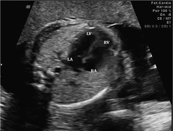
For cardiac screening assessment, the professional needs an axial four-chamber view of the anatomically normal heart. 9 Several structures may be analysed in this view (Fig 1):
-
∙ Cardiac situs: situs is the position the organ occupies in relation to the bilateral body symmetry. Then, the first step is to determine the foetal position, establishing the left and right, anterior and posterior, and superior and inferior aspects of the foetus.Reference Allan, Hornberger and Sharland 11 After that, you determine that the stomach is in fact on the left side of the foetus. There are visceral and cardiac situs. Basically, visceral situs is a condition in which the major lobe of the liver is on the right side and the spleen, stomach, and descending aorta are on the left. For atrial situs, this is the relationship of the atria with respect to each other. This is conventionally established by noting the arrangement of the aorta and inferior vena cava at the level of the diaphragm, because great vessel arrangement is similar to thoracic and atrial situs.Reference Allan, Hornberger and Sharland 11 , Reference Huhta, Smallhorn and Macartney 25 Using the position of the inferior vena cava and the aorta with respect to the spine, it was possible to separate those with situs solitus from the others.Reference Allan, Hornberger and Sharland 11 , Reference Huhta, Smallhorn and Macartney 25 In situs solitus, the aorta lies to the left of the spine and the inferior vena cava lies to the right. The morphology of the atrial appendages in the foetus is not easily visualised by echocardiography.Reference Allan, Hornberger and Sharland 11 , Reference Huhta, Smallhorn and Macartney 25 The diagnosis of isomerism is usually made because of a discordant position between the heart and stomach, the typical intracardiac anatomy, and the arrangement of the aorta and inferior vena cava in the abdomen.Reference Allan, Hornberger and Sharland 11 , Reference Huhta, Smallhorn and Macartney 25
-
∙ Position and the axis of the heart: the usual position is anterior left central chest and the heart apex is normally deviated about 45±20° towards the left side of the foetus – levoposition and levocardia.
-
∙ Size of the heart: the heart occupies about one-third of the chest. We can observe normal and proportional dimensions of the cardiac chambers.
-
∙ Pulmonary venous return: pulmonary veins draining into the left atrium – these structures are part of a more detailed foetal cardiac examination, but can be relatively easily visualised when using colour Doppler.
-
∙ Observation of the atria: the size and interatrial septum; left atrium on the left side with visible pulmonary vein drainage and lamina foramen ovale; right atrium on the right side and visible drainage of the vena cava, as visualised using colour Doppler.
-
∙ The lamina of the foramen ovale with good mobility in the left atrium.
-
∙ Atrioventricular valves: the mitral valve is on the left and the tricuspid valve is on the right. Note the tricuspid valve located slightly more apical than the mitral valve. Observe the mobility of the valves and the existence of valvular insufficiency – with assistance of the colour Doppler.
-
∙ Observation of the ventricles: right ventricle with the typical moderator band at its apex; proportional and normal-sized ventricles and intact ventricular septum.
Four-chamber view of the foetal heart. LA=left atrium; LV=left ventricle; RA=right atrium; RV=right ventricle.

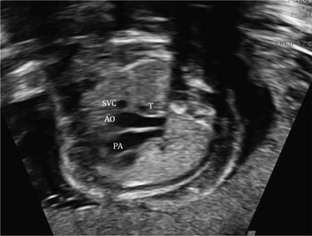
In the same axial plane of the four-chamber view, the ultrasonographist should slightly move the transducer from caudal to the cranial portion of the foetus. With this slight movement, the sonographer can observe normal outflow tracts of the heart and the position of the three vessels with respect to the trachea (Figs 2 and 3). In the normal outflow tracts of the heart, the aorta and pulmonary arteries cross to each other, with the aorta posterior to the pulmonary artery. The sonographer would observe:
-
∙ First, the left ventricular outflow tract: the connection of the left ventricle to the aorta. Here, we can evaluate the interventricular septum integrity, the walls of the ventricles, and the mobility of the aortic valve.
-
∙ Second, right ventricular outflow tract: see the connection of the right ventricle and the pulmonary artery; then, observe the relationship of the pulmonary artery with the right ventricle. It is possible to observe the split in the pulmonary artery – different from that observed in the aorta – that gives rise to the pulmonary branches. The practitioner can note the mobility of the pulmonary valve in this image.
-
∙ Finally, the three vessels and trachea plane may be observed located further anterior to the plane. The sonographer can see the usual relationship of the great vessels of the heart: V-configuration pointing to the posterior spine on the left with the pulmonary artery proportionally larger than the aorta, and the aorta larger than the superior vena cava. The distal aortic arch and ductus arteriosus are to the left to the trachea.
The outflow tracts of the heart. Ao=aorta artery; LV=left ventricle; PA=pulmonary artery; RV=right ventricle.

Three vessels and the trachea (3VT). Ao=aorta artery; PA=pulmonary artery; SVC=superior vena cava; T=trachea.

Colour Doppler scan clearly shows the crossing of the great vessels in the normal heart, aids observation of the interventricular septum integrity, and allows the clinician to check for adequate functionality of the ventriculoatrial valves.
Analyses of the outflow tract and the three vessels and trachea in cardiac screening improved the accuracy of diagnosing congenital heart disease.Reference Kirk, Riggs, Comstock, Lee, Yang and Weinhouse 26 – Reference Vettraino, Lee, Bronsteen and Comstock 29 In 2009, Xu et alReference Xu, Hu, Gu, Yang and Dai 30 conducted a prospective study in 5000 pregnant women – single foetus. Cardiac screenings were performed in accordance with the International Society of Ultrasound in Obstetrics and Gynecology guidelines. Foetal heart defects were observed in 1.5% of all subjects. Of these, 31.5% were detected using four-chamber views alone, whereas 69% were detected using four-chamber views and views of the outflow tract, and the three vessels and trachea.
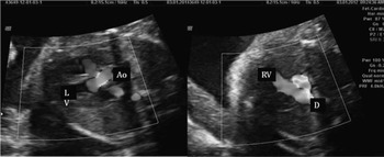
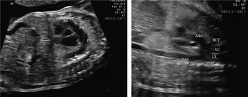
Evaluation of the aortic and ductal arches is not included in the International Society of Ultrasound in Obstetrics and Gynecology guidelines. However, sonographers should be careful to note cases with slight disparities between the cardiac chambers, especially with a predominance of right chambers, because some degree of aortic arch obstruction may indicate congenital heart disease,Reference Hikoro, Mats, Michael, Hana and Helena 31 especially coarctation of the aorta. The heart arches are evaluated by visualising through the sagittal plane of the foetus (Fig 4). The clinician can analyse the following:
-
∙ Ductal arch view: note that the right atrium communicates with the right ventricle, and the right ventricle communicates with the pulmonary artery, which continues with the ductus arteriosus further to the descending aorta.
-
∙ Aortic arch view: the clinician can observe the ascending aorta, which continues with the transverse arch that gives rise to the neck vessels – brachiocephalic trunk, left carotid and left subclavian – and follows the descending aorta.
The aortic and ductal arch views. AAo=ascending aorta; BCT=brachiocephalic trunk; D=ductus arteriosus; DA=descending aorta; LC=left carotid; LS=left subclavian; PA=pulmonary artery; RV=right ventricle.

Associating congenital heart diseases with cardiac planes
We can suspect some heart diseases according to the changes found in each echocardiographic plane.
The following may be seen in the four-chamber view:
-
∙ Cardiac situs: situs inversus and situs ambiguous – heterotaxy syndrome. Situs inversus is a condition in which there is a mirror image of the situs solitus pattern, with the aorta to the right and the inferior vena cava to the left, with both vessels again being symmetrically disposed with respect to the spine.Reference Allan, Hornberger and Sharland 11 , Reference Huhta, Smallhorn and Macartney 25 The term isomerism refers to the symmetrical development of normally asymmetrical organs or organ systems, which is the main feature of heterotaxy syndromes. A combination of cardiac, vascular and visceral abnormalities make up these syndromes. There are many correlates to predict the atrial situs: splenic status, lung lobation, position of abdominal viscera, bronchial morphology, pulmonary venous connection, presence or absence of coronary sinus, pulmonary artery morphology, systemic venous connection, hepatic atrial connection, and position of the inferior cava and the aorta. The position of the inferior vena cava and the aorta alone has been shown reliably to separate those patients with situs solitus from the others situs.Reference Huhta, Smallhorn and Macartney 25 The echocardiographic finding of left isomerism is the identification of the aorta being centrally located and the presence of a vessel between the aorta and the spine: the interruption of the inferior vena cava with azygous continuation or hemiazygos vein into the superior vena cava is almost universal. In the axial plane of the foetus with left isomerism, we can see a vena – azygos vena – between the aorta and the spine.Reference Allan, Hornberger and Sharland 11 , Reference Huhta, Smallhorn and Macartney 25 Many times, these caracteristics are very difficult to see; however, we can differentiate them through the colour Doppler: the direction of the flow in these two vessels is different; then, the colours are different, because the flow of azygous vena is going to the heart and the flow of the aorta is going out of the heart.Reference Allan, Hornberger and Sharland 11 Complete heart block is also found in some patients with left isomerism; in this condition, the pregnancy is often referred for foetal echocardiography because of the detection of a bradycardia.Reference Allan, Hornberger and Sharland 11 The echocardiographic finding of right isomerism is the identification of the inferior vena cava and the aorta together on one or the other side of the spine, with the aorta being posterior.Reference Allan, Hornberger and Sharland 11 , Reference Huhta, Smallhorn and Macartney 25 The pulmonary venous connection should be carefully examined, as by definition it is always anomalous, although the most common connection is for a venous confluence to drain directly to the centre of the atrial mass.Reference Allan, Hornberger and Sharland 11
-
∙ The position and the axis of the heart: dextroposition/mesoposition – the heart is positioned in the right/midline hemithorax; and dextrocardia/mesocardia – the heart apex is positioned to the right side of the chest or midline. There are some cases where we can see dextroposition with the apex still oriented to the left – levocardia. Positional abnormalities can be secondary to left diaphragmatic hernia or presence of space-occupying lesions, such as cystic adenomatoid malformation, foetal lung hypoplasia or agenesis.
-
∙ Size of the heart: cardiomegaly or pericardial effusion.
-
∙ Pulmonary venous return: should be seen draining to the left atrium at least one left and one right pulmonary vein. In cases of abnormal venous return, the clinician can suspect of partial or total anomalous pulmonary venous drainage by seeing the collector draining in a systemic vein or in the right atrium.
-
∙ Observation of the atria: atrial dilatation in cases of atresia, stenosis or atrioventricular valve regurgitation – as seen in Ebstein’s anomaly.
-
∙ Atrial septal tissue: the clinician must use care while moving the lamina of the foramen ovale in cases of restrictive flow and if there is septum primum.
-
∙ Atrioventricular valves: significant regurgitation may be observed, as in cases of valvular dysplasia or as seen in Ebstein’s anomaly. The sonographer can note valvular atresia, which is usually seen with hypoplastic ventricle – hypoplasia of the ventricle corresponding to valve atresia. At this plane, we can also see a common atrioventricular valve – absence of normal “cross” appearance of the heart due to disruption at this point in both the atrial and ventricular septa, know as atrioventricular canal defect (AVCD). 69% of all patients with AVCD have Down’s syndrome.Reference Koenig, Hijazi and Zimmerman 32
-
∙ Anatomy of the ventricles: right ventricular hypoplasia, as seen in some cases of pulmonary or tricuspid atresia, or hypoplasia of the left ventricle, as seen in cases of mitral or aortic atresia, may be observed. The sonographer may see the right ventricle located to the left and the left ventricle located to the right, which is indicative of L loop. It is also known as ventricular inversion or congenitally corrected transposition of the great vessels. As the name suggests, there are discordant connections at both the atrioventricular and ventriculoarterial junctions.
In the images of the outflow tract of the heart and the three vessels and trachea axis, the clinician may observe the following:
-
∙ Left ventricular outflow tract: in this view, stenosis or aortic atresia, ventricular septal defect or two vessels leaving the left ventricle featuring a double-outlet left ventricle may be observed.
-
∙ Right ventricular outflow tract: pulmonary stenosis or pulmonary atresia or two vessels leaving the right ventricle featuring double-outlet right ventricle may be observed.
-
∙ Three vessels and trachea axis: when is seen just two vessels in this view, the clinician should suspect of transposition of the great arteries or common arterial trunk. In the former, there are three vessels however these vessels are in different position, giving an erroneous impression of only two vessels. In the common arterial trunk, there are indeed two vesselsReference Chaoui and McEwing 33 . When the pulmonary artery shows dimension smaller than normal, stenosis or pulmonary atresia should be suspected. A smaller than normal aorta may indicate aortic stenosis or atresia.Reference Chaoui and McEwing 33 When the trachea is seen entrapped between the aortic arch and pulmonary trunk, and the arterial ductus is seen to the right of the trachea, we should be aware of the possibility of right-sided aortic arch.Reference Chaoui and McEwing 33
In the sagittal plane of fetal, the cardiac arches are observed. At this point, it is important to check for coarctation of the aorta and aortic arch interruption. These analyses are very difficult because the ductus arteriosus may hide the aortic arch and mask cardiac abnormalities.Reference Vettraino, Lee, Bronsteen and Comstock 29 , Reference Head, Jowett, Sharland and Simpson 34 – Reference Sharland, Chan and Allan 37 Clinicians should suspect this anomaly if they observe dilatation of the right heart chambers.
New technologies
The early studies of three-dimensional techniques using Spatio-Temporal Image Correlation appeared in 2003. Investigators discussed the difficulties in obtaining an adequate assessment of cardiac structure by using the various two-dimensional imaging techniques.Reference DeVore, Falkensammer, Sklansky and Platt 17 In 2006, International Society of Ultrasound in Obstetrics and Gynecology introduced the guidelines for assessing the foetal heart. Imaging difficulties became more evident because of heart size variation, which is dependent upon gestational age, foetal position and movements during analysis, poly/oligohydramnios, maternal obesity and weeks of gestation at the time of examination. At the same time, 3D/4D technology was introduced and some clinicians advocated the use of this new technology and Spatio-Temporal Image Correlation for image acquisition. Spatio-Temporal Image Correlation offered the advantage of off-line assessment of cardiac structures, connections and functions.Reference Viñals, Poblete and Giuliano 15 – Reference DeVore, Falkensammer, Sklansky and Platt 17
Recent studies have described the advantages of using 3D/4D foetal echocardiography. Several studies have compared the findings of two-dimensional imaging and Spatio-Temporal Image Correlation in a specific type of heart disease in order to identify the benefits of the new methods.Reference Vinãls, Mandujano, Vargas and Giuliano 38 – Reference Pooh and Korai 44
Despite the great efforts made so far, this new technology is still considered an additional method in echocardiography. However, we believe that the new methods will be very useful for tracking in the future. The 3D/4D technology allows for a more comprehensive and practical assessment of congenital heart disease in the foetus.
The impact of foetal cardiac screening on the prevalence of congenital heart diseases
Advances in the methods of screening for foetal heart disease have increased the numbers of diagnoses of these diseases. Thus, the clinician should be prepared for questions from the family regarding the type of heart disease, prognosis, planning for delivery and recurrence in future pregnancies. Families should also be informed about the types of treatment available, post-operative complications and possible associations of congenital heart disease with other diseases or trisomies. A multidisciplinary team prepared to comprehensively attend to these cases is therefore necessary. Such a team should include a foetal physician, a foetal cardiologist, psychologist, geneticist and other specialists, as required for each case. Some families opt for termination of pregnancy, depending on their beliefs and local laws. Termination tends to decrease the prevalence of postnatal congenital heart disease.Reference Carvalho, Mavrides, Shinebourne, Campbell and Thilaganathan 19 – Reference Eggebo, Heien, Berget and Ellingsen 24 , Reference Xu, Hu, Gu, Yang and Dai 30 – Reference Koenig, Hijazi and Zimmerman 32 , Reference Head, Jowett, Sharland and Simpson 34 , Reference Vinãls, Mandujano, Vargas and Giuliano 38 – Reference Garne, Berghold, Johnson and Stoll 45
Conclusion
The purpose of this review was to critically assess some concepts of the International Society of Ultrasound in Obstetrics and Gynecology guidelines for performing foetal cardiac scans. We agree that the guidelines serve as a standard and help to systematise the screening for congenital heart disease, but we think that some topics may be added to design the most appropriate screening method. However, we cannot expand the topics to be evaluated in this examination without good training of sonographers who undergo this screening. We support continuing the education for professionals to expand their knowledge of normal cardiac structures, thus enabling them to perform sequential segmental analyses of these structures. We hope that intrauterine diagnosis rate of congenital heart disease will increase, thereby leading to better family planning and delivery and ultimately reducing infant morbidity and mortality due to congenital heart disease.