Introduction
Glucocorticoids are steroids produced in the adrenal cortex; their levels are under the control of the hypothalamic–pituitary–adrenal axis (Necela and Cidlowski, Reference Necela and Cidlowski2004). Previous studies have shown that stress-induced increases in circulating glucocorticoids or exogenous administration of synthetic glucocorticoid dexamethasone inhibit the functions of the hypothalamus and pituitary, repress transcription of GnRH, and consequently reduce the levels of FSH and LH (Whirledge and Cidlowski, Reference Whirledge and Cidlowski2013; Yuan et al., Reference Yuan, Yang, Hu, Fan, Zhang, Zhou, Wang, Lu and Ma2014; Whirledge and Cidlowski, Reference Whirledge and Cidlowski2017). In addition, Tetsuka et al. (Reference Tetsuka, Takagi, Ambo, Myat, Zempo and Onuma2016) reported that the ovary is a target of natural or synthetic glucocorticoids. Glucocorticoids act directly on the ovaries by inhibiting the production of steroid hormones, and its action depends on binding to the glucocorticoid receptor (GR). In this way, the GR is one of the important mechanisms of regulation, and its expression has already been confirmed in the follicles, corpus luteum and in the epithelium of the ovarian surface of rats and humans (Whirledge and Cidlowski, Reference Whirledge and Cidlowski2010). Although the involvement of glucocorticoids in ovarian function is well documented, its action on bovine follicle development is still not known.
Dexamethasone is a synthetic glucocorticoid (Bavaresco et al., Reference Bavaresco, Bernardi and Battastini2005) that has been widely used in clinical application due to its anti-inflammatory and immunosuppressive functions (De Bosscher et al., Reference De Bosscher, Haegeman and Elewaut2010). However, this hormone can adversely affect the reproductive system and may act directly on the ovary. In cattle, use of dexamethasone for labour induction can lead to placental retention (reviewed by Ganaie et al., Reference Ganaie, Japheth, Ali, Lone, Mir and Malik2018). Kliem et al. (Reference Kliem, Rodler, Ulbrich, Sinowatz, Berisha, Meyer and Schams2013) reported that dexamethasone therapy negatively influenced the initial formation of the corpus luteum, causing a decrease in progesterone levels. In mice, Hułas-Stasiak et al. (Reference Hułas-Stasiak, Dobrowolski, Pawlikowska-Pawlęga, Tomaszewska and Muszyński2017) showed that dexamethasone impaired folliculogenesis and increased follicular atresia by induction of autophagy and apoptosis. Other studies have reported that dexamethasone regulated apoptotic activity in granulosa cells in preovulatory follicles (Sasson et al., Reference Sasson, Tajima and Amsterdam2001; Yuan et al., Reference Yuan, Yang, Hu, Fan, Zhang, Zhou, Wang, Lu and Ma2014) and that high concentrations of dexamethasone impaired oocyte maturation that occurs in parallel with changes in follicle differentiation in cultured rat follicles (Merris et al., Reference Merris, Van Wemmel and Cortvrindt2007). However, other studies have reported the opposite results for early ovarian follicles. For example, dexamethasone maintained primordial follicle ultrastructure in bovine ovarian tissue cultured in vitro (Silva et al., Reference Silva, Ribeiro, Menezes, Barberino, Renato, Dau, Costa, Melo, Bezerra, Donato, Peixoto, Matos, Gonçalves, Van den Hurk and Silva2017). In mouse, dexamethasone augmented the number of growing ovarian follicles and corpora lutea and caused a significant increase in ovarian weight, suggesting that it exerts antiapoptotic action on granulosa cells (Hułas-Stasiak et al., Reference Hułas-Stasiak, Dobrowolski, Pawlikowska-Pawlęga, Tomaszewska and Muszyński2017). Dexamethasone also augmented the levels of anti-Müllerian hormone secreted by granulosa cells, which indicated that it may act as a promoter of preantral follicle development (Yuan et al., Reference Yuan, Yang, Hu, Fan, Zhang, Zhou, Wang, Lu and Ma2014). Dexamethasone also potentiated the effect of FSH on granulosa cell steroidogenesis, showing that this hormone may modulate the effects of FSH on preantral follicle growth and differentiation (Roy et al., Reference Roy, Wang and Yang2003). Therefore, it is very important to know if dexamethasone affects early follicle development in cows, which consequently can have an effect on female fertility. However, the influence of dexamethasone on growth, viability and ultrastructure of bovine secondary follicles during in vitro culture is not yet known.
The objectives of the present study were to evaluate the effects of different concentrations of dexamethasone (1, 10, 100 and 1000 ng/ml) on growth, viability, antrum formation, morphology and ultrastructural integrity of bovine secondary follicles cultured in vitro for 18 days.
Materials and methods
Chemicals
Culture medium and other chemicals used in the present study were purchased from Sigma Chemical Co. (St. Louis, USA) unless otherwise indicated in the text.
Source of ovaries
Bovine ovaries (n = 50) of cycling cows were collected from a local slaughterhouse located in Sobral, Ceara, northeastern Brazil. These ovaries were collected immediately after the death of the cows and were washed in 70% ethanol for about 10 s and twice in sterile 0.9% saline solution. Subsequently, the ovaries were transported within 1 h to the laboratory in tubes containing 0.9% saline solution supplemented with penicillin (100 mg/ml) and streptomycin (100 mg/ml) at 4°C (Paulino et al., Reference Paulino, Cunha, Silva, Souza, Lopes, Peixoto, Peixoto, Brito, Van den Hurk and Silva2018). This study was carried out in accordance with procedures approved by the Ethics and Animal Welfare Committee of the Federal University of Ceara.
Isolation and in vitro culture of preantral follicles
In the laboratory, the ovarian cortex was fragmented (1–2 mm), with the aid of a scalpel, in TCM-199 supplemented with HEPES (0.05 mM/ml), 100 IU/ml penicillin and 10 mg/ml streptomycin. Subsequently secondary follicles (~0.2 mm) were visualized under a stereomicroscopic microscope (SMZ 645 Nikon, Tokyo, Japan; ×40 magnification) and dissected manually from fragments using 25G needles. Follicles with visible oocytes surrounded by granulosa cells, without an antral cavity and with an intact basement membrane were selected for culture (Paulino et al., Reference Paulino, Cunha, Silva, Souza, Lopes, Peixoto, Peixoto, Brito, Van den Hurk and Silva2018, Reference Paulino, Barroso, Silva, Souza, Bezerra, Silva, Donato, Peixoto and Silva2020). Follicles with an extruded oocyte and opaque granulosa cells were considered to be atretic (Paulino et al., Reference Paulino, Cunha, Silva, Souza, Lopes, Peixoto, Peixoto, Brito, Van den Hurk and Silva2018, Reference Paulino, Barroso, Silva, Souza, Bezerra, Silva, Donato, Peixoto and Silva2020). After selection, the follicles were cultured individually in drops of medium under mineral oil in Petri dishes (60 × 15 mm, Corning, USA). The base medium was TCM-199 supplemented with 10 μg/ml insulin, 5.5 μg/ml transferrin and 5 ng/ml selenium (ITS), 3.0 mg/ml bovine serum albumin (BSA), 2 mM glutamine, 2 mM hypoxanthine, 50 μg/ml ascorbic acid and 0.05 IU FSH (oFSH, from sheep pituitary, Sigma). In total, 194 bovine secondary follicles were isolated and distributed randomly into wells containing 100 µl TCM-199+ alone or supplemented with dexamethasone at concentrations of 1, 10, 100 or 1000 ng/ml for 18 days at 38.5°C and 5% CO2 in air. Dexamethasone concentrations were chosen according to Silva et al. (Reference Silva, Ribeiro, Menezes, Barberino, Renato, Dau, Costa, Melo, Bezerra, Donato, Peixoto, Matos, Gonçalves, Van den Hurk and Silva2017). A partial exchange (60 μl) of medium was performed every 2 days. The osmolarity of the medium was measured at each exchange. The experiments were repeated six times.
Evaluation of morphology and development of cultured follicles
On days 0, 6, 12 and 18, follicles were considered morphologically normal when they had intact oocytes, without damage to the basement membrane. Follicles with opaque granulosa cells and/or darkened oocytes, as well as damaged basal membranes were considered atretic. The formation of the antrum was determined by visualizing a translucent cavity between the layers of granulosa cells (Paulino et al., Reference Paulino, Barroso, Silva, Souza, Bezerra, Silva, Donato, Peixoto and Silva2020). The follicle diameter (µm) was calculated only in morphologically normal follicles using two perpendicular measurements from the photographic records of follicles and using an inverted microscope with NIS Elements 2.4 software (Nikon, Nikon Instruments Inc., Japan). To calculate follicle daily growth, the value that represented the difference between follicle diameter at days 0 and 18 was divided by the number of days in culture (18).
Viability evaluation using fluorescence microscopy
After culture, to evaluate the viability of the cells, follicles for each treatment (n = 20) were stained with 4 mM of calcein-AM and 2 mM of ethidium homodimer (Molecular Probes, Invitrogen, Karlsruhe, Germany) in a darkroom at 37°C for 15 min. After exposure to fluorescence markers, follicles were examined under a fluorescence inverted microscopy (Nikon, Eclipse, TS 100, Japan, ×40 magnification). Oocytes and granulosa cells were considered viable if the cytoplasm was positively stained with calcein-AM (Green), while nonviable cells had chromatin marked with ethidium homodimer (red).
Histological analysis of cultured secondary follicles
Uncultured (within ovarian cortex) and in vitro cultured secondary follicles were fixed for histological analysis. After 18 days of culture, the follicles were fixed (n = 8, per treatment) for 24 h at room temperature in 4% paraformaldehyde in phosphate-buffered saline (PBS; pH 7.4). After fixation, follicles were dehydrated in a graded series of ethanol concentrations (70, 80, 90 and 100%), clarified with xylol, and embedded in paraffin (Gomes et al., Reference Gomes, Aragão, Neto, Barroso, Paulino, Silva, Vasconcelos, Silva and Silva2020). For each group of follicles, 5-µm sections were mounted on slides and stained with eosin and haematoxylin. The slides were examined under an optical microscope (Nikon, Tokyo, Japan). Follicles cultured in vitro were classified individually as morphologically normal or atretic, as described previously.
Ultrastructural analysis of cultured bovine secondary follicles
To better examine cell morphology and organization of organelles, transmission electron microscopy was performed to analyze the ultrastructure of secondary follicles before (day 0 – fresh control) and after18 days of in vitro culture. The isolated follicles (n = 6–8 per treatment) were fixed in Karnovsky’s solution (4% paraformaldehyde and 2.5% glutaraldehyde in 0.1 M sodium cacodylate buffer, pH 7.2) for at least 4 h at room temperature (~25°C). After fixation, cultured follicles were embedded in 4% low melting agarose droplets and kept in sodium cacodylate buffer. The specimens were fixed in 1% osmium tetroxide, 0.8% potassium ferricyanide and 5 mM calcium chloride in 0.1 M sodium cacodylate buffer for 1 h at room temperature, washed in sodium cacodylate buffer and stained with 5% uranyl acetate. The samples were then dehydrated through a gradient of acetone solutions and then embedded in epoxy resin (Epoxy-Embedding Kit, Fluka Chemika). Afterwards, semithin sections (2 µm) were cut, stained with toluidine blue and analyzed using light microscopy at ×400 magnification. Subsequently, ultrathin sections (70 nm) were obtained from bovine secondary follicles. The ultrathin sections were counterstained with uranyl acetate and lead citrate and examined under a Morgani-FEI transmission electron microscope.
Statistical analysis
Data were tested for normality using Shapiro–Wilk test, using Statview 5.0 software (SAS Institute, Inc., Cary, NC, USA). Comparisons of follicle diameters after culturing follicles with different concentrations of dexamethasone were performed using analysis of variance (ANOVA) followed by Kruskal–Wallis test. Data concerning follicle survival and antrum formation after in vitro culture in each treatment were compared using chi-squared test, and results were expressed as percentages. Differences were considered significant when P-values were < 0.05.
Results
Effects of dexamethasone on follicle growth, antrum formation and survival
Table 1 shows that, for all treatments, 6-day cultured follicles had significantly higher diameters than those follicles at day 0. However, follicle diameters after 6, 12 or 18 days of culture were not significantly different. When comparisons among treatments were performed after either 6, 12 or 18 days of culture, no significant effect of dexamethasone on follicle growth was observed.
Diameters (mean ± standard error of the mean (SEM)) of bovine secondary follicles after 0, 6, 12 or 18 days of in vitro culture in TCM-199+ alone or supplemented with different concentrations of dexamethasone (DEXA; 1, 10, 100 or 1000 ng/ml)

a,bLowercase letters shows statistical differences among columns, P < 0.05.
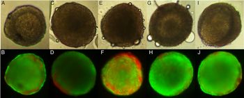
The presence of dexamethasone in the culture medium did not influence the percentages of normal follicles (Fig. 1) and antrum formation after 18 days of culture (Fig. 2). Figure 3 shows that follicles cultured in TCM-199 alone or supplemented with different concentrations of dexamethasone (1, 10, 100 and 1000 ng/ml) were mainly stained with calcein-AM. Only follicles cultured in medium supplemented with 1 or 10 ng/ml dexamethasone had stromal peripheral cells stained with ethidium homodimer.
Percentages of normal secondary follicles cultured for 18 days in TCM-199+ alone or supplemented with different concentrations of dexamethasone. a,b,cLowercase letters show statistical differences among bars, P < 0.05.

Percentages of antrum formation in secondary follicles cultured for 18 days in TCM-199+ alone or supplemented with different concentrations of dexamethasone. a,b,cLowercase letters show statistical differences among bars, P < 0.05.

Viability of bovine secondary follicles cultured for 18 days and evaluated using light microscopy (A, C, E, G, I) (×100 magnification) and after staining with calcein-AM (green) and ethidium homodimer (red) (B, D, F, H, J). Secondary follicle cultured in TCM-199+ alone (control) (A, B); or containing 1 ng/mL dexamethasone (DEXA) (C, D); 10 ng/mL DEXA (E, F); 100 ng/mL DEXA (G, H) or 1000 ng/ml DEXA (I, J).

Morphology of in vitro cultured follicles
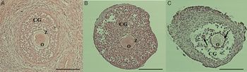
Figure 4 shows the morphology of normal bovine secondary follicles before mechanical isolation (Fig. 4 A), as well as after 18 days of culture in the presence of dexamethasone (Fig. 4 B). The morphology of a atretic follicle is shown in Fig. 4(C). Figure 4(A) shows that, before isolation, secondary follicles had rounded oocytes, visible zona pellucida and well organized follicular cells, without an antral cavity. Figure 4(B) shows an 18-day cultured follicle with normal morphology, i.e. intact oocyte, visible zona pellucida surrounded by granulosa cells, indicating connections between follicular cells and the oocyte. In addition, follicular cells were organized, therefore different from the morphology of the atretic follicle shown in Fig. 4(C). This follicle has poor connections between follicular cells, and low density and disorganization of granulosa cells.
Normal uncultured secondary follicle within ovarian tissue (A), isolated normal follicle after 18 days of culture in the presence of dexamethasone (B), cultured atretic follicle (C). CG, granulosa cells; O, oocyte; Z, zona pellucida. Arrows indicate disorganization of granulosa cells. Scale bars represent 100 μm.

Ultrastructural analysis of cultured secondary follicles
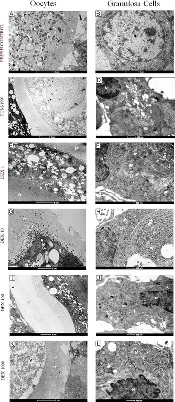
Normal uncultured follicles had oocyte and granulosa cells with mitochondria with normal cristae and well preserved membranes (Fig. 5 A, B). Sparse vesicles were spread throughout the oocyte cytoplasm and a regular cytoplasmic membrane was observed. Oocyte and granulosa cells had good connections (Fig. 5 A). The follicles cultured in control medium (TCM-199+) showed some vacuolization in oocytes and the cytoplasm of granulosa cells, slight detachment of the zona pellucida and decreases in the extensions between oocyte and granulosa cells (Fig. 5 C, D). In the oocyte, it was also possible to verify various organelles, while granulosa cells had mitochondria with no apparent signs of alteration (Fig. 5 D). The follicles cultured in the presence of 1 ng/ml dexamethasone showed oocytes (Fig. 5 E) with retraction of the zona pellucida, but it was still possible to observe the oocyte–granulosa connections. Oocytes had increased numbers of vacuoles, but mitochondria with normal cristae were visible. These follicles also had well preserved granulosa cells (Fig. 5 F). Granulosa cells of follicles cultured in the presence of 10, 100 or 1000 ng/ml dexamethasone were also well preserved, showing normal mitochondria and endoplasmic reticulum, suggesting high metabolic activity (Fig. 5 H, J, L). However, the oocytes of follicles cultured with 10, 100 or 1000 ng/ml dexamethasone had a decrease in the number of microvilli, large vacuolated areas, organelles with signs of degeneration and absence of connections between oocyte and granulosa cells (Fig. 5 G, I, K).
Representative micrographs of bovine secondary follicles in fresh control and cultured follicles for 18 days in the presence or absence of dexamethasone. Uncultured follicles (A, B); and follicles grown in the control medium alone (C, D); or supplemented with 1 ng/ml dexamethasone (E, F); 10 ng/ml dexamethasone (G, H); 100 ng/ml dexamethasone (I, J); 1000 ng/ml dexamethasone (K, L). N, nucleus; M, mitochondria; R, endoplasmic reticulum; V, vacuole; ZP, zona pellucida. Arrow, detachment of ZP; arrowhead, connections between oocyte and granulosa cells.

Discussion
The present study demonstrated that secondary follicles cultured in 1 ng/ml dexamethasone grow normally in vitro and maintain their ultrastructure after 18 days of culture. At the end of culture, these follicles preserved communication through oocyte–granulosa prolongations. In rats, it has been reported that dexamethasone increased the number of granulosa cells in cultured secondary follicles, promoting an increase in the diameter of these follicles (Hułas-Stasiak et al., Reference Hułas-Stasiak, Dobrowolski, Pawlikowska-Pawlęga, Tomaszewska and Muszyński2017). According to Saraiva et al. (Reference Saraiva, Matos, Faustino, Celestino, Silva and Figueiredo2010), this interaction between follicular cells and oocyte cells represented important communication and signalling responses between granulosa cells and oocytes through gap junctions. These transitions occurred through structures known as transient projections, which allow the bidirectional transport of ions and metabolites that regulate growth of oocytes (Mello et al., Reference Mello, Ferreira, Silva, Mello and Palhano2013; Clarke, Reference Clarke2018). In addition, a large numbers of mitochondria in the oocytes of cultured follicles reinforced the maintenance of oocyte integrity (Sun et al., Reference Sun, Wu, Lai, Park, Cabot, Cheon, Day, Prather and Schatten2001).
Dexamethasone did not influence the growth, viability and antrum formation of bovine preantral follicles grown in vitro, however other authors have reported that subcutaneous administration of dexamethasone can cause deficiency in female fertility, as it affects the ovarian reserve of primordial follicles, which consequently affects primary and secondary follicles, decreasing the size of the ovary (Ristić et al., Reference Ristić, Nestorovic, Manojlovic-Stojanoski, Filipovic, Sosic-Jurjevi, Milosevic and Sekulic2008). Probably, in the present study, isolated follicles were not influenced by the effects of dexamethasone due the concentrations used in culture. However, Cikoš et al. (Reference Čikoš, Babeľová, Špirková, Burkuš, Kovaříková, Šefčíková, Fabian and Koppel2019) showed that GRα and GRγ transcripts were relatively expressed in mouse oocytes and that differences in responses to glucocorticoids may be associated with the activation of different GR subtypes (Cikoš et al., Reference Čikoš, Babeľová, Špirková, Burkuš, Kovaříková, Šefčíková, Fabian and Koppel2019). Recently, Silva et al. (Reference Silva, Ribeiro, Menezes, Barberino, Renato, Dau, Costa, Melo, Bezerra, Donato, Peixoto, Matos, Gonçalves, Van den Hurk and Silva2017) reported that dexamethasone added to cultures of follicle enclosed in ovarian tissue maintained the percentage of normal follicles and the follicle ultrastructure, but this hormone was not efficient in promoting follicle activation and development. In addition, Chicaro (Reference Chicaro2009) showed that the effects of dexamethasone depended on culture time and hormone concentration. In addition, it is also important to consider that the effects of glucocorticoids on bovine oocytes and follicles depend on the mechanism of regulation of glucocorticoid activity (Roy et al., Reference Roy, Wang and Yang2003; Tetsuka et al., Reference Tetsuka, Takagi, Ambo, Myat, Zempo and Onuma2016) through enzymes 11βHSD1 and 11βHSD2, making the follicles and oocytes capable of activating or deactivating the action of glucocorticoids, respectively. According to Tetsuka et al. (Reference Tetsuka, Takagi, Ambo, Myat, Zempo and Onuma2016), this enzyme system can be a protective mechanism for cells against high levels of glucocorticoids, when harmful. In pig, granulosa cells from 3–5 mm antral follicles were cultured in vitro in the presence or absence of FSH and/or dexamethasone; it was observed that dexamethasone increased P450scc mRNA levels and progesterone production (Yang et al., Reference Yang, Yu and Li2001).
Histological analysis showed that follicles cultured with dexamethasone had zona pellucida around the oocyte and dense layers of preserved granulosa cells. The preservation of granulosa cells and their communication between oocytes is vital both for oocyte development and for follicle differentiation (Sánchez and Smitz, Reference Sánchez and Smitz2012). However, ultrastructural analysis showed that oocytes of follicles cultured in the presence of 10, 100 or 1000 ng/ml dexamethasone had large vacuolated areas, organelles with signs of degeneration and absence of connections between oocyte and granulosa cells. Previously, Hułas-Stasiak et al. (Reference Hułas-Stasiak, Dobrowolski and Tomaszewska2014) showed that dexamethasone impaired mouse folliculogenesis and enhanced follicular atresia through induction of autophagy and apoptosis. It was also reported that high concentrations of dexamethasone impaired oocyte maturation in cultured rat follicles (Merris et al., Reference Merris, Van Wemmel and Cortvrindt2007).
In conclusion, growth of bovine secondary follicles in vitro is not influenced by dexamethasone. The ultrastructure of follicles cultured in control medium alone or supplemented with 1 ng/ml dexamethasone was well preserved. However, the presence of high concentrations (10, 100 or 1000 ng/ml) of this hormone induced ultrastructure changes in their oocytes. These data are important as they show that low concentrations of dexamethasone have no negative effect on early ovarian folliculogenesis in animals undergoing anti-inflammatory therapies.
Financial support
This research was supported by grants from the National Council for Scientific and Technological Development (CNPq, Brazil) and Coordination for the Improvement of Higher Education Personnel (CAPES). J.R.V. Silva is an investigator for CNPq. P.A.A. Barroso is the recipient of a scholarship from the Coordination for the Improvement of Higher Level Personnel (CAPES), Brazil.
Conflict of interest
The authors declare that there is no conflict of interest that would prejudice the impartiality of this scientific study.
Ethical standards
The authors declare that all procedures were performed according to national and institutional guidelines on the care and use of animals.