Introduction
Schistosoma haematobium and S. bovis are closely related sister taxa within the S. haematobium group (Rollinson & Southgate, Reference Rollinson, Southgate, Rollinson and Simpson1987). Both species utilize freshwater snails of the genus Bulinus and their distribution overlaps in many parts of Africa. Schistosoma haematobium is a major human pathogen, causing urinary schistosomiasis, afflicting ~150 million people in 54 countries in Africa, Madagascar and the Middle East (WHO, 2002). The adult worms usually locate themselves in the venous drainage system of the bladder and the terminal spined eggs work their way through the bladder wall and are excreted in the urine; haematuria is a common sign of infection. It has been estimated that annual mortality due to non-functioning kidneys caused by infections with S. haematobium could be as high as 150,000 (Van der Werf et al., Reference Van der Werf, De Vlas, Brooker, Looman, Nagelkerke, Habbema and Engels2003). Schistosoma bovis causes intestinal schistosomiasis in ruminants, especially cattle, sheep and goats; it occurs in northern, western and eastern parts of Africa, extending southwards to central Angola, southern Zaire and northern Zambia, the Mediterranean region and the Middle East. De Bont & Vercruysse (Reference De Bont and Vercruysse1998) suggest that at least 30% of the entire cattle population living in endemic areas are infected with schistosomes. While most infections are sub-clinical, heavy infections can give rise to significant pathology, leading to severe production losses. Precise identification of S. haematobium and S. bovis in areas where they coexist is important in order to provide accurate transmission data for epidemiological studies and for monitoring control programmes.
Transmission is a dynamic process, with new transmission areas appearing and disappearing due to ecological changes. The emergence of areas of sympatry of these parasites and their snail hosts, in particular in northern Senegal, has been reported to be due to man-made ecological changes, which have also caused the movement of humans and their domestic livestock to new transmission areas (Southgate et al., Reference Southgate, Tchuem Tchuenté, Sène, De Clercq, Théron, Jourdane, Webster, Rollinson, Gryseels and Vercruysse2001).
Schistosome cercariae emerging from wild-caught Bulinus species are difficult to distinguish, although the relative position of sensory receptors is of some value (Bayssade-Dufour, Reference Bayssade-Dufour1982) as are isoenzymes (Wright & Ross, Reference Wright and Ross1980; Brémond et al., Reference Brémond, Sellin, Sellin, Naméoua, Labbo, Théron and Combes1993). Identification of cercariae has often relied upon the laboratory passage of pooled cercarial populations in rodents and the identification of the resulting adult worms. Laboratory passage can lead to selection and genetic bottlenecking of the parasites (Shrivastava et al., Reference Shrivastava, Gower, Balolong, Wang, Qian and Webster2005) and cercarial samples containing a mixture of species may well be missed. Recent developments in molecular biology have increased the number and availability of molecular markers for schistosomes, providing tools to investigate many areas of Schistosoma biology, such as characterizations of new species, population genetics, interactions between species and precise species identification (Rollinson et al., Reference Rollinson, Webster, Webster, Nyakaana and Stothard2009).
In 2000, Barber et al. developed a method based on restriction fragment length polymorphism (RFLP) analysis of the ITS2 polymerase chain reaction (PCR) product, using two restriction enzymes that generated a species-specific banding pattern, to identify S. haematobium from other S. haematobium group species, providing a good nuclear rapid diagnostic test (RDT). More recently, Abbasi et al. (Reference Abbasi, King, Sturrock, Kariuki, Muchiri and Hamburger2007) differentiated S. haematobium from related species by PCR amplification of a nuclear inter-repeat sequence. This provided a good nuclear diagnostic tool for S. haematobium but provided negative reactions for S. bovis. Hamburger et al. (Reference Hamburger, Abbasi, Ramzy, Jourdane and Ruppel2001, Reference Hamburger, Hoffman, Kariuki, Muchiri, Ouma, Koech, Sturrock and King2004) used the Dra1 repeat to detect S. haematobium infections in infested water and snails, providing a useful tool to identify transmission areas and pre-patent snail infections. A drawback of using these approaches in a co-endemic area is that they would not be able to detect mixed populations of S. haematobium and S. bovis.
Here we present a high-throughput one-step multiplex PCR diagnostic method developed on the mitochondrial DNA (mtDNA) barcoding region to detect and discriminate between S. haematobium and S. bovis in all lifecycle stages (miracidia, cercariae and adults). This technique requires only ~3 h of lab time, involving a PCR using one universal forward primer and two species-specific reverse primers to produce species-specific PCR fragments that can be discriminated by gel electrophoresis. The utility of this diagnostic tool is discussed together with the development of sampling techniques needed to study these two important schistosome species.
Material and methods
Schistosoma haematobium and S. bovis adult worms
Total genomic DNA (gDNA) from individual adult worms, preserved in the liquid nitrogen schistosome collection of the Natural History Museum, London (NHM), originating from Senegal and obtained after the first passage in laboratory animals (S. haematobium (NHM571) and S. bovis (NHM342)), was extracted using the DNeasy™ Tissue kit (Qiagen, Crawley, West Sussex, UK) according to the manufacturer's protocol, and the gDNA was eluted in 100 μl. The concentration of the extracted gDNA was determined using a NanoDrop® ND-1000 Spectrophotometer. The gDNA extracts were diluted to obtain a working concentration of 50 ng/μl.
Primer design
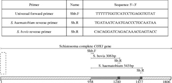
The cytochrome oxidase subunit 1 (COX1) mtDNA sequences of several S. haematobium strains (from Cameroon, Senegal, Kenya, Mali, Malawi, Zanzibar, Zambia, Egypt and Tanzania, unpublished data) and several S. bovis strains (from Uganda, Senegal and Tanzania, unpublished data) were aligned in MacClade 4.08 (Maddison & Maddison, Reference Maddison and Maddison2005). Using MacVector® 9.5.2 (MacVector Inc., Cambridge, UK) with the following primer settings: length 18–30 bp, GC% 30–55, Tm (°C) 55–80 (MacVector takes several features into account including self-duplexing, hairpins, specificity and mismatches), a universal forward primer for both S. haematobium and S. bovis was designed. Species-specific reverse primers for S. haematobium and S. bovis were designed at different distances from the forward primer to produce amplicons of different sizes (S. haematobium 543 bp, S. bovis 306 bp) (fig. 1), enabling accurate discrimination between the PCR products by agarose gel electrophoresis.
PCR primer sequences and their positions, together with the species-specific PCR fragments within the complex COX1 gene.

PCR amplification and visualization
PCR amplifications were performed in a total reaction volume of 25 μl using Ready-to-go PCR Beads (Amersham Pharmacia Biotech, Amersham, Bucks, UK), each containing 1.5 units of DNA Taq polymerase, 10 mm of Tris-HCl (pH 9), 50 mm of KCl, 1.5 mm of MgCl2, 200 μm of each dNTP and stabilizers, including bovine serum albumin (BSA), various combinations of the universal forward primer and specific reverse primers and the S. haematobium/S. bovis gDNA templates (refer to figs 2–4).
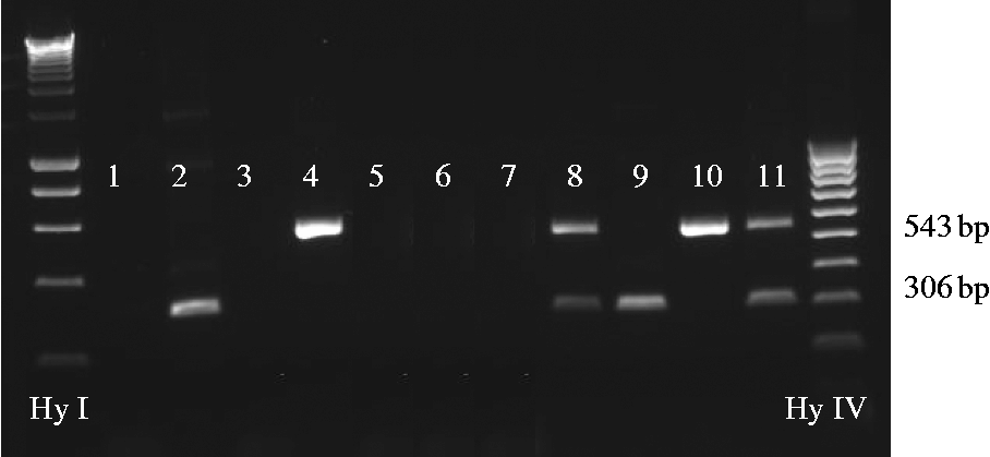
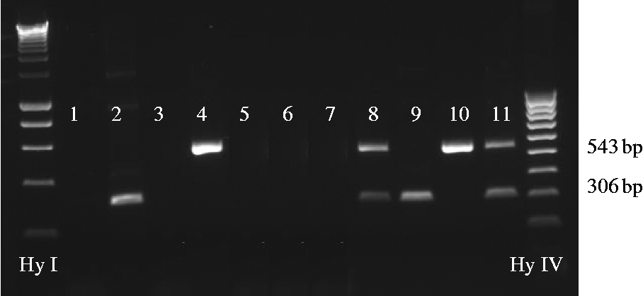
Testing specificity of the primers and multiplex PCRs. Each lane corresponds to a separate multiplex PCR containing S. haematobium (S. h) and S. bovis (S. b) gDNA (50 ng), separately and combined, extracted from adult worms, as below. Schistosome strains were from Senegal. Ten pmol of each primer was used in each assay except in PCR 11, where 5 pmol of each primer was used. Hy, hyperladder.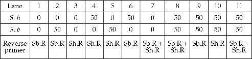
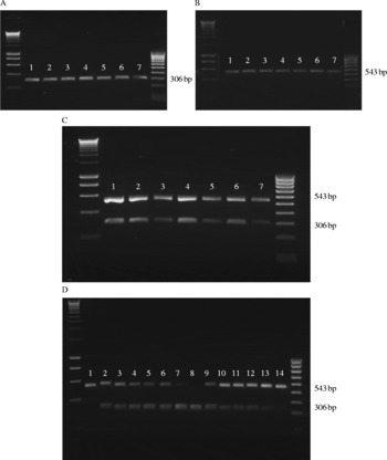
(A–D) Testing sensitivity of the multiplex PCR on individual S. bovis (S. b) (A) and S. haematobium (S. h) (B) and mixed S. bovis and S. haematobium (C and D) DNA templates. Schistosome strains were from Senegal. Genomic DNA extracted from adult worms was diluted and used in each PCR together with 10 pmol of each primer (Shb.F, Sb.R and Sh.R). For (A)–(C), lanes 1–7, respectively, show the amplicons obtained in the assays containing serially diluted gDNA at a concentration equal to 50, 25, 12.5, 6.25, 3.13, 1.56, 0.78 (ng), for each species. For (D) the concentraion (ng) of gDNA for each species used in each PCR (lanes 1–14) is as follows:

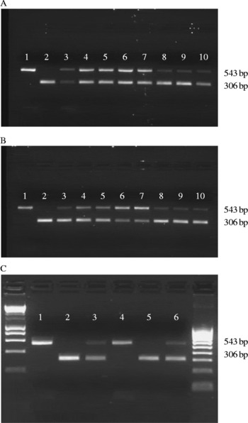
Testing sensitivity of the multiplex PCR on different numbers and mixes of cercariae and miracidia of S. bovis (S. b) and S. haematobium (S. h). For (A) and (B), each multiplex PCR contained DNA extracted from the different cercarial (A) or miracidial (B) mixes, as below: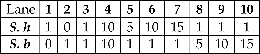 For (C), each lane corresponds to a single multiplex PCR containing one or two Whatman FTA® discs carrying gDNA from an individual cercaria (C) or miracidium (M), as below:
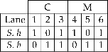 Where there is 0, a blank purified disc was added. For all PCRs 10 pmol of each primer (Shb.F, Sb.R and Sh.R) was used. The S. h cercariae and S. h miracidia were Cameroon and Zanzibar strains, respectively, and the S. b cercariae and miracidia were Ugandan strains.

Thermal cycling was performed in a PerkinElmer 9600 Thermal Cycler (PerkinElmer, Waltham, Massachusetts, USA) and the PCR conditions used were: 5 min denaturing at 95°C: 40 cycles of 30 s at 95°C, 30 s at 58°C, 1 min at 72°C; followed by a final extension period of 7 min at 72°C. Four microlitres of each amplicon were run out on 2% ethidium bromide agarose gel for 30 min at 120 V and photographs were taken using the UVP gel documentation system.
Testing the sensitivity of the multiplex PCR
PCR tests were done using different combinations of adult worm gDNA and species-specific reverse primers to test the specificity of the primers for each species (fig. 2, lanes 1–6). A multiplex PCR was developed incorporating the universal forward primer and both the species-specific reverse primers, and tests were done using different combinations of template gDNA of both species and primers at different concentrations (fig. 2, lanes 7–11). The sensitivity of the multiplex PCR was tested using serial dilutions of the template gDNA and also larval stages from each species, separately or combined (see figs 3 and 4). The concentration of the gDNA used in each PCR was determined accurately using a NanoDrop® ND-1000 spectrophotometer (NanoDrop Products, Wilmington, Detroit, USA).
Validation
To check specificity of the primers against other S. haematobium group species, the multiplex PCR was carried out on gDNA extracts from S. margrebowiei, S. leiperi, S. mattheei, S. intercalatum, S. guineensis and S. curassoni, using the same conditions as described above at increasingly higher annealing temperatures. The primers were also tested against S. mansoni and S. rodhaini, which are also prevalent in Africa, and S. bovis and S. haematobium controls were run at the same time. Any products amplified were sequenced as described later to confirm the identity of the species.
Specificity and sensitivity of multiplex PCR on larval life-stage forms of S. haematobium and S. bovis
Cercariae
Patent laboratory Bulinus wrighti snails infected with S. haematobium from Cameroon and patent laboratory B. productus infected with S. bovis from Uganda were put in separate pots of fresh water and exposed to light to stimulate shedding. The cercariae were poured into a Petri dish and viewed under a binocular microscope. Individual cercariae were collected in 3 μl of water using a Gilson pipette and pipetted into 1.5 ml Eppendorf tubes in various numbers and species combinations, as shown in fig. 4A. Each time the presence of the individual cercaria in the pipette tip was checked prior to being pipetted into the Eppendorf by visualization under the binocular microscope. If the cercariae were shed from the snails on different days, the collected cercariae were frozen at − 20°C until the rest of the cercariae were added or the gDNA was extracted. Genomic DNA was extracted from the cercaria/cercariae mixes (fig. 4A) using the DNeasy™ Tissue kit (Qiagen) according to the manufacturer's protocol, and the gDNA was eluted in 50 μl. The multiplex PCR was carried out using exactly the same conditions as described earlier and 2 μl of the gDNA extracts. The concentration of the gDNA extracted from the single cercaria was determined using a NanoDrop® ND-1000 spectrophotometer.
Miracidia
A laboratory hamster with a patent infection of S. haematobium from Zanzibar and a laboratory mouse with a patent infection of S. bovis from Uganda were culled and the livers containing the schistosome eggs removed. The livers were homogenized separately and pushed through a 212 μm Endecott sieve with excess 0.85% saline. Eggs were separated from the liver tissue in a series of washes and sedimentation in 0.85% saline. Eggs were hatched by placing them in fresh water and exposing them to light. Individual miracidia that hatched from the eggs were collected in various numbers and species combinations (fig. 4B) using the same techniques as those used for the cercariae. Genomic DNA extractions and multiplex PCRs were carried out as described above.
Whatman FTA® preserved miracidia and cercariae
Individual cercariae and miracidia obtained as above were also pipetted on to Whatman FTA® indicator cards (Whatman plc, Maidstone, Kent, UK) in a volume of 3 μl of water and allowed to dry for 1 h. Addition of the sample activates chemicals in the cards that lyse cells, inactivate proteins and immobilize the genomic nucleic acids. For gDNA extraction, a 2.0 mm disc was removed from the card at the centre, where the sample was loaded, using a Harris Micro Punch and incubated for 5 min in 200 μl of FTA® purification reagent (Whatman plc). The FTA® purification reagent was removed and the disc was incubated for a further 5 min in 200 μl of fresh FTA® purification reagent. This process was repeated for a total of three washes and was followed by 2 × 5 min incubations in 200 μl of Tris-EDTA buffer. Samples were air dried at 56°C for 10–30 min and the disc was checked visually to make sure that it was dry. Each disc contained gDNA from one cercaria or miracidium and the multiplex PCR, using the same conditions as described earlier, was carried out using mixes of FTA discs, as shown in fig. 4C.
Sequencing
To confirm identity of the amplicons, selected PCR products were purified or gel purified (in the case of multiplex amplicons) using the Qiagen PCR Purification Kit (Qiagen) according to the manufacturer's protocol. Each fragment was sequenced using the original PCR primers, fluorescent dye terminator sequencing kits (Applied Biosystems, Foster City, California, USA) and the sequencing reactions were run on an Applied Biosystems 377 automated sequencer. The sequences were assembled and edited manually using Sequencher V4.5 (Gene Codes Corp., Ann Arbor, Michigan, USA). Identity of the sequences was confirmed using the Basic Local Alignment Search Tool (BLAST) and submitted to EMBL/GenBank.
Results
Specificity of the multiplex PCR for S. haematobium and S. bovis
The species-specific reverse primers used together with the universal forward primer always generated a clear single band of the expected size for S. haematobium (543 bp) and S. bovis (306 bp), depending on which DNA template was present in the PCR. Two bands of the correct size were generated when a mix of the DNA template was used. The data also show that the reverse primers would not amplify the species (S. haematobium or S. bovis) that they were not designed for (fig. 2). This specificity was confirmed by sequencing the different bands (GenBank accessions: FJ586241 and FJ586242). The primers proved robust, retaining their specificity for different geographical strains, S. haematobium from Senegal, Cameroon and Zanzibar and S. bovis from Senegal and Uganda (see figs 2–4). The primers were designed using sequence alignments of several geographical strains across Africa. The universal forward primer was designed at a position almost identical (one polymorphic position) between all individuals of both S. haematobium and S. bovis. The species-specific reverse primers were designed at 100% conserved positions within the specific species but the S. haematobium specific reverse primer had 29% mismatching base pairs to S. bovis and the S. bovis specific reverse primer had 33% mismatching base pairs to S. haematobium. These mismatches (the majority of which were located at the 3′ end of the primer) enabled the primers to be species specific. As they were designed in regions that are very conserved across all geographical strains, these primers are expected to amplify all S. haematobium and S. bovis strains. PCR/lane 11/fig. 2 also shows that halving the primer concentration does not change the robustness of the primers but the proportions of each primer used would need to be the same.
Sensitivity of multiplex PCR
As can be seen in fig. 3, the multiplex PCR is highly sensitive, amplifying gDNA templates of 0.78 ng, generating bands that are clearly detectable for both S. haematobium and S. bovis. Also, when there is no difference in concentration of the two different templates in the multiplex PCR, there is no biased PCR amplification of the small DNA fragment, even at 0.78 ng of DNA (fig. 3C). In mixed templates where the DNA concentration of one species is sixfold higher than the other species, fragments of the correct size are still produced and visible (fig. 3D). A PCR bias can be seen preferentially amplifying the DNA template that is most abundant in the multiplex PCR, producing a stronger band, but the two bands are still detectable.
Detection ability of the multiplex PCR on larval stages
The multiplex PCR shows 100% ability to detect both fresh and FTA-preserved S. haematobium and S. bovis miracidia and cercariae (fig. 4). The technique is highly sensitive, producing detectable PCR bands from a single miracidium or cercaria. The concentration of the DNA extracted from a single miracidium was 1.6–3.6 ng/μl and for a single cercaria was 3.1–4.5 ng/μl, highlighting the high sensitivity of this multiplex PCR. The multiplex PCR was successful at producing both bands when both species were mixed in equal quantities or when one species was more abundant than the other (fig. 4A and B). Preferential amplification of the smaller S. bovis band was seen when the S. bovis larval stages were more abundant compared to that of S. haematobium and also with the FTA-preserved material; however, both bands were still detectable in all cases (fig. 4).
Discriminatory power of multiplex PCR
All products from fig. 2; PCRs 1+6, fig. 3A–C; PCRs 7+14, fig. 3D; PCRs 4, 7+10, fig. 4A and B; and PCRs 3+6, fig. 4C were sequenced. Sequence data from all the 543 bp bands that were sequenced were correctly identified as S. haematobium and all the 306 bp bands that were sequenced were correctly identified as S. bovis.
Validation
The PCR primers did not amplify S. mansoni, S. rodhaini, S. margrebowiei, S. leiperi and S. intercalatum but for S. mattheei, S. curassoni and S. guineensis both the S. haematobium and S. bovis diagnostic bands amplified up to an annealing temperature of 61°C despite mismatches at the primer annealing sites. At 65°C only the S. bovis band amplified for these species and the control S. haematobium reaction started to become very faint. BLAST searches of the sequences from these species confirmed their identity. The sequences of the two bands from the same species were also identical, as expected. Schistosoma mattheei, S. curassoni and S. guineensis are phylogenetically closely related to S. haematobium and S. bovis (Webster et al., Reference Webster, Southgate and Littlewood2006) and therefore their COX1 sequences are more similar to S. haematobium and S. bovis than those of the other species tested, enabling the primers to amplify.
Discussion
Specificity and validity
The multiplex rapid diagnostic PCR (RD-PCR) incorporating a universal Schistosoma forward primer and two different reverse primers each designed specifically for S. haematobium and S. bovis has provided a useful and robust rapid diagnostic technique for the identification of all larval stages of these two schistosomes. The specific reverse primers were designed from alignments of all available COX1 sequence data from several different geographical locations, it is therefore quite likely that they will still retain their robustness with regard to natural intra-species genetic variation observed across Africa. In this study they performed well in strains from Cameroon, Uganda, Zanzibar and Senegal. Also, because this assay amplifies the universal COX1 ‘barcoding’ fragment, sequence data for barcoding purposes can also be obtained for the samples analysed, if required, providing an extra utility of these primers for population genetic studies. The technique shows clearly that both S. haematobium and S. bovis present a clear, single-band profile, differing in 237 bp for the two species: 543 bp and 306 bp, respectively. A double-banded profile is present for a mix of the two species. The fact that a diagnostic band is always amplified for each species enables the discrimination between false negatives and failed PCRs, which is a potential problem of the diagnostic technique to detect S. bovis presented by Abbasi et al. (Reference Abbasi, King, Sturrock, Kariuki, Muchiri and Hamburger2007).
In relation to the most prevalent and important, S. haematobium, of the other S. haematobium group species examined in this study S. bovis is the most abundant, potentially having the widest geographical overlap. Therefore, compared to the other S. haematobium group species, the discrimination of these two species is of most importance. The primers can also discriminate both species from S. margrebowiei, S. leiperi and S. intercalatum, as the primers will not amplify the latter three species. However, the primers do amplify S. mattheei, S. curassoni and S. guineensis, creating a double-banded profile (at the protocol annealing temperature). In the case of single worms, cercariae or miracidia this actually provides a RD-PCR for these three species but cannot be used to distinguish between them. In the limited areas of Africa where these species occur in sympatry with S. haematobium and S. bovis, further validation of species identity would be required by DNA sequencing of the obtained PCR products.
Sensitivity
Various combinations of mixed DNA templates (originating from all lifecycle stages) in different concentrations proved the sensitivity and specificity of this RD-PCR. DNA concentrations as low as 0.78 ng produced clearly detected PCR bands, as did DNA from a single miracidium and cercaria of both species. With regard to pooled samples, there is no complete bias in preferential amplification of one band over the other, which could create false-positive results leading to one species being missed if it was in the minority. This is also not validated in the method presented by Abbasi et al. (Reference Abbasi, King, Sturrock, Kariuki, Muchiri and Hamburger2007) as no multiplex or mixed DNA PCRs were carried out. The small size of the PCR fragments (306–543 bp) should facilitate amplification of low-quality or low-quantity DNA and, if needed, the fragments can be sequenced using the same primers as those used in the original PCR.
The RD-PCR also worked on Whatman FTA®-preserved specimens. Whatman FTA® cards are a new storage protocol for larval schistosomes, which allow the sampling of large numbers of individual miracidia, eggs and cercariae from the schistosome populations directly from the field. This cuts out the need for the laboratory passage of the parasites, which can cause selection of certain strains (Gower et al., Reference Gower, Shrivastava, Lamberton, Rollinson, Webster, Emery, Kabatereine and Webster2006). This RD-PCR, together with this sampling technique, allows the screening of schistosome populations to identify individual S. haematobium and S. bovis larval stages in areas of sympatry. This sampling technique also cuts out the need for pooling samples, which further validates the use of the RD-PCR, as discussed earlier.
However, pooling can be used to speed up analysis. For example, cercariae from snails can be pooled in order to test quickly whether or not there is a mixed infection of S. haematobium and S. bovis. Also, water samples (from environmental monitoring) or pooled DNA from snails can be tested with this multiplex PCR. This technique shows that bands of the correct size are still produced when the DNA concentration of one species is sixfold higher than the other species. This will be most valuable in areas where S. bovis and S. haematobium are transmitted sympatrically.
Utility
This assay provides a quick (2.5-h PCR followed by 30 min of gel electrophoresis), cost-effective, reliable, robust and sensitive molecular diagnostic tool. It has great potential to provide information on host use and transmission foci for S. haematobium and S. bovis in areas where the species overlap. It is cost-effective and amenable for high-throughput screening of large populations. This facilitates greater data harvesting, providing further insights into the epidemiology of these schistosomes and aiding control of the diseases they cause. Also, low technological demands make this tool a good candidate to be implemented in laboratories with modest resources. As with the Dra1 repeat assay (Hamburger et al., Reference Hamburger, Hoffman, Kariuki, Muchiri, Ouma, Koech, Sturrock and King2004), this diagnostic tool has the potential to facilitate large-scale PCR screening of snail populations, harvesting patent and or pre-patent infections of S. haematobium and/or S. bovis. The RD-PCR can theoretically be used on snail genomic DNA extracts to detect the schistosome DNA within the snail and this could also be advanced to a real-time PCR assay as a quantitative approach. The methods described here could help define interactions between species such as S. haematobium with S. guineensis (Webster et al., Reference Webster, Tchuem Tchuenté and Southgate2007) and S. mattheei (Wright & Ross, Reference Wright and Ross1980) as well as S. bovis and S. curassoni (Rollinson et al., Reference Rollinson, Southgate, Vercruysse and Moore1990), especially when used with nuclear diagnostic markers described by Hamburger et al. (Reference Hamburger, Abbasi, Ramzy, Jourdane and Ruppel2001, Reference Hamburger, Hoffman, Kariuki, Muchiri, Ouma, Koech, Sturrock and King2004) and Barber et al. (Reference Barber, Mkoji and Loker2000).
In conclusion, recent advances in the molecular characterization of schistosomes and their genomes (mitochondrial and nuclear) have provided several molecular diagnostic markers for different species. This mt RD-PCR is a cost-effective and robust assay to differentiate larval stages of S. haematobium and S. bovis, which are widespread throughout much of Africa. Advances in parasite identification can help elucidate transmission patterns, intermediate host specificity and interactions between closely related parasites. This RD-PCR will be a valuable identification tool in places where transmission of human and cattle schistosomiasis occur in close proximity.
Acknowledgements
Work at NHM was funded by the EU-CONTRAST grant (FP6-2004-INCO-DEV-3/PL032203). We are grateful to the many people who helped in various ways with the isolation and collection of parasites, and to Mike Anderson and Jayne King for their help in the laboratory passage of the schistosomes. T.H. was funded by a Marie Curie Fellowship (Meif-ct-2004-501684) and is currently a Postdoctoral Fellow of the Research Foundation – Flanders (FWO-Vlaanderen).
The initial concept and multiplex PCR primers were designed by Tine Huyse. All laboratory work was carried out by Bonnie Webster, who also generated and annotated the figures. Bonnie Webster, Tine Huyse and David Rollinson wrote the text, which was also edited by Russell Stothard.