Introduction
Dysautonomia results from dysfunction of central and/or peripheral components of the autonomic nervous system and may occur as a primary disorder or as a secondary manifestation of systemic disease.Reference Elinos, Veladiz-Gracia, González-Sierra, Rubio-Galicia, Cifuentes and Morales1–Reference Shen, Sheldon and Benditt5 Recognition has increased substantially in recent years because of greater awareness of orthostatic intolerance, expanded availability of autonomic testing, and the emergence of post-acute sequelae of severe acute respiratory syndrome coronavirus 2 infection (long COVID), which has become a major post-infectious contributor to autonomic dysfunction.Reference Blitshteyn, Whiteson and Abramoff6–Reference Bozkurt, Das and Addison11 Autonomic dysregulation is also observed in association with autoimmune, connective tissue, and metabolic disorders, suggesting shared pathophysiologic mechanisms across diverse aetiologies.Reference Blitshteyn2,Reference Miller, Stiles and Sheehan12–Reference Yao, Subramaniam and Raja14 This narrative review synthesises current evidence from paediatric cardiology, neurology, and autonomic medicine to provide a clinically relevant framework for understanding the integrated brain–heart–vascular pathophysiology, diagnosis, and management of post-COVID paediatric dysautonomia.
Epidemiology
The epidemiology of paediatric dysautonomia remains incompletely defined because current consensus statements and diagnostic criteria for orthostatic disorders are derived primarily from adolescents aged 12 to 19 years, with limited paediatric-specific guidance for younger children.Reference Stewart, Boris and Chelimsky3,Reference Shen, Sheldon and Benditt5,Reference Brignole, Moya and De Lange15–Reference Vassiliou, Tsampasian and Luchian17 The incidence appears to increase during puberty, with a female predominance consistently reported in adolescent cohorts with orthostatic intolerance.Reference Stewart, Boris and Chelimsky3,Reference Lau, Fedorowski and Raj18 Hormonal and cardiovascular changes during adolescence may enhance sympathetic activity, reduce plasma volume, and predispose susceptible individuals to orthostatic tachycardia and cerebral hypoperfusion.Reference Coupal, Heeney and Hockin19 The prevalence of post-COVID dysautonomia is particularly difficult to determine because many associated symptoms, including tachycardia, dizziness, and fatigue, overlap with those of established autonomic disorders and are not specific to post-acute sequelae of severe acute respiratory syndrome coronavirus 2 infection.Reference Gross, Carmilani and Stockwell10,Reference Stute, Stickford, Province, Augenreich, Ratchford and Stickford20–Reference Tamariz, Rozenfeld and Iglesias24 Racial and ethnic differences remain poorly characterised, as most published cohorts predominantly include White adolescents, likely reflecting disparities in healthcare access, referral patterns, and diagnostic recognition rather than true differences in disease prevalence.Reference Rhee, Thaweethai and Pant25
Pathophysiology
Dysautonomia reflects disruption of integrated autonomic, cardiovascular, and neurovascular mechanisms that maintain circulatory stability during orthostatic and exertional stress (Figure 1). Dysfunction of the central autonomic network impairs cardiovascular and cerebrovascular regulation, resulting in tachycardia, altered vascular tone, and cerebral hypoperfusion.Reference Trimble, Switzer and Blitshteyn26,Reference Tsai Owens, Biggs and Fahrenkamp27 Peripheral abnormalities, including impaired sympathetic vasoconstriction, excessive venous capacitance, and reduced skeletal muscle pump function, decrease stroke volume and circulatory reserve, leading to compensatory tachycardia, fatigue, cognitive dysfunction, and presyncope.Reference Lau, Fedorowski and Raj18,Reference Weintraub, DePace, Munoz, Kaczmarski, Manno and Colombo28–Reference Jardine, Pointon, Frampton, Wright, Buckenham and Stewart32 Syncope represents the extreme manifestation of failed compensation, in which excessive venous pooling triggers reflex parasympathetic activation, vasodilation, hypotension, and transient cerebral hypoperfusion.Reference Moodley33
Pathophysiology of paediatric dysautonomia and syncope.

Figure 1. Long description
A diagram of the pathophysiology of paediatric dysautonomia and syncope. The diagram represents the integrated brain-heart-vascular system and the reflex syncope cascade. The brain/central autonomic network, heart, and peripheral vasculature are interconnected. The brain/central autonomic network includes the brainstem, hypothalamus, and cortical modulation. The heart is involved in chronotropic response and stroke volume. The peripheral vasculature includes venous capacitance, vascular tone, and the skeletal muscle pump. The reflex syncope cascade starts with excessive peripheral venous pooling, leading to decreased venous return, decreased cardiac ventricular filling, and activation of cardiac mechanoreceptors. This cascade increases parasympathetic tone, decreases vascular tone, increases vasodilation, decreases venous return, decreases cerebral perfusion, and results in syncope. The diagram also shows connections between the brain/central autonomic network, heart, and peripheral vasculature leading to tachycardia, exercise intolerance, fatigue, and cognitive symptoms brain fog.
Recent literature increasingly suggests that postural orthostatic tachycardia syndrome and related orthostatic disorders may reflect abnormalities of central nervous system regulation rather than isolated peripheral autonomic dysfunction.Reference Wells, Malik and Brooks34 One study highlighted the substantial burden of central nervous system symptoms, together with evidence of altered cerebral perfusion, neuroimaging abnormalities, and therapeutic responses to centrally acting interventions, supporting a broader central nervous system-centred model of dysautonomia.Reference Blitshteyn35 Our proposed “brain–heart–vascular” framework integrates central and peripheral mechanisms rather than viewing them as competing explanations. The phrase “Never the Heart, Always the Brain” reflects a shift away from attributing symptoms solely to primary cardiac disease; however, it may oversimplify a complex physiology in which central autonomic circuits, cardiovascular mechanics, vascular regulation, and cerebral perfusion are inextricably interconnected (Figure 2).
Conceptual framework illustrating competing narratives in paediatric dysautonomia.

Post-COVID dysautonomia may exacerbate established autonomic disturbances through post-infectious immune dysregulation, endothelial and microvascular injury, autonomic reflex impairment, hypovolemia, and small-fibre neuropathy.Reference Chu, Chan and Yuen36–Reference Giunta, Giuliani, Sabbatinelli and Olivieri40 Severe acute respiratory syndrome coronavirus 2 may access brainstem autonomic centres through cranial nerve or haematogenous pathways, disrupting central autonomic regulation.Reference Baig, Khaleeq, Ali and Syeda41–Reference Liu, Tan, Wu, Gui, Suo and Li43 Emerging evidence further suggests that persistent viral antigens, inflammatory mediators, and autoantibodies directed against adrenergic and cholinergic receptors may reduce heart rate variability and promote autonomic instability.Reference Peluso, Lu and Tang44–Reference El-Rhermoul, Fedorowski and Eardley48 These mechanisms likely interact with established pathways involved in paediatric dysautonomia, resulting in overlapping clinical phenotypes, including orthostatic intolerance, postural orthostatic tachycardia syndrome, inappropriate sinus tachycardia, orthostatic hypotension, and syncope.Reference Bozkurt, Das and Addison11,Reference Castiglioni, Rampichini and Corti49–Reference Morrow, Villatoro, Kokorelis, Rowe and Malone54 Similar disturbances in autonomic and cerebrovascular regulation have also been described in myalgic encephalomyelitis/chronic fatigue syndrome, highlighting shared pathophysiologic pathways across post-infectious and multisystem disorders.Reference Issa, Lin and Chen55–Reference Campen, Rowe and Visser59
Clinical presentation
Long-COVID has increased recognition of dysautonomia in children and adolescents.Reference Raj9–Reference Bozkurt, Das and Addison11,Reference Vassiliou, Tsampasian and Luchian17,Reference Depace and Colombo38,Reference Delogu, Aliberti and Birritella50–Reference Dani, Dirksen and Taraborrelli52,Reference Morrow, Villatoro, Kokorelis, Rowe and Malone54,Reference Grubb60,Reference Bisaccia, Ricci and Recce61 Many patients experience chronic symptoms such as dizziness, fatigue, nausea, cognitive dysfunction, and visual disturbances without meeting diagnostic criteria for a single autonomic syndrome; in these cases, terms such as orthostatic intolerance or undifferentiated dysautonomia are appropriate.Reference Kanjwal, Sheikh, Karabin, Kanjwal and Grubb62–Reference Ståhlberg, Mahdi, Johansson, Fedorowski and Olshansky64 Current consensus criteria for orthostatic intolerance disorders are derived largely from adolescent postural orthostatic tachycardia syndrome cohortsReference Shen, Sheldon and Benditt5,Reference Brignole, Moya and De Lange15,Reference Raj, Guzman and Harvey16,Reference Raj, Bourne and Stiles65 and may not fully capture the broader and heterogeneous clinical spectrum of paediatric post-COVID dysautonomia.Reference Xie, Shirsath and Scurrah66
Age influences symptom expression.Reference Bozkurt, Das and Addison11,Reference Vassiliou, Tsampasian and Luchian17,Reference Rao, Gross and Mohandas21 Younger children often present with nonspecific manifestations such as reduced appetite, sleep disturbances, or persistent cough.Reference Gross, Thaweethai and Salisbury67 School-aged children commonly report headaches, abdominal pain, and difficulty concentrating.Reference Gross, Carmilani and Stockwell10 Adolescents more frequently describe fatigue, lightheadedness, memory impairment, and altered smell or taste. Post-exertional symptom exacerbation, defined as worsening of symptoms following minimal physical or cognitive activity, is a characteristic feature of Long COVID and may contribute substantially to functional impairment.Reference Gross, Carmilani and Stockwell10
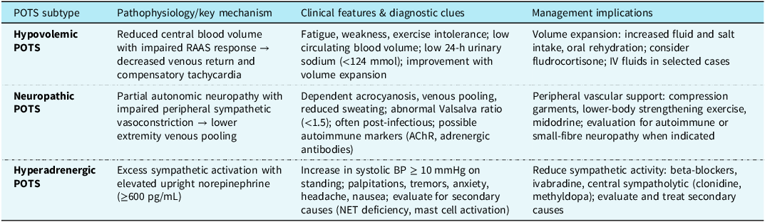
Postural orthostatic tachycardia syndrome is among the most common phenotypes of paediatric dysautonomia. In adolescents, it is defined by chronic orthostatic intolerance accompanied by an orthostatic heart rate increase of at least 40 beats per minute or an upright heart rate exceeding age-specific thresholds in the absence of orthostatic hypotension.Reference Lau, Fedorowski and Raj18,Reference Boris and Moak31,Reference Chen, Du, Jin and Huang68 However, absolute heart rate thresholds are not universally met; some patients instead exhibit inappropriate sinus tachycardia persisting for at least three months, accompanied by gastrointestinal and neurological symptoms that overlap substantially with postural orthostatic tachycardia syndrome.Reference Staples, Thompson and Moodley29,Reference Morrow, Villatoro, Kokorelis, Rowe and Malone54,Reference Raj, Bourne and Stiles65 Table 1 summarises the three major postural orthostatic tachycardia syndrome phenotypes: neuropathic, hypovolemic, and hyperadrenergic.Reference Zhang, Xu and Du69–Reference Boris72
POTS subtypes in children and adolescents (Adapted from Moodley M. “Syncope and postural orthostatic tachycardia syndrome.” In: Swaiman’s paediatric neurology: principles and practice, 2025, pages 963–970, 7th ed. Elsevier)

POTS = postural orthostatic tachycardia syndrome; BP = blood pressure; IV = intravenous; AChR = acetylcholine receptor; NET = norepinephrine transporter; RAAS = renin-angiotensin-aldosterone system.
Neuropathic postural orthostatic tachycardia syndrome is characterised by partial distal autonomic denervation with impaired peripheral vasoconstriction, resulting in excessive lower-extremity venous pooling and compensatory tachycardia.Reference Boris and Moak31 Hypovolemic postural orthostatic tachycardia syndrome, the most common paediatric phenotype, is characterised by reduced plasma volume, impaired renin–aldosterone responses, and physical deconditioning. Patients commonly present with fatigue and exercise intolerance and often improve with fluid and salt supplementation combined with structured exercise rehabilitation.Reference Boris72 Hyperadrenergic postural orthostatic tachycardia syndrome involves exaggerated sympathetic activation with elevated norepinephrine levels, orthostatic tachycardia, and occasionally increased blood pressure, producing symptoms such as palpitations, tremulousness, and anxiety.Reference Shibao, Arzubiaga and Roberts73
Compared with idiopathic postural orthostatic tachycardia syndrome, post-COVID presentations more often demonstrate a clearly identifiable post-infectious onset, greater multisystem symptom burden, more frequent post-exertional symptom exacerbation, and a fluctuating or relapsing clinical course.Reference Stute, Stickford, Province, Augenreich, Ratchford and Stickford20,Reference Brock and Bosmans37,Reference Ståhlberg, Mahdi, Johansson, Fedorowski and Olshansky64 Although postural orthostatic tachycardia syndrome is typically not associated with orthostatic hypotension, some patients exhibit features of both tachycardia and reflex hypotension. Orthostatic tachycardia–hypotensive syndrome has therefore been proposed as a variant of orthostatic intolerance that combines characteristics of postural orthostatic tachycardia syndrome and orthostatic hypotension. In a head-up tilt table testing cohort of patients with orthostatic intolerance (n = 277), orthostatic tachycardia–hypotensive syndrome accounted for approximately 36% of cases and was characterised by an initial orthostatic heart rate increase exceeding 30 to 40 beats per minute followed by hypotension and syncope, suggesting a transitional phenotype between tachycardia-dominant and vasovagal mechanisms.Reference Numan, Eldokla and Butler74
Assessment and diagnosis
Diagnosis is primarily clinical and integrates symptom patterns, orthostatic physiology, and exclusion of alternative disorders.Reference Stewart, Boris and Chelimsky3,Reference Shen, Sheldon and Benditt5,Reference Raj, Guzman and Harvey16,Reference Weintraub, DePace, Munoz, Kaczmarski, Manno and Colombo28 According to the American Heart Association scientific statement, history and physical examination remain the cornerstone of evaluation (Class I, Level of Evidence B).Reference Shen, Sheldon and Benditt5 Clinicians should assess positional symptoms, precipitating factors, hydration status, exertional tolerance, and associated gastrointestinal, cognitive, sleep, and thermoregulatory symptoms.Reference Stewart, Boris and Chelimsky3,Reference Raj, Guzman and Harvey16,Reference Weintraub, DePace, Munoz, Kaczmarski, Manno and Colombo28,Reference Heyer75 Structured symptom instruments, such as the Composite Autonomic Symptom Score-31, may help quantify autonomic symptom burden.Reference Sletten, Suarez, Low, Mandrekar and Singer76 Although the Composite Autonomic Symptom Score-31 has been applied in adolescent and mixed-age paediatric cohorts, it has not been formally validated in younger children.Reference DiCriscio, Wain and Smith4
Figure 3 outlines a practical diagnostic framework for paediatric cardiology clinics, beginning with standardised orthostatic vital signs and progressing to phenotype classification and selective advanced testing in patients with atypical, severe, or refractory symptoms. Repeat orthostatic measurements are often useful because autonomic findings may vary with hydration status, medications, intercurrent illness, symptom severity, and disease course.Reference Fedorowski, Ricci and Hamrefors77 Baseline electrocardiography is recommended to exclude arrhythmia, whereas echocardiography should be reserved for patients with abnormal cardiac examination findings, exertional syncope, or a family history of structural or inherited cardiac disease.Reference Shen, Sheldon and Benditt5,Reference Fedorowski, Fanciulli, Raj, Sheldon, Shibao and Sutton8 Advanced testing, including head-up tilt table testing, may be considered when the diagnosis remains uncertain or symptoms persist despite first-line therapy (Class IIa, Level of Evidence B).Reference Stewart, Boris and Chelimsky3,Reference Miller, Stiles and Sheehan12,Reference Yao, Subramaniam and Raja14,Reference Weintraub, DePace, Munoz, Kaczmarski, Manno and Colombo28,Reference Singer, Sletten, Opfer-Gehrking, Brands, Fischer and Low78–Reference Gąsior, Sacha and Pawłowski82 Near-infrared cerebral oximetry during head-up tilt table testing provides a noninvasive measure of cerebral tissue oxygen saturation and may help identify cerebral perfusion abnormalities.Reference Novak83 Patients with postural orthostatic tachycardia syndrome demonstrate greater reductions in cerebral oxygen saturation during orthostatic provocation than controls, supporting impaired cerebrovascular regulation despite preserved systemic blood pressure.Reference Kharraziha, Holm and Bachus84
Diagnostic and initial management algorithm for paediatric dysautonomia.

Laboratory testing is used primarily to exclude secondary contributors rather than to confirm dysautonomia.Reference Vernino, Bourne and Stiles30 A baseline evaluation typically includes a complete blood count, comprehensive metabolic panel, thyroid function studies, iron studies with ferritin, vitamin D and vitamin B12 levels, and morning cortisol measurement (Class IIa, Level of Evidence C).Reference Shen, Sheldon and Benditt5 Additional testing should be guided by the clinical phenotype. Supine and upright catecholamine measurements may identify hyperadrenergic patterns, whereas renin and aldosterone assessment may help evaluate suspected hypovolemia.Reference Pena, Moustafa, Mohamed and Grubb13,Reference Vernino, Bourne and Stiles30,Reference Fedorowski, Ricci and Hamrefors77,Reference Singer, Sletten, Opfer-Gehrking, Brands, Fischer and Low78,Reference Vernino, Low, Fealey, Stewart, Farrugia and Lennon85–Reference Goldstein and Cheshire88 Genetic testing may be considered in patients with syndromic features, neuropathy, or multisystem involvement.Reference Maltese, Manara, Beccari, Dundar, Capodicasa and Bertelli89 Specialised investigations, including neuroimaging and comprehensive autonomic testing, should be reserved for atypical or complex presentations and are generally performed in referral centres.Reference Peacock, Majot, Bansal, Neshiwat, Dimeff and Prasad90,Reference Jones91 A structured, stepwise approach helps avoid unnecessary investigations while ensuring that clinically meaningful contributors are identified.
Management
Once dysautonomia is diagnosed, clinicians should discontinue potentially exacerbating medications, identify relevant comorbidities, and assess autonomic involvement across cardiovascular, respiratory, renal, gastrointestinal, and musculoskeletal systems to better define underlying mechanisms; similar principles apply to post-COVID presentations.Reference Feigofsky and Fedorowski92–Reference Whiteson, Azola and Barry94 Early expectation-setting is important, with education for patients and families emphasising that dysautonomia reflects autonomic dysregulation rather than structural disease and that recovery may be gradual, particularly following post-infectious illness.Reference Blitshteyn2,Reference Fedorowski, Fanciulli, Raj, Sheldon, Shibao and Sutton8,Reference Boris and Moak31,Reference Fedorowski, Ricci and Hamrefors77 Management includes both nonpharmacologic and pharmacologic interventions tailored to symptom burden, haemodynamic profile, and functional impairment.
Nonpharmacologic therapy
Education, reassurance, and trigger avoidance remain the foundation of treatment (Class I, Level of Evidence B).Reference Shen, Sheldon and Benditt5 Patients should be encouraged to avoid prolonged standing, dehydration, excessive heat exposure, and large carbohydrate-rich meals.Reference Karol, Koseska and Borowicz95,Reference Breier, Paranjape and Scudder96 Increased dietary sodium intake may expand plasma volume, reduce upright norepinephrine levels, and lessen orthostatic tachycardia, including in hyperadrenergic phenotypes.Reference Karol, Koseska and Borowicz95,Reference Breier, Paranjape and Scudder96 Increased dietary sodium intake may expand plasma volume, reduce upright norepinephrine levels, and decrease orthostatic tachycardia, including hyperadrenergic phenotypes.Reference Karol, Koseska and Borowicz95,Reference Garland, Gamboa and Nwazue97 However, long-term safety data remain limited, and aggressive salt supplementation should be used cautiously in patients with renal disease, nephrolithiasis, hypertension, or other cardiovascular risk factors. Increased fluid and sodium intake is recommended for children with orthostatic hypotension or neurocardiogenic syncope (Class IIa, Level of Evidence B).Reference Raj9,Reference Fu and Levine98,Reference Williams, Raj, Schondorf, Shen, Wieling and Claydon99 Management strategies can generally be applied across idiopathic dysautonomia, post-COVID dysautonomia, and dysautonomia associated with systemic disease, although treatment should be individualised according to blood pressure profile, tachycardia burden, post-exertional symptom exacerbation, and relevant comorbidities.Reference Zhang, Xu and Du69,Reference Treadwell, Wagner, Reston, Phillips, Hedden-Gross and Tipton100
Practical targets for adolescents include approximately 2.5 to 3.5 litres of fluid intake and at least 8 g of sodium chloride daily when tolerated. Urine osmolality below 300 mOsm/kg or 24-hour urinary sodium excretion greater than 200 mmol may indicate adequate volume expansion. Intravenous saline may be used transiently during acute decompensation but is not recommended as chronic therapy (Class III, Level of Evidence C).Reference Shen, Sheldon and Benditt5,Reference Bourne, Karalasingham and Siddiqui101 Collectively, these interventions improve venous return and cerebral perfusion, expand plasma volume, and reduce sympathetic activation through baroreflex-mediated negative feedback mechanisms.Reference Stewart102
Graduated exercise rehabilitation is a cornerstone of therapy and should emphasise recumbent or semi-recumbent activities initially, with gradual progression according to symptom tolerance. For patients with post-exertional symptom exacerbation, activity pacing and individualised rehabilitation strategies are preferred over fixed exercise prescriptions.Reference Gross, Carmilani and Stockwell10,Reference Zhang, Xu and Du69
Pharmacologic therapy
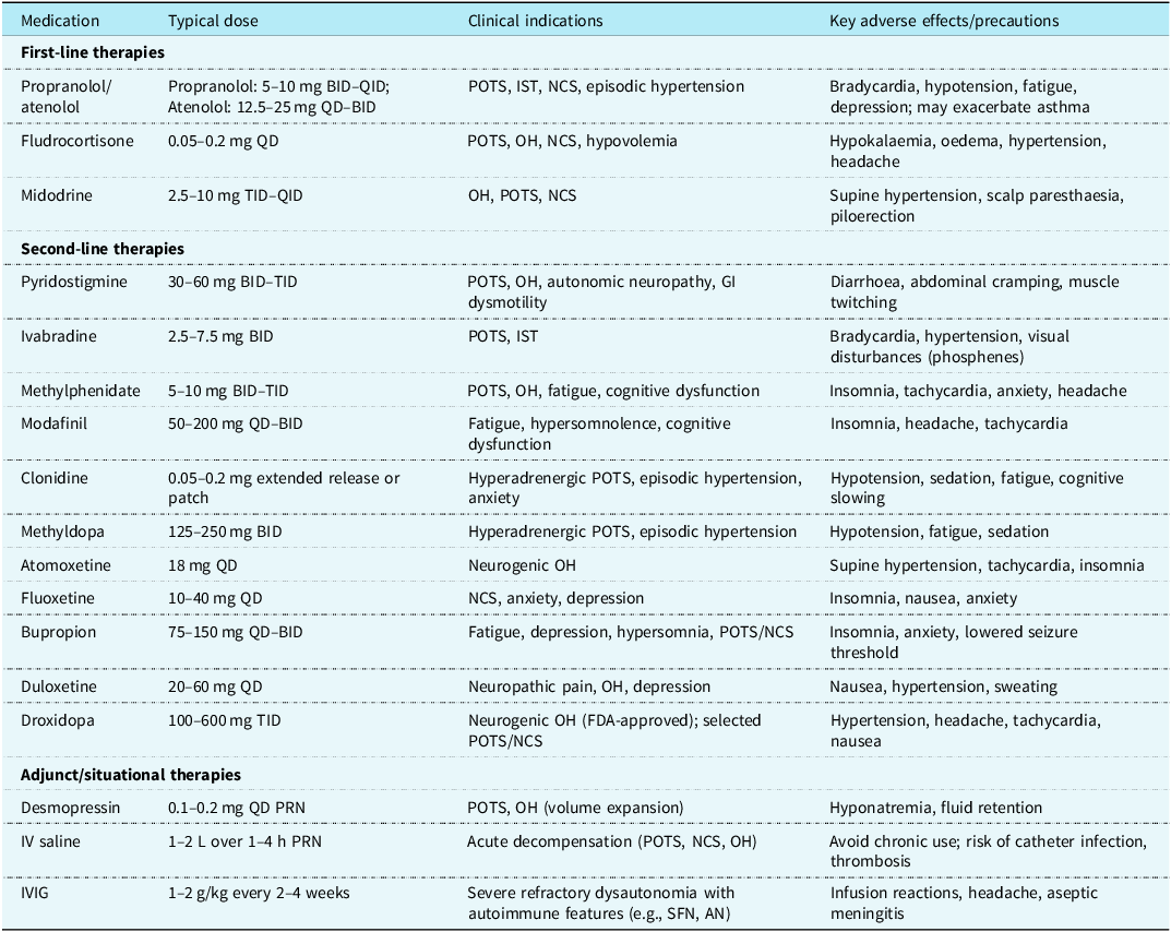
Pharmacologic therapy should be reserved for patients with persistent symptoms despite optimised nonpharmacologic management and should target the dominant physiologic mechanism (Table 2). Beta-blockers and ivabradine may reduce tachycardia in hyperadrenergic or inappropriate sinus tachycardia phenotypes by lowering heart rate, prolonging diastolic filling time, improving cardiac output, and enhancing cerebral perfusion.Reference Stewart102 Fludrocortisone may benefit patients with hypovolemia, whereas midodrine improves peripheral vasoconstriction in neuropathic or hypotensive phenotypes. Pyridostigmine may be useful in selected patients with autonomic neuropathy or gastrointestinal dysmotility.Reference Raj, Guzman and Harvey16 Additional agents, including droxidopa and atomoxetine, may be considered in refractory cases, although paediatric evidence remains limited.Reference Treadwell, Wagner, Reston, Phillips, Hedden-Gross and Tipton100 Symptom-directed therapies may also be helpful; for example, ondansetron can be used for nausea. Melatonin is a safe and commonly used option for insomnia in adolescents with dysautonomia and may improve both sleep quality and autonomic balance.Reference Stewart, Boris and Chelimsky3
Phenotype based pharmacologic therapies for post COVID dysautonomia

POTS = postural orthostatic tachycardia syndrome; IST = inappropriate sinus tachycardia; NCS = neurocardiogenic syncope; OH = orthostatic hypotension; IV = intravenous; IVIG = intravenous immunoglobulin; SFN = small-fibre neuropathy; AN = autonomic neuropathy; BID = twice daily; TID = three times daily; QD: once daily; PRN: as needed.
Evidence supporting clonidine in paediatric dysautonomia remains limited. Its use is based primarily on physiological rationale, particularly its central sympatholytic effects in hyperadrenergic postural orthostatic tachycardia syndrome, together with extrapolation from adult studies.Reference Lin, Kote and Andersson103 In selected adolescents with hyperadrenergic phenotypes, long-acting clonidine may reduce sympathetic outflow and orthostatic tachycardia, although its use may be limited by sedation, fatigue, and hypotension.
Stimulant medications may be considered in adolescents with prominent fatigue or cognitive dysfunction, supported by observational paediatric autonomic data and expert consensus statements.Reference Kanjwal, Saeed, Karabin, Kanjwal and Grubb104,Reference Boris and Bernadzikowski105 Immunotherapy should be reserved for selected patients with suspected immune-mediated dysautonomia or systemic autoimmune disease.Reference Blitshteyn, Funez-Depagnier, Szombathy and Hutchinson106 Several studies describe improvement with intravenous immunoglobulin, plasma exchange, or corticosteroids, although evidence remains limited and randomised paediatric trials are lacking.Reference Schofield and Chemali107–Reference Vernino, Hopkins, Bryarly, Hernandez and Salter110
Lifestyle modification & exercise rehabilitation
Lifestyle modification is a cornerstone of dysautonomia management. Symptom improvement is often achieved through optimisation of sleep hygiene, increased hydration, higher dietary sodium intake, structured meal patterns, and regular physical activity.Reference Speer, Naumovski, Semple and McKune111,Reference Cavigli, Fusi and Focardi112 Because these interventions can be challenging to maintain, some patients may benefit from cognitive-behavioural support aimed at improving adherence, enhancing coping strategies, and promoting functional recovery.Reference Anderer113,Reference Caliman-Sturdza, Gheorghita and Lobiuc114 Although a recent trial in patients with cognitive long COVID found no significant differences among online cognitive training, structured cognitive rehabilitation, and transcranial direct current stimulation, supportive behavioural interventions may still help patients adapt to chronic symptoms and participate more effectively in rehabilitation programmes.Reference Luyer, Habes, van Hak and Buurman115
A balanced diet may complement autonomic management by supporting overall cardiovascular and metabolic health. Emphasis should be placed on regular meals that include fruits, vegetables, whole grains, and lean protein sources while limiting excessive caffeine intake, sugar-sweetened beverages, and highly processed foods that may exacerbate symptoms.Reference Breier, Paranjape and Scudder96
Structured exercise training is recommended for children and adolescents with orthostatic intolerance and physical deconditioning (Class IIa, Level of Evidence B).Reference Shen, Sheldon and Benditt5,Reference Nerli, Selvakumar and Cvejic116 Exercise programmes should begin with recumbent or semi-recumbent aerobic activities, such as rowing, recumbent cycling, or swimming, and gradually progress to upright endurance and resistance training. This approach improves skeletal muscle pump function, stroke volume, and circulatory reserve while minimising symptom exacerbation during early rehabilitation.Reference Trimble, Switzer and Blitshteyn26,Reference Fu and Levine117,Reference Berry, McKinley and Bayes118 Controlled studies have demonstrated improvements in haemodynamic parameters and functional capacity with structured exercise training,Reference Trimble, Switzer and Blitshteyn26 and one study reported superior functional outcomes compared with beta-blocker therapy.Reference Fu, Vangundy, Shibata, Auchus, Williams and Levine119 For patients with post-exertional symptom exacerbation, individualised pacing strategies and gradual progression of activity are preferred over rigid exercise prescriptions.Reference Gross, Carmilani and Stockwell10
Psychological support and school reintegration
Behavioural health concerns increased substantially during the post-COVID era, particularly among female adolescents and young adults.Reference Auro, Holopainen, Perola, Havulinna and Raevuori120 Psychological assessment and supportive interventions may improve outcomes in paediatric dysautonomia by addressing common comorbidities, including anxiety, depressive symptoms, sleep disturbances, pain amplification, and maladaptive avoidance behaviours that contribute to disability regardless of the underlying physiologic mechanism.Reference Vernino, Bourne and Stiles30,Reference Kizilbash, Ahrens and Bruce121–Reference Miller, Sheehan, Bourne, Feeley and Arnold123 Psychological support may further enhance recovery by improving adherence to rehabilitation programmes, strengthening coping skills, and facilitating school reintegration.Reference Anderer113
Interdisciplinary rehabilitation programmes have demonstrated improvements in functional disability, psychological distress, and cognitive symptoms among adolescents with orthostatic intolerance.Reference Attard, Attard and Stanniland124 Collaboration among healthcare providers, families, and schools is essential to maintain academic participation while physiological stability is restored.Reference Sherby, Kalb and Coller125 Individualised accommodations may include flexible scheduling, unrestricted access to hydration, reduced standing requirements, temporary workload modification, scheduled rest periods, and graded return-to-school plans. Cognitive-behavioural approaches incorporating behavioural activation, relaxation training, and cognitive restructuring have also been associated with reduced functional impairment and improved functional participation in adolescents with orthostatic intolerance.Reference Attard, Attard and Stanniland124
Conclusions
The phrase “Never the Heart, Always the Brain” reflects growing recognition that post-COVID paediatric dysautonomia is neither solely a primary cardiac disorder nor exclusively a central nervous system disorder. However, this framing may oversimplify a complex physiology involving tightly interconnected autonomic, cardiovascular, and neurovascular mechanisms. Post-COVID dysautonomia is best conceptualised as a distributed brain–heart–vascular network disorder in which tachycardia often represents a compensatory response to impaired circulatory regulation rather than intrinsic cardiac disease. Accurate diagnosis requires standardised orthostatic assessment, careful clinical phenotyping, and exclusion of structural heart disease and arrhythmia. Management should follow a mechanism-guided, multidisciplinary approach that emphasises patient education, optimisation of hydration and sodium intake, individualised rehabilitation, pacing strategies when post-exertional symptom exacerbation is present, psychological support, school reintegration, and phenotype-directed pharmacologic therapy for persistent functional impairment. Persistent gaps in paediatric disease definitions, mechanistic understanding, and evidence-based treatment underscore the need for prospective paediatric studies to improve diagnostic precision, inform individualised therapeutic strategies, and enhance long-term outcomes.
Financial support
None.
Competing interests
The authors declare that they have no known competing financial interests or personal relationships that could have appeared to influence the work reported in this paper.
Ethical standard
None.