Introduction
Radiation therapy (RT) is one of the most important methods for cancer treatment, with an estimated 50% of patients having received external RT in 2025 in EU-28 (Ref. Reference Borras1). Technology, especially in the medical imaging and accelerators domains, has been the main driver of RT advances. Despite the remarkable improvements in tumour dose conformation achieved (Ref. Reference Bernier, Hall and Giaccia2), adverse side effects of RT are still common. Normal tissue tolerances keep being the main limitation in RT and compromise an effective treatment of radioresistant tumours.
In recent years, RT is becoming a biology-driven discipline. The ever increasing evidence of the importance of the so-called ‘non-targeted’ effects on the biological response to radiation (Ref. Reference Mothersill, Rusin and Seymour3), such as cell signalling effects (Ref. Reference Rodel4), the role of the vascular (Ref. Reference Park5), stromal and immunological changes (Ref. Reference Weichselbaum6) induced in the tumour and its microenvironment by the radiation, is leading to a paradigm shift in RT. The impact of those ‘non-targeted’ effects on the biological response to radiation starts to be considered as key mechanisms themselves to improve the therapeutic index in RT.
Standard (external) RT has been restricted to the same few physical parameters (Refs Reference Steel, McMillan and Peacock7, Reference Murshed8): the same few temporal schemes (the most common being one session of 2 Gy/day during several weeks), low dose rates (<10 Gy/min), beam particle type (photons are used in 90% of the treatments) and spatial distributions (homogeneous dose distributions, mostly using large beam sizes of several square centimetres or millimetres in the case of stereotactic radiosurgery leading to homogenous dose distributions). Interestingly, the close bond existing in RT between physics and biology may allow an activation/modulation of the aforementioned ‘non-targeted’ effects by tuning the physical parameters (dose delivery method) of irradiation. To cite a few recent examples:
1. Temporal schemes using very high-dose radiation in one fraction could transform the immunosuppressive tumour microenvironment (TME) resulting in an intense CD8 T-cell tumour infiltrate (Ref. Reference Filatenkov9).
2. Very high-dose rates (>40 Gy/s), as those employed in FLASH therapy, appear to prevent both activations of the TGF-β/SMAD cascade and acute apoptosis in blood vessels, among other effects, resulting in a significant gain in normal tissue tolerances (Ref. Reference Friedl10).
3. The use of protons instead of photons has been considered advantageous mainly because of a more localised release of dose, in the so-called Bragg peak region. However, currently, there are clues that proton radiation might also have distinct biological properties (Ref. Reference Girdhani, Sachs and Hlatky11), such as an enhanced ability to inhibit tumour angiogenesis or an increased sensitivity to T-lymphocyte killing of tumour cells (Ref. Reference Gameiro12).
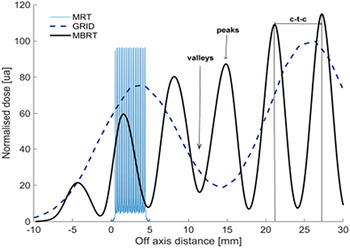
4. Finally, the utilisation of distinct spatial distributions, such as in spatially fractionated radiotherapy (SFRT) also activates different biological mechanisms from those involved when direct damage by ionizing radiation takes place. SFRT uses a combination of spatial fractionation of the dose and narrow beams (Refs Reference Mohiuddin13–Reference Dilmanian16). The dose profiles in SFRT consist of regions of high dose or ‘peaks’ and regions of low dose or ‘valleys’. This contrasts with the flat profiles in standard RT (Refs Reference Prezado17, Reference De Marzi18) (see Fig. 1). The beams may be spatially fractionated in one or two dimensions. SFRT does not attempt to treat the whole tumour like the conventional approaches. Instead, SFRT allows for the delivery of high radiation doses only in some areas within tumours, especially bulky tumours. This avoids the induction of detrimental toxicities to surrounding tissues.
Lateral profiles in SFRT and comparison of several forms of SFRT. Taken from (Ref. Reference Prezado17).

There are four main types of SFRT: GRID therapy (Ref. Reference Mohiuddin13), LATTICE therapy (LRT) (Ref. Reference Amendola19), microbeam radiation therapy (MRT) (Ref. Reference Slatkin15) and minibeam radiation therapy (MBRT) (Ref. Reference Dilmanian16). While a significant number of patients have already been treated with the first two (Refs Reference Amendola19–Reference Duriseti21), MRT and MBRT are still at preclinical stage. A remarkable reduction in normal tissue toxicity compared to standard RT was observed (Ref. Reference Yan22). All four types have been mainly explored with X-rays thus far. However, the domain is rapidly moving towards the use of charged particles instead. More details will be given in ‘Forms of spatially fractionated radiotherapy’ section.
Forms of spatially fractionated radiotherapy
As described in the Introduction, there are four main types of SFRT: GRID therapy, LRT, MRT and MBRT. One of the main differences among them is the geometry and size of the beamlets (hot spots). Table 1 summarises the main features of each of them. However, there are numerous indications that the distinction among the techniques is more profound than a simple difference in geometry or scale. Likely, not all the biological mechanisms playing a role in one of the techniques will be acting or have the same relative weight in the others. One example is the prompt vascular repair observed in normal tissues after MRT (Ref. Reference Bouchet23), an effect which has never been observed in any of the other techniques. Further details will be presented in the next subsections.
Summary of the main features of the different techniques in SFRT

GRID therapy
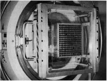
GRID therapy originated only a few years after the birth of RT itself. A. Köhler proposed in 1909 (Ref. Reference Laissue, Blattmann and Slatkin14) the use of a perforated screen (grid collimator) at the exit of the X-ray tube to split the irradiation field in a series of small pencil beams. The aim was to reduce the skin toxicity when deep-seated tumours were to be treated with the orthovoltage machines existing at that time. The technique was commonly and successfully used until the 1950s (Ref. Reference Marks24). The arrival of the medical linear accelerators (LINACS), providing beams with higher penetration and better skin sparing, destined Grid to oblivion for several decades. GRID therapy was rediscovered in the 70s using Co-60 units and in the 90s using LINACS (Ref. Reference Meyer25) (see Fig. 2). Photon GRID therapy is currently delivered mainly with brass collimators but also with multileaf collimators (Ref. Reference Billena and Khan20). The beamlet sizes range between 1 and 2 cm. New developments include TOMOGRID, which uses helical tomotherapy to improve the dose conformity of GRID (Ref. Reference Zhang26). Peak-to-valley dose ratios (PVDRs) range from 2 to 5 and prescription doses between 10 and 20 Gy in one fraction.
GRID collimator in the tray holder of a Philips SL75/5. Taken from Mohiuddin et al. (Ref. Reference Gameiro12).

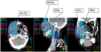
A compilation of most clinical GRID studies reported thus far can be found in Billena et al. (Ref. Reference Billena and Khan20). Those include mainly palliative treatments of head and neck cancers, soft tissue sarcomas and some other bulky tumours. Figure 3 shows a treatment plan example of a head and neck cancer case (Ref. Reference Penagaricano27).
Axial, sagittal and coronal views of the dose distribution of a GRID treatment of a head and neck patient. Taken from Penagaricano et al. (Ref. Reference Meyer25).

The results of available clinical data on GRID therapy indicate a significant increase in therapeutic index. However, the lack of a control arm, the presence of a heterogeneous population, an uncontrolled combination with standard RT and a dose prescription point constitute some important limitations of the reported studies. In particular, the dose prescription is usually made at the surface or at the depth of the maximum dose deposition in the peak region. Consequently, the same GRID geometry and prescription will result in very different doses both to the target and organs-at-risks depending on the specific anatomical context.
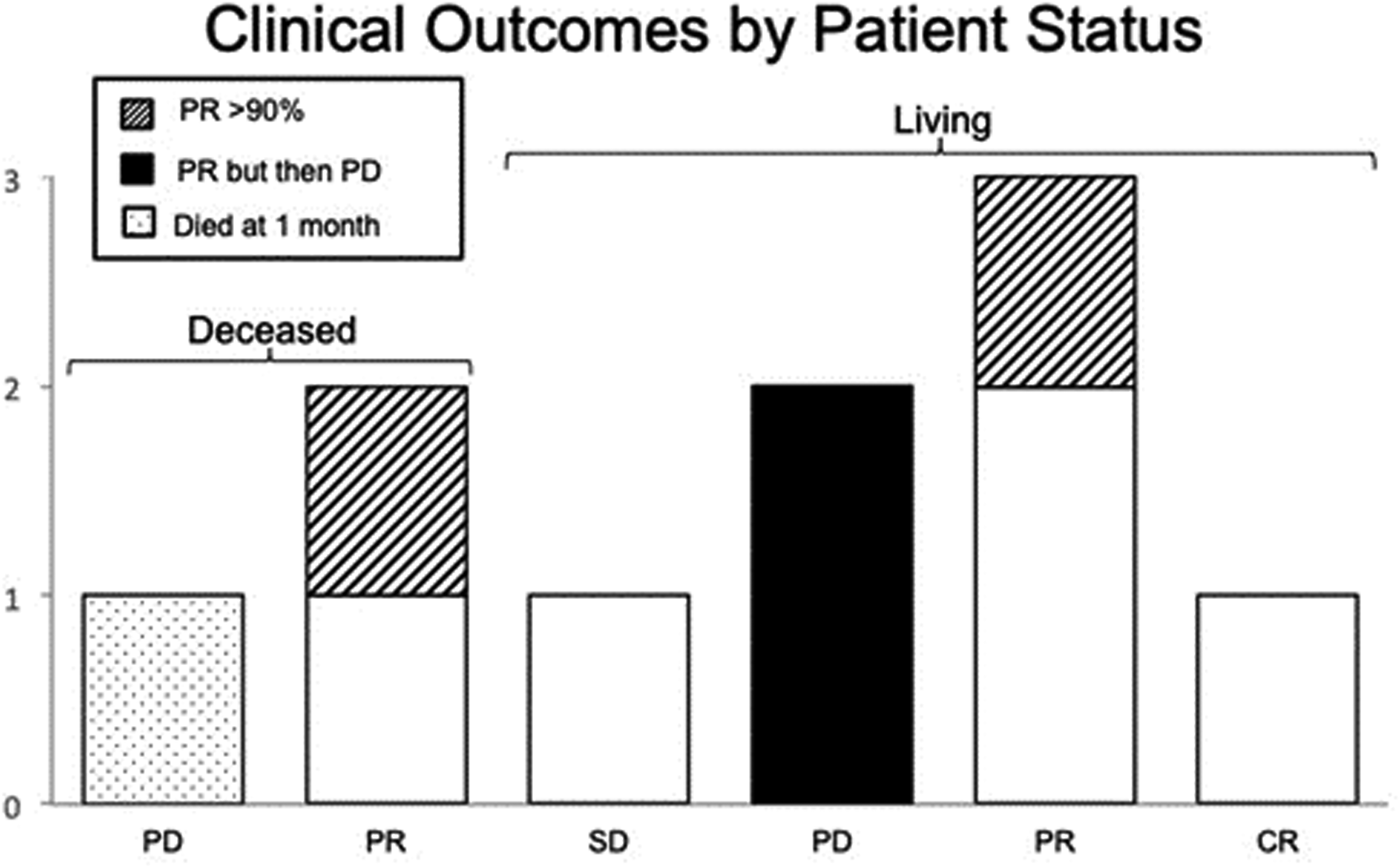
A recent evolution is proton GRID therapy (Refs Reference Henry28, Reference Gao29), proposed a few years later than proton minibeam radiation therapy (pMBRT) (Ref. Reference Prezado and Fois30). Like photon GRID therapy, proton GRID therapy uses beamlets of ~1 cm diameter spaced by 2–4 cm. Thus, in order to have a characteristic pattern of peaks and valleys, the technique is limited to the treatment of bulky tumours (mainly palliative treatments). The use of protons results in a more uniform dose within the tumour (lower PVDR) while helps sparing normal tissues located beyond the tumour. Negligible dose is deposited after the Bragg peak position. These advantageous characteristics may benefit patients who could be treated with photons neither with standard broad beam (BB) irradiations nor with photon GRID, due to the dose tolerance of normal tissues. The treatment of 10 patients (large tumours) proton GRID therapy (1 cm-wide beams) with palliative intent has been reported (Ref. Reference Mohiuddin31). The median time to last follow-up was around 6 months. At last follow-up, seven were alive and three had died (see Fig. 4). A percentage of 60% of the treated patients suffered from pain, 30% of the patients experienced a decreased range of motion. Bleeding and hydronephrosis were observed in 20 and 10% of the patients, respectively.
Clinical outcomes by patient status. CR, complete response; PD, progressive disease; PR, partial response; SD, stable disease. Taken from (Ref. Reference Gao29).

LATTICE therapy
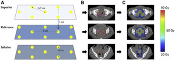
LRT was proposed in 2010 (Refs Reference Wu32, Reference Xiaodong Wu33). LRT (Refs Reference Amendola19, Reference Wu32) is a concept that has evolved from 2D grid to 3D distribution of high-dose volumes (‘vertices’) or lattice in the tumour volume. In contrast to GRID therapy, the spatial fractionation is only present in the target, where multiple localised high-dose small spheres (1–2 cm size) called vertices are produced within the tumour. Their separation ranges from 2 to 6 cm. The dose is diminished in the surrounding normal tissues (Ref. Reference Duriseti21) with respect to conventional irradiations (see Fig. 5). Some proposed techniques for delivering LRT include intensity-modulated radiation therapy or volumetric-modulated arc technique (Ref. Reference Teoh34), multiple focused non-coplanar beams with a robotic-controlled linear accelerator, and proton/heavy ion beams with spot scanning nozzles (Ref. Reference Wu32).
(A) Geometric representation of sphere placement. (B) Axial computed tomography slices of a target with the spheres outlined. (C) Dose distributions after volumetric are therapy planning for the target. Taken with permission from (Ref. Reference Mohiuddin31).

More than 150 patients with late-stage bulky tumours have received LRT (Refs Reference Amendola19, Reference Amendola35–Reference Pollack39). Responses to LRT varied depending on clinical conditions and a summary can be found in (Ref. Reference Wu32). LRT was applied either alone or in combination with conventionally fractionated RT, chemotherapy or immunotherapy (Ref. Reference Jiang40). The majority of cases received LRT with palliative intent as partial ablative tumour debulking or as boost treatment. In the latter case, complete remission of a stage IIIA non-small lung cell cancer in one patient has been reported (Ref. Reference Amendola35). Additionally, complete remission of one metastatic lesion has also been reported in one case report after LRT combined with anti-PD1 immunotherapy (Ref. Reference Jiang40).
Finally, the use of charged particles and LRT has also been started to be explored. A recent treatment plan evaluation study has proven the feasibility of LRT using proton and carbon ion pencil beam for sinonasal adenoid cystic carcinoma (Ref. Reference Yang41).
Microbeam radiation therapy
MRT is the most extreme form of SFRT (Ref. Reference Slatkin15). It uses an array of 25–100 μm-wide beams spaced by 200–400 μm. It originated at large synchrotrons (Refs Reference Slatkin15, Reference Slatkin42), facilities which provide beams with suitable features for this technique: very high flux, kilovoltage (kV) energies and negligible divergence (Ref. Reference Gil43). Numerous small animal experiments have shown that MRT spares normal tissues at extremely high doses (300–600 Gy peak dose in one fraction) (Refs Reference Bouchet23, Reference Bouchet44–Reference Brauer-Krisch49). MRT has also proven to control highly aggressive tumour models (Ref. Reference Fernandez-Palomo50), when very high doses (300–600 Gy) are deposited. While most of the studies have used unilateral irradiations and one single-fraction, a few recent studies have showed the possible gain of either a temporal fractionation in MRT using a crossfire geometry (Ref. Reference Fernandez-Palomo51) or of the use of multiple beam arrays (Refs Reference Fernandez-Palomo51, Reference Eling52). At large synchrotrons, such as the European Synchrotron Radiation Facility (ESRF), the available dose rates (~10 000 Gy/s) fall within the FLASH effect regime (Refs Reference Favaudon53, Reference Mazal54). However, thus far, no cumulative FLASH and SFRT effects have been observed. Similar sparing effect was found after MRT irradiations at ultra-high-dose rates versus MRT irradiations at dose rates precluding FLASH both in vivo (Ref. Reference Smyth55) and in vitro (Ref. Reference Steel56). This might be due to a possible existence of an upper dose limit for the activation of FLASH effect (40–50 Gy (Ref. Reference Vozenin57)), significantly lower than the doses usually employed in MRT.
MRT requires the use of kV beams (inadequate for deep-seated tumour treatments) (Ref. Reference Prezado58). Higher beam energies would reduce too much the spatial dose modulation and thus, the benefits of MRT. The combination with charged particles is not favourable either (Ref. Reference Martinez-Rovira, Fois and Prezado59). The use of micrometric beams imposes the need for very high-dose rates to avoid that the cardio synchronous pulsations result in the loss of the characteristics peak and valley pattern (Ref. Reference Manchado de Sola60). For more than two decades, the required beam flux has only been available at a few synchrotron facilities worldwide. Recent technical developments to produce more compact sources, such as carbon nanotube systems (Ref. Reference Hadsell61) and line-focus X-ray tube (Ref. Reference Winter62), may allow for the first systematic and comprehensive radiobiology experiments and facilitate the translation of the technique to clinical trials.
MRT is one of the most intriguing and fascinating preclinical RT techniques but the most complex one as well. Despite 30 years' existence, no patients have yet been treated with MRT. This is explained by the confinement to large synchrotrons offering limited beamtime, a small scientific community, together with the high complexity of this technique both technically and dosimetrically (Ref. Reference Prezado JFA63). Finally, in September 2021, the first veterinary (canine) patient was treated at the ESRF (personal communication R. Serduc). This is an important step forward towards the first human patient.
The current challenge is to develop safe protocols allowing to investigate the diverse range of potential clinical applications of MRT, having identified and explored through in vivo studies. Among others MRT could be used as a boost for conventional RT (Ref. Reference Bouchet64), as a primer for drug delivery thanks to a transient vascular permeability in the tumour induced by MRT (Ref. Reference Sabatasso65) or to alleviate the symptoms of neurological conditions. In this context, MRT could be used to deliberately transect key neuronal pathways or ablate highly localised regions of the brain in order to modulate or suppress the networks responsible for abnormal movement as well as to treat epilepsy (Refs Reference Serduc66–Reference Pouyatos69).
Minibeam radiation therapy
MBRT was proposed in 2006 to overcome the main limitations of MRT while still providing a very high therapeutic index (Ref. Reference Dilmanian16). The beam widths range from 0.3 to 1.0 mm, which enable its implementation outside synchrotrons into low-cost equipment, such as small animal irradiators (Refs Reference Prezado70, Reference Bazyar71). MBRT also allows the use of higher beam energies with respect to MRT (Ref. Reference Prezado58).
The beams used in MBRT remain narrow enough to achieve a significant increase of normal tissue tolerances (Refs Reference Dilmanian16, Reference Prezado70, Reference Deman72, Reference Prezado73). A significant tumour control effectiveness has also been observed in glioma-bearing rats (Refs Reference Deman74–Reference Sotiropoulos76). Thereby, MBRT is a technique that could offer a good compromise between the thick beams used in GRID and LRT, which impose a limit in normal tissue dose tolerances, and the extreme conditions of MRT.
Some first treatments in veterinary patients (dogs) suffering from brain tumours have been performed with promising results (Ref. Reference Kundapur77). All dogs showed almost complete response on the follow-up MRI at the time of the report (Ref. Reference Kundapur77). Four of seven dogs had a pathological complete response and one dog was still on follow-up. Fewer long-term pathologically correlated toxicities were observed with respect to dogs treated with standard RT.
The high therapeutic index of MBRT could potentially be further increased by combining it with the superior relative biological effectiveness of charged particles, their more selective depth dose deposition as compared with photons along with their negligible dose deposition after the Bragg peak. Moreover, there are some indications that heavy ions, such as carbon, neon or argon, can potentiate the immune response in a more effective way than photons or even proton beams (Ref. Reference Tinganelli and Durante78). This might result in a highly effective synergic effect with the potential immune activation in SFRT (Ref. Reference Potez79).
Therefore, the alliance of charged particles and MBRT may allow to fully profit from the advantages of the spatial fractionation of the dose, while paving a direct avenue for optimal future patients' treatments. Different approaches have been explored in the recent years.
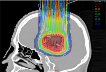
pMBRT represents the most evolved experimental form of SFRT with charged particles (Refs Reference Prezado and Fois30, Reference Zlobinskaya80). It allies the more localised dose distribution in the Bragg peak of protons, with the possibility of achieving simultaneously a homogeneous dose distribution in the tumour and a pronounced spatial fractionation of the dose in normal tissues and all with a single field of minibeams (Refs Reference De Marzi18, Reference Prezado and Fois30) (see Fig. 6). This contrasts with X-ray MBRT, where multiple interlacing fields are usually needed to achieve target dose homogenisation, which increases the technical challenges.
Does distributions in pMBRT. Taken from (Ref. Reference Prezado17).

A series of radiobiological studies have already demonstrated the remarkable normal tissue reduction (skin and brain) provided by pMBRT as compared to standard proton therapy (Refs Reference Zlobinskaya80–Reference Prezado83). A higher inflammatory response and cell death were observed in a three-dimensional (3D) human skin model after standard PT irradiations as compared to pMBRT (Ref. Reference Zlobinskaya80). Standard PT also resulted in higher degree of oedema, erythema or desquamation in mouse ears as compared to pMBRT irradiation at the same average dose (60 Gy) (Ref. Reference Girst81).
pMBRT also leads to a remarkable reduction of neurotoxicity (Ref. Reference Prezado83). Long-term follow-up of rats receiving whole-brain irradiation with either standard PT (25 Gy in one session) or pMBRT (peak doses of 58 Gy/one fraction, average dose of 25 Gy) revealed no significant damage after pMBRT treatments while extensive brain damage, including necrosis, demyelination, astrogliosis and microglial activation, was observed in conventional PT irradiated group (Ref. Reference Prezado83). It has also been demonstrated that pMBRT has no significant impact on cognitive, motor or emotional processes (Ref. Reference Lamirault82).
In addition, the tumour control effectiveness of pMBRT equals or is better than that of standard RT in glioma-bearing rats, avoiding the extensive adverse effects observed in the latter and thus widening the therapeutic window (Refs Reference Lamirault84–Reference Prezado86). High percentages of long-term survivals (> 6 months, free of tumour), up to 80%, were achieved in different pMBRT configurations (Refs Reference Prezado86, Reference Bertho87). Tumour sterilisation was achieved even in cases where large areas of the tumour received low (non-tumouricidal) doses (Ref. Reference Prezado85). This contradicts the classical paradigm of RT requiring the deposition of a lethal dose in every tumour cell for tumour ablation. Recent evaluations point at a determinant role of the immune system in the anti-tumour response of pMBRT (Ref. Reference Annaig Bertho88). Moreover, the fact that large areas (or even whole brain) could be irradiated without significant side effects would allow overcoming one of the major difficulties to be faced in gliomas treatment: its infiltrative nature.
The biological data gathered so far in pMBRT encourages the preparation of phase I clinical trials. Along that line, a first treatment plan evaluation study in two different brain tumours, glioma and meningioma, revealed that pMBRT plans can meet the criteria recommended for clinically acceptable dose-volume histograms in organs at risks. Satisfactory homogeneity was achieved in the target while a similar level of spatial dose modulation in normal tissues as in preclinical studies was obtained (Ref. Reference Lansonneur89).
With the objective to reach clinical trials in the next few years, important efforts have concentrated on the optimisation of the minibeam generation at clinical centres. Some important milestones, like the design of a scanning dynamic collimator for spot-scanning proton minibeam production (Ref. Reference Sotiropoulos and Prezado90) and of a new nozzle design for magnetically focussed minibeams (Ref. Reference Schneider91), were accomplished. The latter approach removes the need for any mechanical collimation, which improves irradiation efficiency and flexibility as well as the degree of spatial fractionation in healthy tissue. Moreover, a recent conceptual design study has showed that this new minibeam nozzle in combination with proton LINAC accelerators such as LIGHT (Advanced Radiotherapy Plc) may provide an optimal implementation of pMBRT by allowing the generation of magnetically focussed minibeams with clinically relevant parameters (Ref. Reference Schneider92). It could furthermore be used for conventional pencil beam scanning. In addition, the high irradiation efficiency of magnetically focussed minibeams could pave the way towards a combination of pMBRT and proton FLASH therapy at a clinical proton accelerator (Ref. Reference Schneider92).
In addition, the reduced multiple Coulomb scattering and higher relative biological effectiveness of heavier ions (Helium to Oxygen) as compared to protons might be advantageous for SFRT. So far, mainly dosimetry studies have been carried out, both theoretical (Monte Carlo (MC) simulations) and experimental dosimetry (Refs Reference Dilmanian, Eley and Krishnan93–Reference Martinez-Rovira97). The favourable dosimetry revealed in those investigations encourages the realisation of biological experiments. In the only biological experiment published up to date (Ref. Reference Dilmanian98), one rabbit brain was irradiated with several arrays of 130 MeV/nucleon carbon 300 μm-wide beams interlacing at the target, showing a reduced impact on the non-targeted tissues. Biological experiments are warranted to confirm the clinical potential of this approach.
Finally, another innovative approach is the combination of MBRT with very heavy ions, such as Neon or Argon. Their reduced oxygen enhancement effect benefits the treatment of hypoxic tumours, which remains one of the major challenges in RT. Indeed, their effectiveness was demonstrated in the past in clinical trials, which were discontinued due to important late tissue results, causing the use of those beams to be discontinued (Ref. Reference Castro99). Nevertheless, the gain in tissue sparing that might be provided by the use of MBRT might allow profiting from the remarkable effectiveness of those heavy ions, as firstly proposed by Peucelle et al. (Ref. Reference Peucelle, Martinez-Rovira and Prezado100). The advantageous dose distributions obtained in the first MC studies (Refs Reference Gonzalez and Prezado96, Reference Peucelle, Martinez-Rovira and Prezado100) encouraged the further exploration of this avenue: extremely high PVDR values (>100), very narrow penumbras and low valley doses were achieved in the first centimetres as well as in the fragmentation tail, helping in the sparing of proximal normal tissues. Although the yield of secondary nuclear products increases with atomic number, the actual dose being deposited by the secondary nuclear fragments in the valleys becomes dominant only at the Bragg peak (tumour) position (Ref. Reference Peucelle, Martinez-Rovira and Prezado100).
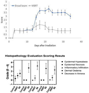
The first in vivo experiment was carried out at HIMAC-NIRS (Japan). Among the different ion species, Ne ions were selected since they were shown to offer the best balance between having a high PVDR and peak-to-valley-linear energy transfer ratio in normal tissues and high linear energy transfer values (close to 100 keV/μm) in the target region (Ref. Reference Gonzalez and Prezado96). Mice legs were irradiated with either standard BB irradiation or with MBRT (20 Gy average dose at 5 mm depth in one fraction) with 230 MeV/A Ne ions and skin reaction evaluated. A percentage of 75% of the animals in the standard BB group reached the endpoints (ulceration, necrosis) two weeks after irradiated and were sacrificed. In contrast, in the MBRT groups, only (reversible) dermatitis was observed (score <2). This suggests a clear gain of Ne MBRT in terms of normal tissue sparing (Ref. Reference Prezado101). Figure 7 depicts the evolution of the skin damage scores as a function of the time and irradiation mode as well as the histology results.
Up: Evaluation of average skin damage scores as a function of follow-up time and irradiation mode. Mean and standard deviation values are represented in the plot. Down: Histology evaluation scoring results showing mean values and standard deviations for the different irradiation modes. Taken from (Ref. Reference Prezado101).

Biological mechanisms in SFRT
All the aforementioned observations point at distinct biological mechanisms put into action in SFRT. Most mechanistic evaluations have been performed in MRT or MBRT studies, as the beam sizes are the most suitable for small animal experiments. Although the full picture is missing, all the experiments show that there is a differential effect between the tumour and normal tissues. The main potential mechanisms described in the literature are the following:
1. Differential vascular effects
2. Cell signalising effects (bystander-like effects)/abscopal effects
3. Inflammation and immunomodulatory effects
4. Cell migration
5. Free radical production and diffusion covering the valley regions in the tumours.
In the next subsections, details on each of them will be provided.
Differential vascular effects
A differential vascular effect in the normal versus tumoural tissues has been observed in MRT experiments (Refs Reference Bouchet23, Reference Sabatasso102). Concerning normal tissues, MRT does not modify any structural or functional parameters such as blood volume, vascular density and perfusion (Refs Reference Serduc46, Reference Serduc103). The endothelial cell lining of the vessels in the microbeam paths remains intact (Ref. Reference Bouchet23). MRT exhibits a preferential damaging effect on the immature vessels, while mature microvasculature is preserved (Ref. Reference Sabatasso102). This has led to the hypothesis that immature blood vessels in the tumour will be more sensitive to MRT while the healthy tissue mature blood vessels will be resistant to MRT. Several studies have reported altered blood vessel integrity, decrease in the number of tumour vessels and significantly reduced blood perfusion, leading to a decrease in perfusion and to tumour hypoxia (Refs Reference Bouchet44, Reference Bouchet104, Reference Griffin105). The effect might vary depending on the tumour type but as well on the beam width (Ref. Reference Bronnimann106), spacing and doses (Ref. Reference Bouchet23). No systematic and robust evaluation has been done on the effects of MBRT on the vasculature. The only published data (Ref. Reference Bronnimann106) report vascular damage after MBRT at doses corresponding to the MRT domain (>300 Gy peak dose) and much higher than the therapeutic doses in MBRT (50–100 Gy).
In GRID therapy, only indirect observations on its impact on vasculature have been obtained. Enhanced sphingomyelinase activity and ceramide levels were only observed in patients with complete or partial response to GRID therapy (Ref. Reference Sathishkumar107). Ceramide has been linked to microvascular endothelial cell sensitisation to radiation-induced apoptosis (Ref. Reference Garcia-Barros108). Similar observations were made in an in vivo study in which Lewis lung carcinoma bearing mice were treated with LRT. Their serum exhibited increased acid sphingomyelinase levels (Ref. Reference Kanagavelu109).
Cell signalising effects (bystander-like effects)/abscopal effects
Bystander-like effects have been considered to play a role in SFRT. The cells in the valley regions receiving low doses are referred to bystander cells in SFRT although strictly speaking the term cohort effect would be more adequate (Ref. Reference Wang110).
In preclinical murine models using SFRT, Asur et al. (Ref. Reference Asur111) found that bystander (GRID-adjacent) cells exhibited an increased expression of genes involved in DNA repair, cell cycle arrest, heat shock protein (HSP) and apoptosis immediately after exposure or 4 h after exposure. Some other studies reported indirect observations in GRID and LRT: a significant increase of TNFα was observed in the serum of patients treated with GRID (Ref. Reference Sathishkumar112) and an in vivo study using mice (Ref. Reference Kanagavelu109) with allogenic Lewis lung carcinoma receiving LRT. The involvement of TNF-α in the bystander signalling pathway has been reported (Refs Reference Wang110, Reference Shareef113) by different authors.
Bystander-like effects have also been evidenced in MRT, both in vivo and in vitro (Refs Reference Lobachevsky114–Reference Smith116), with some dependence on the distance from the high-dose region and p53-status of the tumour.
Concerning normal tissues, the activation of some positive bystander-like effects has been hypothesised (Ref. Reference Dilmanian117) and some experimental indirect evidence was reported: increased levels of HSP 71 turnover (Ref. Reference Smith116), known to protect against all of the neurological disorders, were found in the non-irradiated brain hemisphere of rats.
Abscopal effects have also been observed in several experiments (Refs Reference Kanagavelu109, Reference Fernandez-Palomo118–Reference Ventura120). One of the most explicit ones is the study of Kanagavelu et al. (Ref. Reference Kanagavelu109), in which xenograft A549 lung adenocarcinoma was implanted in the two flanks of mice. One of the two tumours was irradiated with LRT. The growth of both tumours was reduced, and the untreated tumour then also responded more than expected with 2 Gy fractionated RT. There was an increase in the number of infiltrating CD3þ T cells in both the irradiated site and the distant site. This correlated with the deceleration of tumour growth.
Ventura et al. (Ref. Reference Ventura120) evaluated abscopal effects in mice irradiated with either MRT or BB. They found an increased number of clustered DNA lesions, double-strand breaks, apoptotic cells as well as local and systemic immune responses in a variety of non-targeted tissues. No significant difference was observed between MRT and BB irradiations, observation which is in agreement with observations made in other studies (Ref. Reference Fernandez-Palomo118). Accumulating evidence now suggests that the immune system is a major determinant in regulating the abscopal effect (Ref. Reference Grass, Krishna and Kim121). Along this line, increasing evidence is cumulating on the important role of immunomodulation in SFRT, as it will be described in the next section.
Immunomodulation
Inflammation and immune activation seem to play an important role in the anti-tumour response of SFRT (Ref. Reference Griffin122).
Bouchet et al. (Ref. Reference Bouchet123) evaluated the early transcriptomic responses (6 h after irradiation) of normal brain and glioma tissue in rats after MRT irradiation. They observed that half of the genes were common in both healthy and tumour tissues and were predominantly associated with immunity or inflammation. They also identified several pathways related with natural killer cells or CD8+ T lymphocytes, and changes in genes such as HMGB1, toll-like receptors, C-type lectins and CD36. These genes can trigger the activation of innate or adaptive immune cells. Similar conclusions were obtained by Sprung et al. (Ref. Reference Sprung124). In addition, Potez et al. (Ref. Reference Potez79) observed an enhanced production of monocyte-attracting cytokines as well as a higher tumour infiltration by CD8+ lymphocytes and natural killers in MRT-irradiated (B16-F10) melanoma bearing mice with respect to BB-irradiated mice and non-irradiated controls.
In addition, in mice with tumours implanted in both limbs and that received spatially fractionated irradiation, evidence of abscopal immune responses was observed in contralateral tumours. Those presented markedly enhanced infiltration of both antigen-presenting cells and activated T cells, which were preceded by increased systemic IFN-γ production and led to eventual tumour growth delay (Ref. Reference Johnsrud125). T lymphocytes have also been shown to play a central role in the anti-tumour response in pMBRT (Ref. Reference Annaig Bertho88). Recent studies demonstrated that combined ablative dose with low dose of radiation could lead to the reprograming of the immunosuppressive TME to become more immunogenic and synergistically augment the anti-tumour response (Ref. Reference Savage, Pandey and Guha126).
Finally, some recent studies also showed that an antitumor immune response can also be evoked by partial-volume single-dose radiation in murine models (Ref. Reference Markovsky127).
In conclusion, the observations gathered so far suggest that the combination of SFRT and immunotherapy could be a winning alliance. The encouraging preclinical results combining MRT and GMIMPR (gene-mediated immunoprophylaxis) (Ref. Reference Smilowitz128) or the results presented in a case report study (Ref. Reference Jiang40) combining LRT and immune checkpoint inhibitors support further advancement in this direction.
Further experiments where the immune response and pathways within healthy tissue and/or tumour are studied in response to SFRT are necessary to fully understand the mechanisms behind SFRT.
Cell migration
Dilmanian et al. (Ref. Reference Dilmanian129) formulated the hypothesis that the sparing of normal tissue could be explained by the migration and proliferation of stem cells in the valley regions to repair the tissue regions under the peak (high) doses, which would result heavily damaged. Some experimental evidence already exits. First Crosbie et al. (Ref. Reference Crosbie130) showed different migration patterns between tumour and healthy tissues. In this study, inoculated EMT-6.5 tumour and normal mouse skin tissues were harvested at defined intervals post-MRT. Gamma-H2AX was used for precise localisation of irradiated cells. Within 24 h of MRT, peak and valley-irradiated zones were indistinguishable in tumours because of extensive cell migration between the zones. This was not seen in MRT-treated normal skin, which appeared to undergo a coordinated repair response. A more recent study by Fukunaga et al. (Ref. Reference Fukunaga131) showed, for the first time, that the survival and potentially migration of the non-irradiated germ cells in the irradiated testes are required for the tissue sparing effect for spermatogenesis.
Chemical mechanism
Exposure to ionizing radiation produces oxygen-derived free radicals in the tissue environment. These include hydroxyl radicals, superoxide anion radicals and other oxidants such as hydrogen peroxide. This may occur either directly by interaction of irradiation with critical organic biomolecules, resulting in the ejection of electrons leading to the formation of an organic-free radical (R•) or indirectly by the ionisation of H2O molecules leading to the formation of hydroxyl radicals (•OH) in close enough proximity to abstract an H• from a critical biomolecule again forming R• (Ref. Reference Steel, McMillan and Peacock7). If O2 were present, R• could react to form ROO•, which was thought to fix the damage in a relatively unrepairable condition (Ref. Reference Omar Desoukya and Zhoub132).
A recent simulation study (Ref. Reference Riccardo Dal Bello133) has suggested the distribution of hydrogen peroxide as the candidate to interpret the MRT and MBRT efficacy. This molecule is a product of water radiolysis, it is stable, and it rapidly reaches the steady state in production versus removal. Moreover, the H2O2 has a strong oxidizing capacity leading to damage of proteins, lipids, and DNA. Although, most ROS have a short half-life and cause damage locally, hydrogen peroxide (H2O2) has relatively longer half-life. The calculations showed that the H2O2 produced in the dose-peaks diffuses to the dose-valleys during beam-on leading to a homogeneous ROS distribution over the target. The model was tested on three previous independent photon micro-beam and proton mini-beam animal experiments. In addition, there is a wealth of evidence that ROS contribute to the bystander effect extracellularly and also intracellularly through a continuous cascade of events (Ref. Reference Azzam134); therefore, both effects might be linked. Recent studies suggest that the ROS generation in proton beams is more important than that of photon radiation, with levels constantly increasing during the first 20 h after radiation (Ref. Reference Alan Mitteer135). Thus, the potential effects might be more important in proton MBRT than in X-rays MBRT. Further experimental evidence is nevertheless needed to confirm the validity of this model.
Discussion and conclusions
The advancement of both technology and radiobiology knowledge of SFRT (Ref. Reference Yan22) is generating a renewed interest in SFRT at many centres worldwide, with newly formed working groups actively working on guidelines and protocols (Ref. Reference Zhang136). A growing number of clinical trials for palliative SFRT are ongoing or being planned. In addition, the domain of SFRT is rapidly moving towards the use of charged particles due to inherent advantages of this combination, which can enable curative treatments for large and/or radioresistant tumours. This could potentially be achieved with techniques like pMBRT (Ref. Reference Prezado and Fois30). The biological data collected thus far encourage the preparation of phase I clinical trials in pMBRT.
An adequate dose prescription remains one of the main priorities in SFRT. Some recent guidelines and protocols (Ref. Reference Zhang136) propose to keep using peak doses as the main efficacy surrogate. However, the correlation of peak dose and tumour response has never been soundly established, but rather contradicted by recent studies (Ref. Reference Rivera137).
As previously indicated, the biological response and mechanisms in SFRT are not completely understood. Radiobiological experiments support the participation of radiation-induced bystander effects, vascular alterations, and immunologic interactions. However, currently, the relative weight and interrelation of those possible contributors and how these effects are translated when the spatial dose distribution is modified are still unclear. No systematic evaluation of the influence of the beam width and spacing on tissue response to spatially fractionated RT has ever been performed. Further biological experiments are urgently needed to parametrise the relationship between the irradiation parameters (beam width, spacing, PVDR, peak and valley doses) and the radiobiology. And, as previously indicated, the utilisation of charged particles may also modify the underlying mechanisms and their relative weights with respect to photons. For example, there are ever increasing pieces of evidence that charged particles are more effective activating the immune system (Ref. Reference Tinganelli and Durante78).
SFRT promises a high therapeutic index in difficult-to-treat cases today. While its clinical use to date has been limited to a few centres, most commonly for palliative or recurrent cases, SFRT does have potential in treating primary cancers such as head and neck, lung, breast, gynaecologic and sarcoma cases with curative intent. Clinical trials incorporating SFRT as a boost or as the primary RT component, in combination with immunotherapy, are warranted.
It might be the time to abandon the old paradigm in RT and embrace new modalities using radically different ways of depositing the dose, such as SFRT and FLASH therapy, and even in the future the combination of both (Ref. Reference Schneider92). The lesson being taught by SFRT and FLASH therapy is that the exploration of the vast ‘terra incognita’ on how the physical parameters of the irradiation modify the biological response can offer enormous opportunities for optimal patient treatments.
Acknowledgements
Y. Prezado warmly thanks the work of the current and former members of her team ‘New Approaches in Radiotherapy’, the personnel of the Orsay proton therapy centre and the Experimental Radiotherapy platform of Institut Curie. Y. Prezado acknowledges the funding from European Research Council (ERC) under the European Union's Horizon 2020 research and innovation programme (grant agreement No 817908).