Introduction
Rhinophyma is a progressive, disfiguring soft tissue hypertrophy of the tip of the nose.Reference Redett, Manson, Goldberg, Girotto and Spence1, Reference Fincher and Gladstone2 Histologically, it is due to hyperplasia of the cells of the sebaceous glands.Reference Shatokhina, Chumakov and Deriugina3 It is a widely accepted theory that rhinophyma represents the end-stage of chronic acne rosacea.Reference Redett, Manson, Goldberg, Girotto and Spence1 The condition can cause visual obstruction, inflammation and bleeding, in addition to cosmetic disfigurement.
Case reports
Case one
A 69-year-old man with rhinophyma complained of cosmetic deformity, infection and bleeding. He had previously been treated with a prolonged course of antibiotics.
The patient's rhinophyma was coblated under general anaesthetic, using an EVac® 70 wand to (Arthrocare, Stockholm, Sweden), a depth of approximately 3 mm. There was minimal bleeding, easily arrested with a short burst of diathermy from the coblating wand. A petroleum jelly gauze dressing was applied. The procedure took 20 minutes.
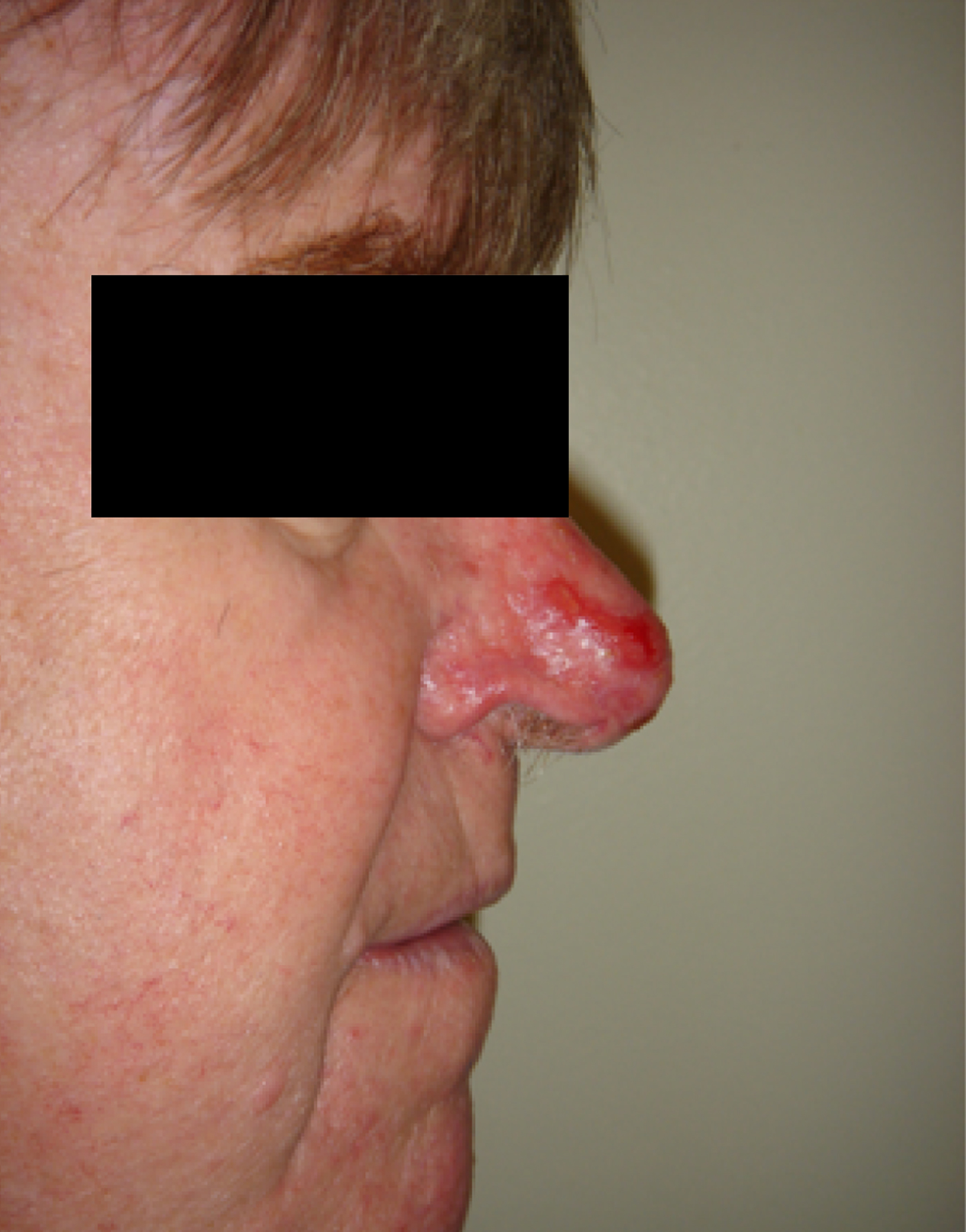
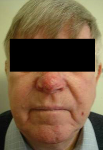
Three days later, the wound was granulating well but there was some mild inflammation. The patient was given a course of amoxicillin. Subsequently, the coblated tissue healed, giving a good cosmetic result (Figures 1 and 2).
Case one, one week post-operatively.
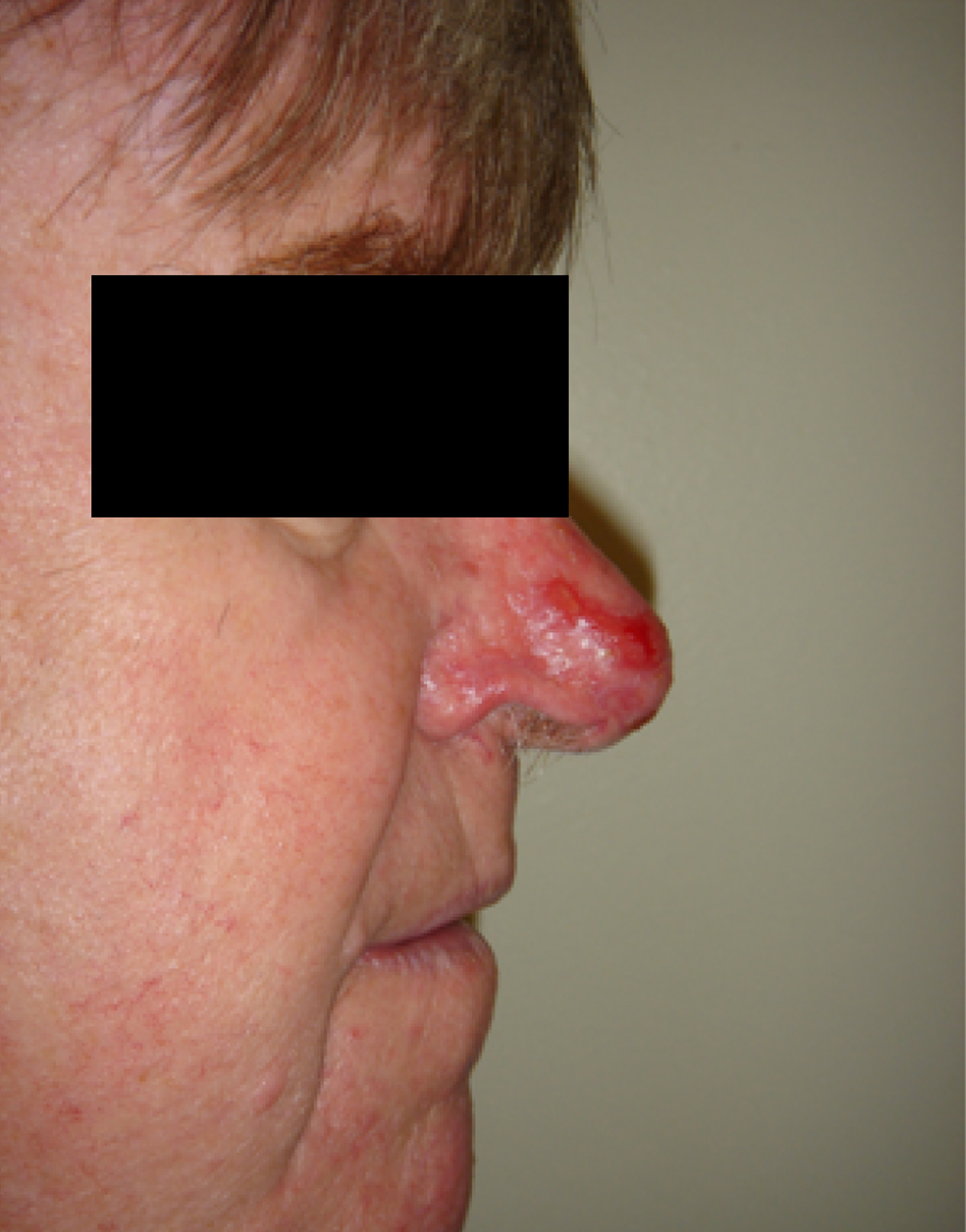
Case one, two weeks post-operatively.

Case two
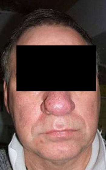
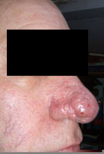
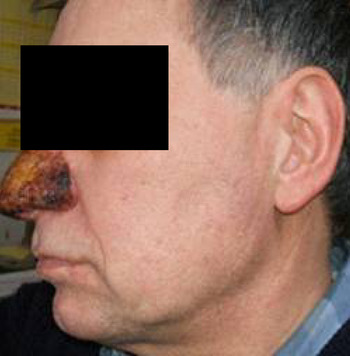
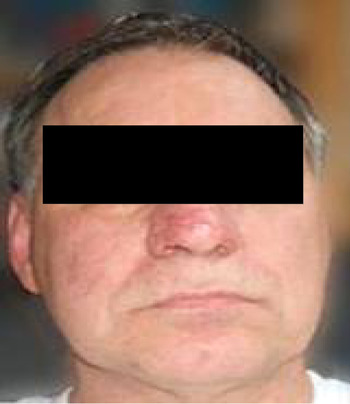
A 59-year-old man presented with a 10-year history of rhinophyma and rosacea which had been treated with oxytetracycline, topical metronidazole and Dalacin T® cream (topical clindamycin, Pharmacia, Sandwich, UK). Despite this treatment, growth of the soft tissue of the nose had ensued, leading to the development of rhinophyma (Figures 3 and 4).
Case two, pre-operatively.

Case two, pre-operatively.

Coblation was performed under general anaesthetic, using the EVac® 70 wand. Tri-Adcortyl ointment (topical clindamycin, Pharmacia, Sandwich, UK) was applied to the nose following surgery. Treatment with oxytetracycline was continued.
Three weeks later, the coblated area was healing well, in an external to internal direction. A 2 × 2 cm area of sloughing was apparent at the tip of the nose, but the surgical margins 1 cm around this area had healed (Figure 5). Some slight bleeding was reported, and treatment with Tri-Adcortyl ointment was continued.
Case two, one week post-operatively.

Four weeks later, the tissue had healed (Figures 6 and 7) and the patient stopped using oxytetracycline. Six months later, he was extremely pleased with the result of the surgery.
Case two, four weeks post-operatively.

Case two, four weeks post-operatively.

Case three
A 54-year-old pilot presented with rhinophyma and rosacea. He had previously been treated with lymecycline, Aknemycin Plus® (topical Erythromycin Ph Eur 4% w/w and Tretinoin BP 0.025% w/w, Uxbridge, UK), erythromycin (500 mg twice daily) and topical metronidazole cream.
On examination, the patient had soft tissue swelling over the tip of his nose.
Coblation debulking was performed under general anaesthetic, using an EVac® 70 wand. The procedure took 30 minutes, and Tri-Adcortyl ointment was applied post-operatively. The patient continued with his treatment with erythromycin and topical metronidazole cream immediately post-operatively.
Nine days later, the patient's nose was healing rapidly, and he was very pleased with the outcome.
Three months later, the patient's skin had cleared and he had discontinued treatment with both erythromycin and topical metronidazole.
Case four
A 61-year-old man presented with a two-year history of rhinophyma and marked facial rosacea. He had had some improvement with lymecycline cream and azelaic acid cream.
The patient underwent coblation-assisted shaving under local anaesthetic, using the EVac® 70 wand. Tri-Adcortyl ointment was applied following surgery.
Two months post-operatively, the patient was reviewed in clinic; the appearance of his nose had much improved. He was discharged to the care of the dermatology team.
Case five
A 67-year-old man presented with an obvious rhinophyma.
He underwent coblation under general anaesthetic, which took 15 minutes to complete. The full nasal triangle was coblated, with feathering, using the EVac® 70 wand. The patient was treated with Tri-Adcortyl ointment for two weeks post-operatively.
Three months post-operatively, the patient's nose had a good shape, and there was little scarring or erythema. The patient was happy with the result.
Case six
A 61-year-old man presented with a large, right-sided rhinophyma and a long-standing history of rosacea. He had been treated unsuccessfully with lymecycline 408 mg once daily.
The patient underwent coblation of the rhinophyma under general anaesthetic. The rhinophyma tissue was reduced down to the epidermal level. The procedure took 18 minutes. Tri-Adcortyl ointment was applied post-operatively for two weeks.
The coblated area healed well, and the patient was referred back to the dermatology team for follow up.
Results
The six reported cases were treated over a two-year period. Patient follow up ranged from one month to over a year. All six patients were happy with the procedure and its results. There were no complaints of excessive post-operative pain. Any milder pain was effectively dealt with using simple analgesia.
One patient was treated with oral antibiotics as the coblated area appeared inflamed.
Several dressings were used in this series. Tri-Adcortyl ointment was found to be effective and easy to use.
We initially performed the coblation procedure under general anaesthetic. However, we found that it was well tolerated when performed under local anaesthetic.
Discussion
Numerous treatment methods have been developed for rhinophyma. Oral antibiotics have shown some success in the early stages.Reference Aferzon and Millman4 The use of systemic isotretinoin has been reported to have some success when combined with sharp excision.Reference Sorhage and Glowania5 Rhinophyma is a fibrotic condition and may be amenable to tamoxifen, a synthetic non-steroidal antioestrogen which can down-regulate transforming growth factor β, associated with fibrotic conditions.Reference Payne, Ko, Ansoaugh, Wheeler, Wright and Robson6
When surgical treatment of rhinophyma is required, the use of a scalpel, together with adrenaline and bipolar diathermy, allows accurate removal of the diseased tissue while preserving the underlying sebaceous glands for spontaneous re-epithelialisation. Sharp dissection procedures include excision with primary sutures, extirpation with plastic covering of the defect by free transplants, subcutaneous resection, decortication and peeling of the proliferations,Reference Payne, Ko, Ansoaugh, Wheeler, Wright and Robson6 dermal abrasion and dermal shaving.Reference Rohrich, Griffin and Adams7, Reference Payne, Ko, Ansoaugh, Wheeler, Wright and Robson6 Dermaplaning and dermabrasion are reported to offer good reproducibility of results.Reference Rohrich, Griffin and Adams7 The microdebrider has also been used (having the same effect as scalpel shaving but allowing more precise contouring), in conjunction with FloSeal®, a gelatin thrombin mixture used to achieve haemostasis.Reference Kaushik, Tahery, Malik and Jones8
Electrosurgical treatment methods use radiofrequency electricity to generate heat in the tissue. This leaves a smooth-edged incision, in a similar fashion to a scalpel.Reference Aferzon and Millman4 Electrocautery is generally accepted to have greater heat dispersion, compared with laser methods; the depth of tissue destruction beyond the operative field approaches 1 mm, compared with 0.5 mm with a laser.Reference Aferzon and Millman4 Scarring has been reported following the use of electrocautery.Reference Jung9
The carbon dioxide laser allows accurate removal of diseased tissue to the desired level without exposing underlying osteocartilagenous tissue. This ensures rapid re-epithelialisation, minimising the scarring which may occur following delayed healing.Reference Jung9 Treatment can be performed under local anaesthetic. The laser creates a black eschar which is wiped off. Re-epithelialisation occurs spontaneously.Reference Jung9 The depth of penetration is 0.1 to 0.2 nm, and a bloodless field is created. Total eradication of diseased tissue is only possible when tissue is excised down to the loose areolar tissue overlying the osteocartilaginous framework.Reference Jung9 The resulting defect must be covered by a skin graft. Cervelli et al. Reference Cervelli, Giudiceandrea, Pigliucci, Maggiulli, Grimaldi and Cervelli10 have reported the case of a patient who received a full thickness dermo-epidermic graft; however, this did not leave a normal cosmetic result.Reference Jung9, Reference Szymanska-Skrzypek, Burduk and Betlejewski11
Subtotal excision using ‘cold’ surgical excision, electrocautery, dermabrasion or laser relies on the retained deep glandular elements for re-epithelialisation.
Other surgical treatment methods for rhinophyma include cryosurgery, ultrasonic scalpel, argon laser and YAG laser.Reference Fincher and Gladstone2, Reference Lim12
Electrocautery is useful for debulking and controlling bleeding, but there is a risk of scarring caused by thermal injury, and the contouring of tissues is less precise and manageable compared with the scalpel.Reference Lim12, Reference Lim13 Rhinophyma treatment with electrocautery and laser has been reported to lead to scar formation, but this has been found to be related to the depth of tissue removed rather than the removal method.Reference Lim14, Reference Lim15
When laser is compared to cold surgical methods of rhinophyma treatment, there is no improvement in operating time, preservation of normal appearance, complications or post-operative pain.Reference Gupta, Handa, Saraswat and Kumar15, Reference Har-El, Shapshay, Bohigian, Krespi and Lucente16 However, laser has been found to cause less bleeding and to be generally a simpler procedure.Reference Lim12
Coblation uses radiofrequency energy to excite electrolytes in a conductive medium, in this case sodium ions in saline. This creates a plasma with enough energy to dissolve tissue at relatively low temperatures, e.g. −60°C. Hence, the risk of thermal tissue damage is decreased. The coblation wand enables the tissues to be contoured to give a good cosmetic result.
Conclusion
Coblation of rhinophyma has the benefit of enabling contouring of the nose in a virtually bloodless field, which is impossible with a scalpel. There is no black eschar, as there is with laser and electrocautery. Due to the lower level of thermal energy required, the depth of tissue destruction beyond the operative field is less than that created by carbon dioxide laser and electrocautery. The procedure is not lengthy in experienced hands (approximately 20 minutes). No adjuncts are needed, and additional haemostatic agents or skin grafts are not required.
In our series, one case was performed successfully under local anaesthetic; this was straightforward and beneficial for both patient and hospital staff. HetheringtonReference Hetherington17 reports three cases of rhinophyma successfully treated with coblation under local anaesthesia plus intravenous sedation, with an operative time of 2 hours.
We conclude that coblation of rhinophyma has many advantages over conventional surgical methods, and produces a good result.