Introduction
The complement system participates in inflammatory processes through several proteins, mainly C3a and C5a anaphylatoxins (Frank & Fries, Reference Frank and Fries1991). Parasites display a variety of mechanisms for effective evasion of the humoral and cellular immune responses of the host. Parasites have developed a large number of specific strategies to overcome immune defence, among others by avoiding either recognition or eradication by complement. Schistosoma mansoni releases the glycocalyx coat, a strong activator of the alternative pathway (AP) of the complement system (Marikovsky et al., Reference Marikovsky, Parizade, Arnon and Fishelson1990). Naegleria fowleri uses membrane vesiculation to remove the membrane attack complex (C5b-9) (Toney & Marciano-Cabral, Reference Toney and Marciano-Cabral1994). Paramyosins of helminth parasites, such as Taenia solium and S. mansoni, induce complement inhibition at the C1 level, being directed at the initiation of the classical pathway (CP) (Laclette et al., Reference Laclette, Shoemaker, Richter, Arcos, Pante, Cohen, Bing and Nicholson-Weller1992). A C9 cross-reactive protein from Trypanosoma cruzi forms pores in the membrane, which contributes to the infection process (Andrews et al., Reference Andrews, Abrams, Slatin and Griffiths1990). Mannan-binding-lectin (MBL) bound specifically to the components of crude extract and excretory-secretory (ES) products of Trichinella spiralis muscle larvae (Gruden-Movsesijan et al., Reference Gruden-Movsesijan, Petrovic and Sofronic-Milosavljevic2003). Carter et al. (Reference Carter, Sumiya, Reilly, Ahmed, Sobieszczuk, Summerfield and Lawrence2007) observed a role for MBL-A in the development of humoral immunity against Brugia malayi and its deficiency renders mice more susceptible to the infection. MBL also interacts with different parasites, such as Leishmania spp. (Peters et al., Reference Peters, Kawakami, Kaul, Ilg, Overath and Aebischer1997), T. cruzi (Kahn et al., Reference Kahn, Wleklinski, Ezekowitz, Coder, Aruffo and Farr1996) and S. mansoni (Klabunde et al., Reference Klabunde, Berger, Jensenius, Klinkert, Zelck, Kremsner and Kun2000). Schistosoma complement C2 receptor inhibitor trispanning (CRIT; formerly, trispanning orphan receptor), like C1-inhibitor, inhibits C1-mediated cleavage of C2, so preventing formation of the CP C3 convertase, C4b2a (Inal, Reference Inal2005). Inal (Reference Inal2005) found a CRIT from Trypanosoma closely related to homologous molecules in human, rat and cod. The use of complement may facilitate parasite survival. Thus, sequestration of host factor H in the hydatid cyst wall inhibits C3b deposition by Echinococcus granulosus (Díaz et al., Reference Díaz, Ferreira and Sim1997).
Anisakis simplex is a nematode parasite which is acquired by human patients when eating raw or undercooked fish or seafood that contains third-stage larvae (L3), producing a disease known as anisakiosis. Anisakis L3 can be invasive, penetrating the host's stomach or the intestinal wall, provoking inflammation (Morris & Sakanari, Reference Morris and Sakanari1994). Anisakiosis is presently one of the most prevalent nematode diseases in Spain; the high prevalence of the parasite in Madrid is related to consumption of raw or poorly cooked fish (Puente et al., Reference Puente, Anadón, Rodero, Romarís, Ubeira and Cuéllar2008). Our previous studies revealed that larval products of A. simplex L3 (crude extracts (CE) and ES products) cause a dose-dependent inhibition of complement haemolytic activity, both for the AP and for the CP. The consumption of complement factors was demonstrated in the assays shortening the preincubation period compared with the standard time of 30 min at 37°C. In this previous work, a possible activation of the complement by incubating appropriate sera dilutions with A. simplex CE and ES concentrations for 0 and 15 min was studied (García-Hernández et al., Reference García-Hernández, Rodero and Cuéllar2007). When the effect of A. simplex larval products on the C3 complement component was tested using C3-deficient sera, a significant decrease in the inhibitory percentages produced by A. simplex products on the haemolysis mediated by the activation of the complement pathways was observed only in the CP, but not in the AP. AP and CP activities were determined using a modified version of the microassay described previously (Halkes et al., Reference Halkes, Beukelman, Kroes, van der Berg, van Dijk and Labadie1997). This fact could indicate that A. simplex larval products also exhibit activity on components that are exclusive of CP (García-Hernández et al., Reference García-Hernández, Rodero and Cuéllar2007). Our findings on the activity of A. simplex larval products on the complement system led us to examine a possible interference with various components of the complement cascade (García-Hernández et al., Reference García-Hernández, Rodero and Cuéllar2007). When the assays were performed using C5-, C6-, C7-, C8- and C9-deficient sera, A. simplex larval products had no impact on the complement system, whereas CE and ES products influenced the C2 complement component (García-Hernández et al., Reference García-Hernández, Rodero and Cuéllar2009). In this case, among early complement components, A. simplex larval products only produced a dose-dependent inhibition of lysis when C2-deficient serum was tested. Besides, A. simplex ES products showed a stronger inhibition than CE. Thus, the inhibitory percentages achieved with ES products in the C2-deficient serum assay were similar to those obtained with the control test. These results were in agreement with the loss of haemolysis inhibition activity by A. simplex larval products using C3-deficient sera on the CP, because the protein C2 is exclusive of this pathway.
The aim of our study was to evaluate C3 and C4 levels in human sera from patients positive for A. simplex antibodies compared to a control group of non-A. simplex-positive subjects. The anti-A. simplex antibody levels were evaluated with respect to the increase or decrease of C3 and C4 complement fractions.
Materials and methods
Serum samples
Up to 309 samples of sera from residents of Madrid (n = 149) or Valencia (n = 160), Spain (age 20–85 years, mean 48.4 years) were randomly selected. Prior signed consent was obtained from all participants in the study. This research was approved by the Clinical Ethics Committees of the hospitals (Central de la Defensa, Madrid and Arnau de Vilanova, Valencia, Spain).
Antigens and specific antibody levels
For the preparation of the CE antigen, A. simplex L3 were extracted from blue whiting (Micromesistius poutassou) and homogenized following sonication and extraction in phosphate-buffered saline (PBS) as described previously (García-Palacios et al., Reference García-Palacios, González, Esteban, Mirabent, Perteguer and Cuéllar1996).
Each well of 96-well microtitre plates (Costar, Corning, New York, USA) was sensitized by the addition of A. simplex CE antigen. Duplicate dilutions of sera at 1/100 in PBS-Tween, containing 0.1% bovine serum albumin (BSA) were added and incubated. Horseradish peroxidase (HRP)-conjugated goat anti-human immunoglobulins, IgM, IgG or IgA (Biosource International, Camarillo, California, USA) were used (Daschner et al., Reference Daschner, Cuéllar, Sánchez-Pastor, Pascual and Martín-Esteban2002; Gutiérrez & Cuéllar, Reference Gutiérrez and Cuéllar2002). For the IgE determination, test sera at 1/2 dilution were added in duplicate. A murine monoclonal antibody against an epsilon human IgE chain (IgG1κ, E21A11, INGENASA, Madrid, Spain) was added and incubated, followed by a goat anti-mouse IgG1 (gamma) HRP conjugate (CALTAG Laboratories, Burlingame, California, USA) (Gutiérrez & Cuéllar, Reference Gutiérrez and Cuéllar2002; Martínez de Velasco & Cuéllar, Reference Martínez de Velasco and Cuéllar2003). Values higher than the mean (X) of the optical density (OD) of 309 subjects obtained for each immunoglobulin and antigen plus once or twice their respective standard deviation (SD) were considered as positive in order to do the statistical treatment to compare the results with C3 and C4 levels.
Measurement of C3 and C4 complement
The quantitative measurement of C3 and C4 was performed using the turbidity assay from VITROS Chemistry Products (Ortho-Clinical Diagnostics, Inc., Rochester, New York, USA) according to the manufacturer's instructions.
Data analysis
The statistically significant relationship between specific anti-A. simplex antibody levels and serum levels of C3 and C4 were studied. The Student's t-test or Mann–Whitney U was used to compare means of variables. Contingence tables were drawn up and odds ratio (OR) and its 95% confidence intervals (CI) were calculated as a measure of association. The Pearson's test was used to study correlation. The level of significance was taken as P < 0.05.
Results and discussion
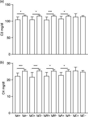
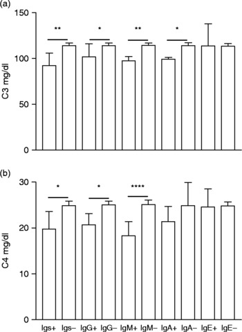
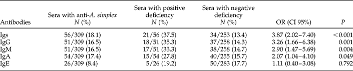
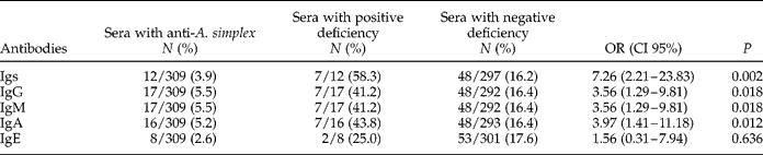
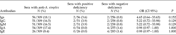
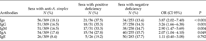
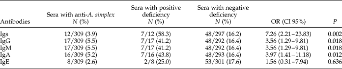
The main objective in our study was to measure levels of C3 and C4 in randomly selected human sera and to correlate these concentrations with the presence of anti-A. simplex specific antibodies. Normal values of C3 and C4 levels are 90–180 mg/dl and 10–40 mg/dl, respectively, according to the test used. In a first step, C3 and C4 levels were significantly lower in subjects who presented anti-A. simplex antibodies, with the exception of IgE (figs 1 and 2). Secondly, in order to compare the obtained results, values higher than the mean of the OD of the 309 subjects obtained for each anti-A. simplex immunoglobulin plus once or twice their respective standard deviations were considered as positive. Antibody prevalences expressed in percentages are shown in tables 1 and 2. Differences in C3 and C4 values with respect to anti-A. simplex negative samples were studied (tables 1, 2 and 3). The presence of an association between anti-A. simplex elevated levels and C3 deficiency (tables 1 and 2) has been demonstrated. The association between anti-A. simplex antibody levels and C4 deficiency was not significant because of the low number of subjects with C4 deficiency (table 3). By means of the Pearson test a negative correlation that was statistically significant between C3/C4 levels expressed in mg/dl and anti-A. simplex antibodies expressed in OD was observed, with the exception of IgE. The highest inverse correlation was obtained in the case of anti-A. simplex IgM.
(a) C3 and (b) C4 levels in sera with positive and negative anti-Anisakis simplex antibody levels ≥ X+SD. Results are expressed as mean and confidence intervals of 95%. *P < 0.05, ***P < 0.005.

(a) C3 and (b) C4 levels in sera with positive and negative anti-Anisakis simplex antibody levels ≥ X+2SD. Results are expressed as mean and confidence intervals of 95%. *P < 0.05, **P < 0.01, ****P < 0.001.

Deficiency in C3 (<90 mg/dl) in sera with positive and negative anti-Anisakis simplex antibody levels≥X+SD.
Igs, immunoglobulins; N, number; OR, odds ratio; CI 95%: 95% confidence interval.
Deficiency in C3 (<90 mg/dl) in sera with positive and negative anti-Anisakis simplex antibody levels≥X+2SD.
Igs, immunoglobulins; N, number; OR, odds ratio; CI 95%, 95% confidence interval.
Deficiency in C4 (<10 mg/dl) in sera with positive anti-Anisakis simplex antibody levels≥X+SD.
Igs, immunoglobulins; N, number; OR, odds ratio; CI 95%, 95% confidence interval.
In the case of immunoglobulins, the probability that a subject with anti-A. simplex antibodies has C3 deficiency was 3.8 times higher than a subject who lacks such specific antibodies (table 1). Likewise, although the number of C4-deficient individuals was low, C4 levels were lower in A. simplex positive subjects. The suggestion of the present investigation could be that a C4 deficiency increased susceptibility to A. simplex infection to a similar extent as a C3 deficiency and could support the hypothesis that the CP, and not the AP, is primarily involved in an antibody-independent humoral immunity to this parasite. The confirmation of these findings could also demonstrate the participation of the complement system in A. simplex third-stage larvae destruction as was demonstrated in Angiostrongylus cantonensis meningoencephalitis (Padilla-Docal et al., Reference Padilla-Docal, Dorta-Contreras and Bu-Coifiú Fanego2009). Leventhal & Soulsby (Reference Leventhal and Soulsby1977) observed that, with time, C3 levels remained markedly depressed (95% depletion) in animals treated with cobra venom factor (CVF), suggesting that the C3 depletion caused by CVF allowed increased eosinophilic infiltration in guinea-pigs infected with Ascaris suum larvae.
Increased C3 and C4 consumption has been demonstrated for other digestive pathogens; for example, Dore et al. (Reference Dore, Fastame, Tocco, Negrini, Delitala and Realdi2005) observed lower levels of C3 and/or C4 complement fractions in Helicobacter pylori-infected subjects with respect to controls. On the other hand, Torcal et al. (Reference Torcal, Navarro-Zorraquino, Lozano, Larrad, Salinas, Ferrer, Roman and Pastor1996) observed that patients with hydatidosis showed a significant increase of IgE and a decrease of C3 when cysts were large and multiple, and Leventhal & Soulsby (Reference Leventhal and Soulsby1977) demonstrated that the third component of complement can bind to the surface of A. suum larvae. The involvement of the complement system by these and other agents, such as A. simplex, should cause an increased consumption of complement components such as C3 and C4. In our study the lower concentration of C3 and C4 in sera with high anti-A. simplex antibody concentration can best be explained by an increased consumption of C3 and C4 components. Anisakis simplex may produce complement consumption and evade the host immune system. Our previous work (García-Hernández et al., Reference García-Hernández, Rodero and Cuéllar2007) suggested that the consumption of complement factors represents a mechanism of action of A. simplex larval products, since shortening of the preincubation period (0 and 15 min), compared with the standard time of 30 min, at 37°C did result in a significant decrease of the inhibitory effect on the lysis of A. simplex products in the CP.
According to Da Costa et al. (Reference Da Costa, Brockman, Alicot, Ma, Fischer, Zhou, Knipe and Carroll1999), the expression of complement inactivators by the parasite might represent an important protective mechanism to avoid host detection and induction of a vigorous IgG memory response. On the other hand, there is an important relationship between human parasite infection induced by A. simplex and urticaria (Daschner et al., Reference Daschner, Cuéllar and Valls2008) and hypocomplement urticarial vasculitis presents as atypical urticaria where hypocomplementaemia is the characteristic feature, with reduced C3 and C4 (Frot et al., Reference Frot, Barbarot, Poignant, Guyot and Stalder2008).
In conclusion, the presence of an association between high levels of anti-A. simplex antibodies and C3/C4 deficiency has been proved. This study demonstrates serological evidence for the involvement of the complement system in the pathophysiology of human A. simplex infection. To our knowledge, this is the first study analysing specific immunoglobulin levels against larval antigens in this common fish-borne disease in relation to complement levels in humans. Additional studies are necessary to ascertain the clinical significance of these modifications.