Introduction
Urban development is expected to rise worldwide, with potential impacts on habitat fragmentation, biodiversity, trophic interactions and ecosystem functions, among other concerns (McKinney, Reference McKinney2006; Seto et al., Reference Seto, Guneralp and Hutyra2012). The conversion of wild ecosystems into urban environments is likely to have consequences for wildlife ecology and the transmission of infectious diseases (Bradley and Altizer, Reference Bradley and Altizer2007; Becker et al., Reference Becker, Streicker and Altizer2015; Liccioli et al., Reference Liccioli2015). Several factors determine how urbanization affects parasite dynamics to reduce or increase rates of parasite transmission (Becker et al., Reference Becker, Streicker and Altizer2015; Birnie-Gauvin et al., Reference Birnie-Gauvin2016). Urban development may alter natural home ranges, foraging behaviour and territoriality among wildlife, leading to changes in nutritional status, body condition, immune defense and stress, which can in turn lead to changes in exposure and/or susceptibility to infectious diseases (Padgett and Glaser, Reference Padgett and Glaser2003; Ezenwa, Reference Ezenwa2004; Oro et al., Reference Oro2013; Murray et al., Reference Murray2015b).
A frequent reason that urban environments alter parasite dynamics is that they increase food availability, particularly of anthropogenic food sources (e.g. garbage, compost piles, pet/bird food) (Marzluff, Reference Marzluff, Marzluff, Bowman and Donnelly2001; Oro et al., Reference Oro2013). These anthropogenic resources may increase host density over short time scales (e.g. by attracting more animals to clumped resources) and longer time scales (e.g. via higher birth and recruitment rates, and lower mortality). Concentrated food resources in urban areas were correlated with higher densities of both raccoons (Procyon lotor; Prange et al., Reference Prange, Gehrt and Wiggers2003) and red foxes (Vulpes vulpes; Recio et al., Reference Recio2015), and may apply to most urban-exploiting animals (Prange et al., Reference Prange, Gehrt and Wiggers2003; Oro et al., Reference Oro2013; Recio et al., Reference Recio2015). The lack of predators in urban areas further increases the potential for aggregation at resource patches and heightened population densities.
The concentration of urban-adapted wildlife around a resource is likely to facilitate the transmission of parasites by exposing hosts to anthropogenic foods contaminated with pathogens and parasites, and/or by altering the density of susceptible hosts and increasing rates of infection-prone behaviour (Kellner et al., Reference Kellner2012; Becker et al., Reference Becker, Streicker and Altizer2015; Murray et al., Reference Murray2016). Concentrated anthropogenic resources that occur in areas of high human density also increase the probability of contact between humans (and their pets) and wildlife, potentially increasing zoonotic transmission of pathogens and parasites. Examples of these zoonotic agents include the rabies virus, plague (Yersinia pestis), hantavirus, Sarcoptes scabiei mite, Borrelia spp., and helminths (Bradley and Altizer, Reference Bradley and Altizer2007; Becker et al., Reference Becker, Streicker and Altizer2015).
Among these zoonotic parasites, the tapeworm Echinococcus multilocularis appears to be increasing in North America (Catalano et al., Reference Catalano2012; Massolo et al., Reference Massolo2014) and may pose a health concern to humans (Deplazes et al., Reference Deplazes2004; Davidson et al., Reference Davidson2012). Echinococcus multilocularis has a complex life cycle and is transmitted primarily through trophic interactions between definitive hosts, which include several wild carnivores (coyotes, foxes, raccoons), and intermediate hosts, usually rodents (Eckert and Deplazes, Reference Eckert and Deplazes2004). When a competent rodent ingests the infective tapeworm eggs, multivesicular hydatid cysts develop (Eckert and Deplazes, Reference Eckert and Deplazes2004). If humans ingest infective tapeworm eggs, hydatid cysts develop in internal organs, especially the liver, resulting in a disease called alveolar echinococcosis (AE). If untreated, the case-fatality rate of AE is > 95% (Craig et al., Reference Craig, Rogan and Allan1996). Historically, human AE in North America was very rare, with only two documented cases, one in Manitoba, Canada (James and Boyd, Reference James and Boyd1937) and the other in Minnesota, USA (Gamble et al., Reference Gamble1979). However, nine cases have recently been confirmed in Alberta, Canada (K.T. Kowalewska-Grochowska and S. Houston, University of Alberta, personal communication, 2018). Alveolar echinococcosis is considered to be a significant emerging zoonotic disease both globally and within North America (Craig et al., Reference Craig, Rogan and Allan1996; Davidson et al., Reference Davidson2012; FAO/WHO, 2014).
Recent studies in Alberta indicated that up to 30% of urban coyotes are infected with E. multilocularis and were shedding eggs (i.e. patent infections) at the time of necropsy (Catalano et al., Reference Catalano2012; Liccioli et al., Reference Liccioli2012, Reference Liccioli2013, Reference Liccioli2014). A similar rate (37%) was detected recently in central British Columbia (Gesy et al., Reference Gesy2013). Here, we investigate the level of parasitic infection among urban coyotes in Edmonton, Alberta, which was previously identified as having an unusually high rate of E. multilocularis infection (62.5%), but from a sample of only eight coyotes (Catalano et al., Reference Catalano2012). We analysed the parasite communities of 23 urban coyotes killed in collisions with vehicles or shot by wildlife management personnel. We report on the prevalence and intensity of helminth infections, including E. multilocularis and other zoonotic parasites. Our goal was to confirm the high rate of infection previously reported in this area, to help assess the rate at which this potential zoonotic parasite is spreading in urban areas in Canada, and perhaps more generally in North America.
Materials and methods
A total of 23 coyote carcasses were frozen at either −40°C (2016–2017) or −80°C (2017–2018) for at least 48 hours and stored frozen until examination, to reduce biohazard risks (Veit et al., Reference Veit1995). The majority of the animals died as a result of collision with a vehicle, and a few were shot as part of a human–wildlife conflict management programme. Coyotes were weighed and their body lengths (nose to terminus of tail) were measured. We made a cursory examination for external parasites and any abnormalities and superficial wounds or injuries. Sex and age class (juvenile vs adults) were recorded.
In preparation for necropsies, carcasses were partially thawed for 2–3 hours at room temperature. For 14 of the carcasses (2016–2017), viscera, including the respiratory system (trachea, lungs) and heart, diaphragm, liver, gall bladder and pancreas, were removed and immediately examined for parasites. The oesophagus, stomach and intestines were removed and the small intestine was divided into the anterior, middle and posterior thirds. Intestinal sections were flushed with water and the entire volume of the intestinal wash was inspected under a stereomicroscope (Eckert et al., Reference Eckert2001). Specific inspections of organs for parasites included examination of the interior surface of the trachea for cysts or adults of Oslerus ossleri (Filaroides), the heart for Dirofilaria immitis, the diaphragm for Trichinella, and the stomach for Physaloptera sp. For the remaining nine carcasses (2017–2018), the intestines were removed, bagged, and frozen at −20°C, and later thawed for inspection as described above. Platyhelminth parasites were preserved in AFA (alcohol–formalin–acetic acid); voucher specimens were stained with Semichon's acetocarmine, cleared with methyl salicylate, and mounted on slides with Permount™. Nematodes were preserved in glycerin–alcohol and slide mounted for identification. Although recent work has distinguished species of Echinococcus via DNA (Santa et al., Reference Santa2018; Schurer et al., Reference Schurer2018), we did so via morphology, including hooks in the scolex (Jones and Pybus, Reference Jones, Pybus, Samuel, Pybus and Kocan2008) to distinguish E. multilocularis from the potentially cohabiting E. canadensis via a random sample of slide-mounted specimens (n = 100) (Rausch, Reference Rausch1968). Voucher specimens have been submitted to the University of Alberta Parasite Collection (accession numbers UAPC-11588–11594).
In cases where E. multilocualris intensities exceeded 500 worms, we diluted samples and counted worms in a subsample to support an estimate of the total number. After manually removing the larger worms, the remaining contents were subject to a sedimentation and decanting procedure, which was repeated a few times until the supernatant was clear. After mixing, a 10% sample was extracted with a large bore pipette. At least two subsamples were taken and counted separately for tapeworms. A third subsample was counted if the first two counts were not within 10% of each other (Eckert et al., Reference Eckert2001); we report the average of these counts.
Statistical analyses were performed using RStudio Version 1.0.136 (R Development Core Team, 2016). Where parasite counts were the dependent variable, a generalized linear model (GLM) with a negative binomial error distribution was used to examine the effect of potential explanatory variables. A binary response variable of infection (0 or 1) was analysed with GLM and binomial error distribution. We implemented backwards deletion and model selection (χ2 test, P > 0.05) to arrive at the minimal model; deviance (~ sum of squares) and P-values are reported.
Results
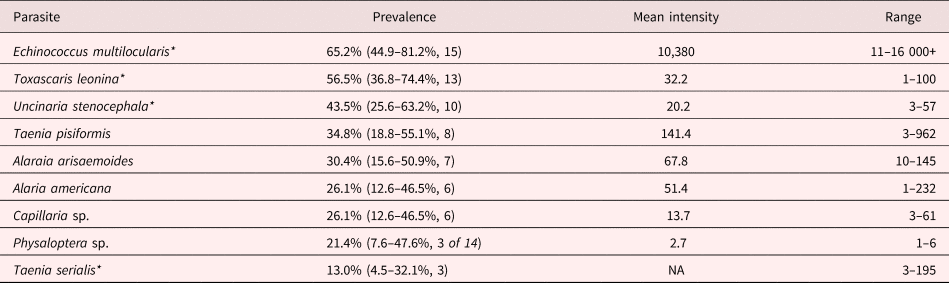
Among the 23 urban coyotes examined, all were infected with at least one of the nine species of helminths identified in this study (table 1). Individual coyotes were infected with an average of three (range 1–6) species of helminths, with E. multilocularis being the most prevalent parasite (65.2%, table 1). The distribution of E. multilocularis among infected coyotes in this study was highly aggregated (dispersion parameter, k = 0.097), ranging from 11 to 100,000 worms (median = 1000). Of the coyotes killed by a car (N = 13), 53.8% were infected with E. multilocularis, whereas 71.4% of those shot for wildlife management (N = 7) were infected, but cause of death was not a statistically significant predictor of infection (deviance = 0.85, P = 0.36). Sex was not a significant predictor of infection (deviance = 0.22, P = 0.64) even though the infection prevalence was slightly higher among male coyotes (N = 13, 61.5%) than in female coyotes (N = 7, 57.1%; sex of three carcasses was undetermined). In cases where age could be determined, 63.6% of the juveniles (N = 11) and 50% of the adults (N = 8) were parasitized; age did not influence the probability of being infected (deviance = 0.18, P = 0.67). Neither body mass (mean 11.0 ± 2.03 kg, deviance = 0.0009, P = 0.98) nor body length (mean 116.1 ± 11.06 kg, deviance = 1.69, P = 0.19) affected infection status.
Prevalence and intensity of gastrointestinal helminths in urban coyotes (Canis latrans) from Edmonton, Canada. Numbers in parentheses represent 95% confidence intervals and number of infected coyotes out of 23 carcasses (unless otherwise indicated).

*Zoonotic disease potential
All of the coyotes infected with E. multilocularis (N = 15) were also co-infected with at least one other helminth, most often with Toxascaris leonina (N = 8 cases) and Uncinaria stenocephala (N = 8 cases, table 1). Seven of the 10 juvenile coyotes were infected with U. stenocephala, whereas only one of eight adults harboured the dog hookworm; age class was a statistically significant predictor of hookworm infection (deviance = 6.48, P = 0.011). Female coyotes were significantly more likely to be infected by hookworms (deviance = 4.83, P = 0.028). Of the female coyotes sampled, 83% were infected with the hookworm, whereas only 31% of the males were infected. Neither age (deviance = −1.36, P = 0.176) nor sex (deviance = 0.05, P = 0.83) was a significant predictor of infection by the roundworm T. leonina. One coyote was infected with a single Oslerus ossleri in its trachea, but we did not recover Dirofilaria immitis from the hearts or Trichinella sp. from the diaphragms of any of the carcasses. Three coyotes were infected with sarcoptic mange.
Discussion
Echinococcus multilocularis was the most prevalent (65.2%) parasite among urban coyotes in Edmonton, Alberta, Canada, which is comparable to the value (62.5%) previously reported for coyotes from the Edmonton area (Catalano et al., Reference Catalano2012). This prevalence of infection is much higher than those reported in other studies of coyotes in North America (range 0–44.4%, reviewed by Massolo et al., Reference Massolo2014) as well as that found 300 km south in a metropolitan area of Calgary, Alberta, where urban coyotes were infected at rates of 20.5% (N = 83) in one study (Catalano et al., Reference Catalano2012) and 29.5% (N = 61) in another (Liccioli et al., Reference Liccioli2012). The intensity of infection (among infected individuals) was also higher in Edmonton (median = 1000, range = 11–100,000) compared to Calgary (median = 20.5, range = 1–1400, Catalano et al., Reference Catalano2012). In contrast to the Calgary studies, in which the prevalence of infection was male-biased (Catalano et al., Reference Catalano2012; Liccioli et al., Reference Liccioli2012), we did not detect a difference by sex in Edmonton coyotes. Similarly, we did not find a statistically significant difference in infection between juveniles and adults, whereas Liccioli et al. (Reference Liccioli2012) reported higher infection prevalence among juvenile coyotes in Calgary. The lack of statistically significant sex- or age-biased infection in our study may be attributable to our smaller sample size, although the trend was in the same direction as in the two Calgary studies (Catalano et al., Reference Catalano2012; Liccioli et al., Reference Liccioli2012).
The second and third most prevalent parasites in our study were T. leonina and U. stenocephala, both of which were commonly found in association with E. multilocularis. Despite having the second-highest prevalence in our study, at 56%, other studies have consistently reported even higher rates of T. leonina infection, of 80–100% (Seesee et al., Reference Seesee, Sterner and Worley1983; Wirsing et al., Reference Wirsing2007; Thompson et al., Reference Thompson, Kutz and Smith2009; Liccioli et al., Reference Liccioli2012). In contrast to Liccioli et al. (Reference Liccioli2012), we did not find infection of T. leonina to be more prevalent among juveniles.
The northern dog hookworm, U. stenocephala, replaces the more common form of the dog hookworm, Ancylostoma caninum, at northern latitudes (Levine, Reference Levine1980). The infection prevalence we observed (44%) was higher than previously reported (29.5%) in Alberta (Liccioli et al., Reference Liccioli2012). Adult worms probably persist for only 4–6 months in the small intestine and infective eggs are shed directly into the environment, probably around dens, but trans-placental or trans-mammary transmission has not been reported (Dow et al., Reference Dow1961; Kalkofen, Reference Kalkofen1987). The increased risk of exposure to infective stages around dens may account for the female- and juvenile-biased infections. The high prevalence of U. stenocephala among urban coyotes may present a risk to domestic animals such as dogs. Further studies are needed to determine the ecological and epidemiological significance of these co-infections in urban coyotes.
Our results showed that urban coyotes in Edmonton host several intestinal parasites, some of which have the potential to be zoonotic (Deplazes et al., Reference Deplazes2004; Strickler, Reference Strickler2005; Rausch and Fay, Reference Rausch and Fay2011; Seguel and Gottdenker, Reference Seguel and Gottdenker2017). Most notable is our measure of the prevalence of infection by E multilocularis at 62%, which supports an earlier similar estimate of 65% by Catalano et al. (Reference Catalano2012). Both estimates are over twice as high as the reported prevalence for this zoonotic parasite in Calgary (above) and 50% higher than the top end of the range reported for all of North America (reviewed by Massolo et al., Reference Massolo2014). Although the sample size in our study is relatively small, these results suggest that Edmonton may be a ‘hot spot’ for E. multilocularis in coyotes, which could increase the risk of transmission to domestic dogs and humans. Such transmission appears to have greater consequences for public health when it involves the European strain of this tapeworm, relative to the strain previously identified from central North America, which appeared to have little zoonotic potential (Hildreth et al., Reference Hildreth2000). Indeed, only two cases of human alveolar echinococcosis (AE) had been reported in North America until quite recently, and none from Alberta until 2013 (Massolo et al., Reference Massolo2014). However, in the last several years nine cases of AE have been documented in Alberta (K.T. Kowalewska-Grochowska and S. Houston, personal communication, 2018). Furthermore, there is increasing evidence of establishment and local transmission of a European-type strain of E. multilocularis in Canada (Gesy et al., Reference Gesy2013; Massolo et al., Reference Massolo2014), which appears to be more virulent and more likely to result in AE than the central North American strain (Jenkins et al., Reference Jenkins2012).
Urban coyotes have the potential to spread zoonotic diseases (Craig et al., Reference Craig, Rogan and Allan1996; Deplazes et al., Reference Deplazes2004; Liccioli et al., Reference Liccioli2012, Reference Liccioli2015), and are considered the primary definitive host of E. multilocularis in urban areas in North America (Leiby et al., Reference Leiby, Carney and Woods1970; Holmes et al., Reference Holmes, Mahrt and Samuel1971; Eckert and Deplazes, Reference Eckert and Deplazes2004). Moreover, populations of coyotes have increased in urban areas throughout North America (Gehrt et al., Reference Gehrt, Anchor and White2009), which has resulted in increasing rates of human–coyote conflict (Poessel et al., Reference Poessel, Mock and Breck2017). Rates of reported conflict are rising in both Edmonton (Murray et al., Reference Murray2015a) and Calgary (Alexander and Quinn, Reference Alexander and Quinn2011). Taken together, these findings indicate a pressing need for increased monitoring of both coyotes and E. multilocularis in urban and peri-urban environments, especially in Alberta. Further studies are necessary to identify factors that increase the likelihood of infection by E. multiloccularis in each of coyotes, dogs and humans. With this information, wildlife managers might identify and mitigate food sources, habitat or other factors that promote parasite transmission or disease, to support coexistence between people and urbanizing wildlife.
Acknowledgements
We wish to thank D. Sanderson at MacEwan Univerisity and S. Sugden at the University of Alberta for assisting with the coyote necropsies. We also thank the anonymous reviewers for their constructive comments and suggestions.
Financial support
This work was supported by NSERC Discovery Grants (LTL, grant number 435245), (CCSC, grant number 05915).
Conflict of interest
None.
Ethical standards
The authors assert that all procedures contributing to this study were conducted in accordance with biohazard requirements at the University of Alberta and MacEwan University.