Introduction
Fontan operation has been applied to patients with single ventricle physiology as the final palliative surgery. Despite acceptable survival in patients after Fontan operation, there are concerns about late complications affecting the major extracardiac organs including the liver and the digestive system. Reference Ohuchi1 However, there is little information regarding renal complications. We herein report two cases of adults after Fontan operation who develop focal segmental glomerulosclerosis.
Case report
Case 1
A-34-year-old man was admitted to our hospital because of dyspnoea on exertion and proteinuria. The patient had undergone a staged repair of unbalanced atrioventricular septal defect and transposed great arteries to Fontan operation at the age of 13 years. He was also complicated with type 2 diabetes mellitus at the age of 34 years. He had been hospitalised frequently for acute exacerbations of chronic heart failure. His medical history included renal infarction at the age of 30 years, arterial thrombosis in the leg at 34 years, and cerebral infarction at 34 years. He was treated with furosemide, digoxin, enalapril, febuxostat, cilostazol, apixaban, aspirin, pitavastatin, and metformin. Systemic oxygen saturation and blood pressure were 92% and 125/88 mmHg, respectively. Chest X-ray showed a cardiothoracic ratio of 0.61, and electrocardiography showed normal sinus rhythm without arrhythmia.
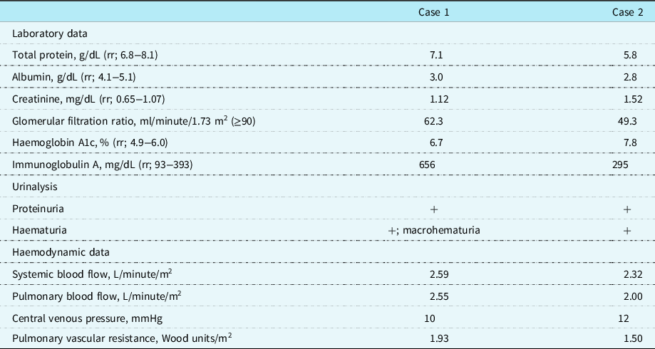
Urinalysis showed macrohematuria and proteinuria. Daily proteinuria was estimated as urine protein per gram of urine creatine, 1.9 g/g· creatinine. Laboratory data showed decreases in serum albumin level and an estimated glomerular filtration ratio and increases in serum immunoglobulin A (Table 1). Haemoglobin A1c was 6.7% (reference range: 4.9−6.0). Further immunological examination revealed negative anti-nuclear antibody, myeloperoxidase-anti-nuclear antibody, nor proteinase-3-anti-nuclear antibody. His haemodynamic data showed a decreased in cardiac output, suggesting circulatory failure.
Laboratory and haemodynamic data in the presented cases

Values given in brackets means reference ranges. Haemodynamic data were measured based on the last cardiac catherisation.
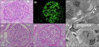
On a suspicion of glomerulonephritis, we performed open renal biopsy under general anaesthesia. Among a total of 74 golomeruli, 40 glomeruli showed mesangial proliferation with mesangial nodular expansion with congestion, 17 glomeruli showed global or segmental mesangial sclerosis, and 12 glomeruli showed hypertrophy with thickening of glomerular basement membrane. Depositions of immunoglobulin A in the mesangial space were found, although no deposition of complements was shown. There were no active lesions of immunoglobulin A nephropathy such as endocapillary hypercellularity, fibrocellular crescent, and cellular crescent. These histopathological findings were compatible to focal segmental glomerulosclerosis (Figure 1a–c).
Histopathological findings in Case 1 ( a − c ) and Case 2 ( d − f ) are shown. ( a ) Periodic acid Schiff (PAS) staining shows mesangial proliferation and nodular expansion with congestion. ( b ) Immunofluorescent staining shows accumulation of immunoglobulin A in the glomeruli. ( c ) Electronic microscopy shows thickening of glomerular basement membrane and effacement of foot process (arrow heads). ( d and e ) PAS staining shows mesangial proliferation and segmental sclerosis. ( f ) Electronic microscopy shows effacement of foot process (arrow heads).

Case 2
A 24-year-old man was admitted because of oedema on the face and lower extremities. The patient had undergone a staged repair of double outlet ventricle and transposed great arteries to Fontan operation at the age of 2 years. He also suffered from type 2 diabetes mellitus and obstructive sleep apnoea at the age of 24 years. He was regularly followed up with medications of furosemide, spironolactone, digoxin, telmisartan, febuxostat, aspirin, and warfarin. On admission, his systemic oxygen saturation and blood pressure were decreased to 88% and 84/51 mmHg, respectively. Chest X-ray showed a cardiothoracic ratio of 0.49, electrocardiography showed normal sinus rhythm without arrhythmia, and echocardiography showed ventricular dysfunction and moderate atrioventricular regurgitation.
Urinalysis showed haematuria and proteinuria with protein per gram of creatine of 1.76 mg/g· creatinine. Laboratory findings are shown in Table 1, which revealed decreases in serum protein level and an estimated glomerular filtration ratio. Haemoglobin A1c was 7.8%. Anti-nuclear antibodies were negative. Contrast-enhanced CT showed heterogeneous enhancement pattern of the liver, which was a compatible finding of Fontan-associated liver disease. His haemodynamic assessment showed a decrease in cardiac output.
Further, we performed open renal biopsy under general anaesthesia on a suspicion of glomerulonephritis. Histological findings showed 17 glomeruli exhibit segmental mesangial hypercellularity, 3 glomerulus show global sclerosis, 5 glomeruli were segmental sclerosis, and one collapsed glomerulus among a total of 95 glomeruli (Figure 1d−f). Immunofluorescence staining showed no remarkable findings. These histopathological findings were compatible to focal segmental glomerulosclerosis.
Discussion
Focal segmental glomerulosclerosis is characterised by sclerosis, hyalinosis, foam-cell infiltration, vacuolisation of podocytes, and podocyte precursor proliferation, Reference Han and Kim2 which causes idiopathic, genetic, or secondary to viral infection, drug, congenital anomaly, systemic hypertension, and acute or chronic vaso-occlusive process. Reference Campbell and Tumlin3
In patients with chronic heart failure, cardiorenal syndrome encompasses a spectrum of disorders involving the heart and kidneys. Reference Rangaswami, Bhalla and Blair4 Previous reports have shown that renal dysfunction is associated with poor prognosis in patients after Fontan operation. Reference Khuong, Wilson and Grigg5-Reference Ohuchi, Negishi, Hayama, Miyazaki, Shiraishi and Ichikawa7 The reasons for the development of renal disease among them are considered as the follows. First, glomerular congestion owing to an increase in central venous pressure can lead to not only glomerular hypertrophy and thickening of the basement membrane but also an increase in renal arterial tone and hypertrophy of the vascular smooth musculature. Reference Gupte, Vaideeswar and Kandalkar8,Reference Broda, Sriraman and Wadhwa9 These alterations in the glomerular circulation can result in glomerulosclerosis. Second, hypoxia and hyperviscosity can be causative factors for the development of focal segmental glomerulosclerosis. Adults with cyanotic CHD occasionally have cyanotic nephropathy characterised by histopathological findings of glomerulomegaly, glomerular capillary congestion, hilar arteriolar dilatation, glomerulosclerosis, and interstitial fibrosis, Reference Gupte, Vaideeswar and Kandalkar8 which overlaps those found in our present cases.
Diabetic patients also manifest focal segmental glomerulosclerosis as either diabetic nephropathy or non-diabetic renal disease. Although the prevalence of focal segmental glomerulosclerosis is estimated as 13% of diabetic patients, focal segmental glomerulosclerosis is frequently found as non-diabetic renal disease, which is related comorbidities such as obesity and hypertension. Reference Sharma, Bomback and Radhakrishnan10 In addition, duration of diabetes is reported to be significantly shorter in patients with non-diabetic renal disease than those with diabetic nephropathy. Reference Sharma, Bomback and Radhakrishnan10 Therefore, we supposed that diabetes had less impact on the development of focal segmental glomerulosclerosis in the presented patients because of the short duration of diabetes and the low prevalence of focal segmental glomerulosclerosis in diabetic nephropathy.
In conclusions, we presented two notable cases who developed focal segmental glomerulosclerosis late after Fontan operation. Focal segmental glomerulosclerosis can be a renal complication associated with Fontan operation. Regular urinalysis should be warranted to detect this pathological feature among them.
Acknowledgements
We are grateful to Dr. Miwa Takeichi and Dr. Mamie Watanabe for helpful in the clinical management, and also to Dr.Satoshi Hisano and Dr.Eisuke Katafuchi for histopathological assessments.
Financial support
This work received no specific grant from any funding agency.
Conflicts of interest
The authors have no conflicts of interest to disclose.