Introduction
Adenoidectomy is generally performed in children, for conditions including nasal obstruction, recurrent otitis media with effusion and obstructive sleep apnoea. Traditional adenoidectomy is carried out by ‘cold steel’ curettage. The main problem with this technique is persistent bleeding, which may require electrocautery or nasopharyngeal packing for control. It has been suggested that suction diathermy is the most appropriate method for adenoidectomy in children.Reference Skilbeck, Tweedie, Lloyd-Thomas and Albert 1
Current evidence on the safety and efficacy of suction diathermy adenoidectomy is adequate to support its use provided that normal arrangements are in place for clinical governance, consent and audit. 2 This procedure requires specific training in the use of diathermy for adenoidectomy, due to the risk of thermal damage to surrounding tissues (e.g. the eustachian tube and cervical ligaments); rarely, this can cause Grisel's syndrome (subluxation of the atlantoaxial joint).
Background
Suction diathermy adenoidectomy primarily aims to remove the adenoids while minimising intra-operative blood loss and the risk of secondary haemorrhage. It utilises heat generated by an electric current to ablate or liquefy adenoid tissue, which is removed using suction. The procedure is performed with the patient suitably draped and positioned (a shoulder bag is usually used to extend the neck). The mouth and lips can be moistened with saline to reduce trauma from friction. A Boyle–Davis gag with a Doherty blade is inserted over the tongue and deployed; Draffin rods are used to secure the position. A nasal catheter is then passed into the nose and used to elevate the soft palate by fixing it to the gag using Burkitt's forceps.
In most departments, visualisation of the adenoid is achieved using a dental mirror. The suction diathermy probe is passed into the mouth and applied to the adenoid tissue in the nasopharynx. The procedure is considered to be complete when the choanae are visible and the nasopharynx has a smooth contour.
Difficulty in demonstrating the nasopharynx to a person other than the operating surgeon is a commonly encountered problem. Teaching suction diathermy adenoidectomy represents a challenge for both trainer and trainee, due to the limited anatomical view obtained by both participants, in addition to the requirement to work with a mirror.Reference Chisholm, Lew-Gor, Hajioff and Caulfield 3 Trainees need to be aware of the relevant anatomy in order to prevent injury to the eustachian tubes, which are in close proximity and in danger of thermal damage if the operative field is not clearly delineated.
On review of the literature, techniques to address this problem involve the use of a trans-oral, 45° endoscope connected to a monitor. This allows the surgical trainee to perform the procedure under direct supervision of the consultant trainer.Reference Lo and Rowe-Jones 4 However, the practicalities of making this equipment regularly available for routine surgery sessions represent a considerable limitation of this method, despite its merits. The pressures of ensuring the availability of the correct equipment in operating theatres which are not used solely for ENT procedures, and which thus may have other types of surgery running concurrently, are considerable, and can sometimes make it difficult to obtain an extra headlight, let alone an endoscope and light source. The use of an endoscope will also add another technical element to the procedure, as well as requiring much repositioning throughout.
Our suggested alternative provides more economy of movement and mimics the usual operation more closely.
Suggested technique
Firstly, the trainer scrubs and performs the initial part of the adenoidectomy, while questioning the trainee and discussing the relevant anatomy and method.
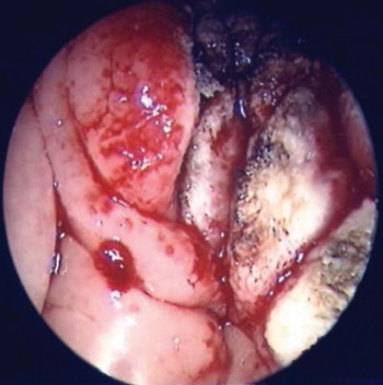
Secondly, the trainer performs the suction diathermy technique on one side only (Figure 1). This serves as a template for the trainee to reproduce on the opposite side.
Nasendoscopic view following suction diathermy performed on the right by the trainer.

Thirdly, the trainee assumes the operating position and completes the adenoidectomy on the opposite side. Thus, they are able to acquire the necessary skills to perform the procedure while being able to directly compare their work to the opposite side. Although this method does not eliminate the difficulty in orientation, it assists the trainee to understand the boundaries of the operative field. By having a template available, the trainee can be confident in their orientation, and can therefore focus on acquisition of motor skills and familiarity with the relevant anatomy.
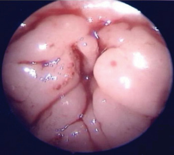
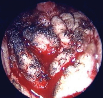
Figures 1, 2 and 3 are used as a guide to show trainees what they will see in the mirror, using the described training technique. (Note that the training technique had been trialled a number of times before these endoscopic images were obtained, and the images are from a case in which the described procedure was followed.)
Pre-operative nasendosopic view of adenoids, obtained with a 0° nasendoscope introduced via the nose prior to the procedure. The picture has been rotated by 180° to give the view obtained when looking down at the patient on the operating table.

Nasendosopic view showing the remnant of the nasopharyngeal adenoid after performance of suction diathermy adenoidectomy by the trainee (left side of the figure).

Unfortunately, Figures 1 to 3 fail to demonstrate the eustachian tube orifices, which are sometimes difficult to delineate from adenoidal tissue. Nonetheless, this fundamental anatomical area can be demonstrated to the trainee due the outline created. Unlike curettage, in suction diathermy adenoidectomy a visual analysis is made of the surgical field, which obviously benefits the procedure. During revision procedures, the band of adenoids abutting the eustachian tube orifices is often still present if cold steel adenoidectomy has been performed. It is important not to leave tissue abutting the eustachian tube orifices, while at the same time avoiding thermal damage to them; many trainees find this difficult to do.
Conclusion
The described approach to suction diathermy adenoidectomy training does not require new pieces of equipment in the operating theatre. This method familiarises trainees with the relevant anatomy and enables them to acquire the skills needed to work via a reflected image. The provision of a completed, contralateral adenoidectomy supplies a comparison enabling the trainee to gain confidence in completing a competent procedure. We believe that, in light of the current emphasis on competency-based training, any means of assisting the acquisition of safe, proficient surgical skills should be embraced.