INTRODUCTION
A prolonged state of oxidative stress has been one of the contributory reasons for triggering lifestyle diseases such as cancer, cardiovascular diseases and diabetes (Yagi, Reference Yagi1994). The result of the imbalance between the production of free radicals and antioxidant defences (Halliwell, Reference Halliwell1994) in favour of free radicals (generated during parasitic infections, inflammation and aerobic metabolism) implicates parasitic diseases (Oliveira and Cecchini, Reference Oliveira and Cecchini2000; Chikibova and Sanikidze, Reference Chikibova and Sanikidze2006) to be a contributory cause of oxidative stress. Studies have shown that protozoan or helminth parasites secrete enzymes that generate free radicals such as superoxide and other intermediate products of free radical activity such as hydrogen peroxide in mammalian tissues (Clark et al. Reference Clark, Hunt and Cowden1986). Various studies have also reported on the presence of oxidative stress in serum and liver tissues of subjects infected with parasites (Dermici et al. Reference Dermici, Delibas, Altuntas, Oktem and Yonden2003; Kamalvand and Ali-Khan, Reference Kamalvand and Ali-Khan2004) and the antioxidant defence mechanism between parasites and their mammalian host (Turrens, Reference Turrens2004). However, most of the evidence is based on studies conducted in developed countries and little is known about interlink between oxidative stress and parasitic infection (especially intestinal parasites) in the Malaysian population.
The present study is aimed to investigate the differences in some of the metabolites secreted in our body in relation to free radical activities and oxidative stress between the normal healthy individuals and the subjects with intestinal parasitic infection. We used urine samples to carry out this study. Generally, urinary metabolites act as a good indicator to measure the average amount of damage excreted from the entire body over a broad span of time (Shioji, Reference Shioji2005). This will be useful in determining the amount of oxidative activity in the body.
MATERIALS AND METHODS
Sample collection
In this study, fresh urine samples (first morning specimen) were collected from age-matched normal healthy individuals (n=95) and humans with intestinal parasitic infection (n=75). Healthy subjects, who serve as controls, consist of students and volunteers from Klang Valley, Malaysia. Samples of the subjects with intestinal parasitic infection were obtained from medical camps in Klang Valley. All samples were collected in sterile cytology containers. Stool examination was carried out for the determination of parasitic infection namely Entamoeba histolytica, Giardia lamblia, Balantidium coli, Blastocystis hominis, Cryptosporidium, Microsporidia, Dientamoeba fraglis, Ascaris lumbricoides, Trichuris trichiuria, hookworm and Taenia sp. Patients positive for 1 or more of these parasites were categorized as positive for intestinal parasites.
Urinalysis using a dipstick method (Combur Test UX, obtained from Roche Diagnostics) was carried out to test for the levels of glucose, bilirubin, nitrite, protein, ketone bodies, urobilinogen and blood. None of the samples (both controls and subjects with parasitic infection) showed any urinary abnormalities. The study was approved by the Ethical Committee of University Malaya Medical Centre (UMMC). Informed consent was obtained from all the study subjects.
Biochemical assays
Assays for three oxidative indices, namely H2O2, AOPP, and LP were carried out according to methods previously established and further modified in our laboratory. All the assays were done on freshly collected urine samples. Urinary hydrogen peroxide (H2O2) which was postulated to be a biomarker of oxidative stress in previous studies was measured using the ferrous ion oxidation xylenol orange version-2 (FOX-2) method (Banerjee et al. Reference Banerjee, Madhusoodanan, Nayak and Jacob2003). This method is based on the oxidation of ferrous ions by the sample oxidizing agents containing H2O2 to ferric ion, which bind with xylenol orange to give a coloured complex. This coloured complex was measured spectrophotometrically and the results were expressed in μmol/l, using H2O2 as the standard.
Advanced oxidation protein products (AOPP) are formed as a result of oxidative stress by the action of chlorinated oxidants, mainly hypochlorous acid and chloramines. AOPP was measured spectrophotometrically according to the method of Witko-Sarsat et al. (Reference Witko-Sarsat, Friedlander, Capeillere-Blandin, Nguyen-Khoa, Nguyen, Canterloup, Drayer, Jungers, Drueke and Deschamps-Latscha1998). The AOPP concentrations were calculated from the standard curve prepared using chloramine T and expressed as μmol/l of chloramine T equivalents.
Lipid peroxidation levels were determined according to the modified method of Ratty and Das (Reference Ratty and Das1986), based on thiobarbituric acid reaction with malondialdehyde (MDA, an end-product of lipid peroxidation). Thiobarbituric acid reactive substances (TBARS) is a well-established assay for screening and monitoring lipid peroxidation. The standard absorption curve for MDA quantification was prepared using 1,1,3,3-tetraethoxypropane and the values were expressed as μmol/l.
Statistical analysis
Data of this study were analysed using SPSS version 13. All data are expressed as mean±s.e.m. and the significant differences between the subject groups were analysed using Student's t-test. Correlations between the parameters for both control and parasite-infected subjects were identified by Pearson's correlation coefficients test and differences were considered significant when P<0·05.
RESULTS
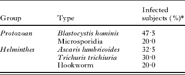
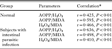
The percentages of infected subjects according to types of parasitic infection are shown in Table 1. These subjects were infected with Blastocystis hominis (47·5%) and microsporidia (20·0%), Ascaris lumbricoides (32·5%), Trichuris trichiuria (30·0%) and hookworm (20·0%). Table 2 depicts the levels of H2O2, AOPP and MDA in normal and intestinal parasite-infected subjects. All these biomarkers were significantly higher (P<0·001, P<0·05 and P<0·05 respectively) in the parasite-infected subjects compared to the normal subjects. Table 3 depicts the level of parameters tested according to 3 main categories of parasitic infections which are helminth infection, protozoa infection and the combination of both helminth and protozoa infections. From this table, all the oxidative indices showed no significant difference among the subject groups. Positive correlation exists between AOPP and H2O2, AOPP and MDA, and H2O2 and MDA for both subject groups in this study (Table 4).
Percentage of infected subjects according to type of parasitic infection
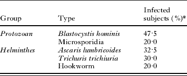
* Percentage of infected subjects is inclusive of those with single and multiple parasitic infections.
Comparison of AOPP, H2O2 and MDA levels between normal and intestinal parasite infected subjects
(Data are given as mean±s.e.m.)

* P<0·05, *** P<0·001 compared with controls by Student's t-test.
Comparison of AOPP, H2O2 and MDA levels among subjects infected with helminth, protozoa and combination of both helminth and protozoa
(Data are given as mean±s.e.m. Degree of significance, P among the subject groups was analysed using ANOVA (SPSS Version 13.0).)

Correlation analysis of AOPP, H2O2 and MDA levels in the study groups
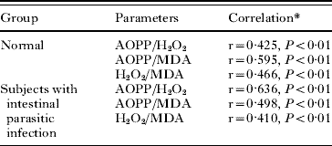
* P<0·01 compared by Pearson correlation coefficient analysis.
DISCUSSION
Parasites have different physiological functions for their survival in certain environments of the host. Most of the parasites adapt to the lack of oxygen conditions within the host by utilizing systems other than oxidative phosphorylation for ATP synthesis. Mitochondrial complex II of the parasite acts as a source for anaerobic energy metabolism (Kita et al. Reference Kita, Hirawake, Miyadera, Amino and Takeo2002). In general, intestinal parasites (lumen-dwelling parasites) are anaerobic parasites. They can be divided into 2 major groups namely protozoa and helminths or worms. When a host's immune system is triggered by an infection of parasites, a massive production of ROS or oxidative burst is activated by macrophages that are associated with the inflammatory system (Rosen et al. Reference Rosen, Pou, Ramos, Cohen and Britigan1995). This provides a first line of defence against these parasites. Macrophage or phagocyte activation causes the release of reactive species that lead to lipid peroxidation, protein damage and DNA strand breaks (Oshima and Bartsch, Reference Oshima and Bartsch1994). These damaged metabolites can be transported systemically throughout the host's body. A range of oxidation products are found in urine and considered to reflect local and systemic oxidative stress (Kirschbaum, Reference Kirschbaum2001).
In the present study, stool examination showed that most of the parasite-infected subjects were found positive for Blastocystis hominis (47·5%) (protozoan) followed by Ascaris lumbricoides (32·5%) (helminth). Although the percentage indicated is inclusive of those subjects with single and multiple parasitic infections, the prevalence of these intestinal parasites in the subjects of this study was higher than that reported by various other researches. However, in the present study, due to the small sample size associated with multiple types of parasitic infections, the parasite-infected subjects could only be segregated into 3 main groups, namely subjects with: helminth, protozoa and both helminth as well as protozoa. However, the statistical analysis for this did not show any significant difference. The burden of parasites for each type of infection (e.g. Trichuris sp., hookworm, Blastocystic hominis, etc.) could be the reason for insignificance. Nevertheless, we have successfully identified significant differences in the parameters measured between the normal and the overall subjects infected with intestinal parasites.
In this study, elevated levels of H2O2, AOPP and MDA in subjects with parasitic infection indicate that their overall oxidative stress level was higher compared to the normal healthy individuals. The formation of H2O2 in the body involves a number of enzymatic reactions especially superoxide dismutase (SOD) and its decomposition involves enzymes mainly catalase (CAT) and glutathione peroxidase (GPx). CAT decomposes H2O2 into water and oxygen molecules. GPx scavenges H2O2 to form water molecules by simultaneously oxidizing tripeptide glutathione (GSH) to oxidized glutathione (GSSG) (Valko et al. Reference Valko, Rhodes, Moncol, Izakovic and Mazur2006). H2O2 alone is not harmful but it can convert into a hydroxyl radical (•OH) when exposed to ultraviolet or ferrous ions (Halliwell, Reference Halliwell2000). In the current study, the level of urinary H2O2 was measured using FOX-2 assay which has been used previously by Long et al. (Reference Long, Evans and Halliwell1999) and Chandramathi et al. (Reference Chandramathi, Suresh, Anita and Kuppusamy2008). Some researchers have modified it to be more specific for H2O2 by considering the difference between the assay done with and without catalase enzyme which destroys H2O2. However, the results obtained by them did not show much difference between the classical and the modified method (Banerjee et al. Reference Banerjee, Madhusoodanan, Nayak and Jacob2003). Studies have demonstrated that reducing agents such as thiols, uric acid and glutathione do not interfere in the acidic environment of the FOX reagent (Nourooz-Zadeh, Reference Nourooz-Zadeh and Packer1999). Thus, Banerjee et al. (Reference Banerjee, Kumar, Kumar, Madhusoodanan, Nayak and Jacob2002) have reported recovery of 100% of the H2O2 from human urine by use of the FOX method. Furthermore, in this study a standardized FOX assay was carried out on all the subjects where the difference should be normalised by the control group. To the best of our knowledge, the study is the first to report on the level of urinary hydrogen peroxide in parasite-infected human subjects. The elevation of H2O2 in these subjects lead to the speculation that parasitic infection may contribute to a higher level of •OH-induced oxidative stress compared to the controls. Our results can be supported by the findings of previous studies done on other classical parameters of •OH-induced oxidative stress such as elevated levels of GPx and SOD in the blood of mice infected with Trichinella spiralis (Derda et al. Reference Derda, Wandurska-Nowak and Hadas2004).
AOPP in plasma and serum have been extensively used as a marker of free radical-induced protein damage. AOPP in the urine has been shown to be elevated in the absence of microvascular or macrovascular complication or renal failure. The low molecular weight AOPP (carbonyl) compounds are possibly excreted under stress conditions (Kalousova et al. Reference Kalousova, Skrha and Zima2002). Studies have reported that the level of advanced glycation end-product (AGE) which is known to be analogous with AOPP (Witko-Sarsat et al. Reference Witko-Sarsat, Friedlander, Capeillere-Blandin, Nguyen-Khoa, Nguyen, Canterloup, Drayer, Jungers, Drueke and Deschamps-Latscha1998) was elevated in the spleen and liver tissues of mice infected with cysts from tapeworms (Kamalvand and Ali-Khan, Reference Kamalvand and Ali-Khan2004). Thus, in this study, the increase of urinary AOPP levels in parasite-infected subjects indicates that they have higher degree of oxidant-mediated protein damage.
Measurement of the MDA level using the TBARS assay, which reflects the lipid peroxidation (damage to lipid), is used as an indicator for oxidative stress in the diseased state (Saygili et al. Reference Saygili, Konukoglu, Papila and Akcay2003). Although much controversy has appeared in studies concerning the specificity of TBARS toward composites other than MDA, the rapidity of the assay qualifies it as one of the broadly-used assays to determine urinary lipid peroxidation (Siciarz et al. Reference Siciarz, Weinberger, Witz, Hiatt and Hegyi2001). In the present study, high levels of urinary MDA in the subjects with parasitic infection compared to the normal individuals may be due to the over production of ROS (during parasitic infection) which causes cellular injuries. Previous studies have reported on increased levels of serum MDA in humans with intestinal parasitic infection such as giardiasis (Dermici et al. Reference Dermici, Delibas, Altuntas, Oktem and Yonden2003) and blastocystosis (Kilic et al. Reference Kilic, Yazar and Saraymen2003).
The correlation analysis among the four indices shows almost similar trait of results in both the subject groups. The positive correlation that existed between H2O2, MDA and AOPP indicates that highly reactive hydroxyl radicals react with a wide variety of organic substrates causing oxidation of lipids and proteins.
In conclusion, the present study provides evidence that oxidative stress (which can be measured non-invasively) is elevated in humans infected with intestinal parasites. This study may influence future researchers to consider the free radical-related pathway to be a target for the intervention of new drugs against parasitic infection diseases.
The study was funded by grant no. FP027/2004A.