


INTRODUCTION
Notoedric mange is a potentially fatal syndrome of contagious dermatitis with systemic complications caused by obligately parasitic, burrowing mites of the genus Notoedres, family Sarcoptidae (Guaguere et al. Reference Guaguere, Prelaud, Craig, Scott, Guaguere and Prelaud1999; Scott et al. Reference Scott, Miller, Griffin, Scott, Miller and Griffin2001; Gross, Reference Gross, Gross, Ihrke, Walder and Affolter2005). These mites are closely related to mites in the genera Sarcoptes, Psoroptes, Chorioptes and Otodectes and include at least 41 named species in the genus (Klompen, Reference Klompen1992). Most Notoedres species have been described from small mammals or bats with few associated clinical, epidemiological, or ecological data. However, severe and sometimes fatal notoedric mange has been described in tree squirrels (Sciurus spp.) infested with Notoedres centrifera, formerly Notoedres douglasi (Lavoipierre, Reference Lavoipierre1964; Klompen, Reference Klompen1992), and felids infested with Notoedres cati (Cornish et al. Reference Cornish, Linders, Little and Vander Haegen2001; Bowman et al. Reference Bowman, Hendrix, Lindsay, Barr and Bowman2002; Riley et al. Reference Riley, Bromley, Poppenga, Uzal, Whited and Sauvajot2007; Serieys et al. Reference Serieys, Foley, Owens, Woods, Boydston, Lyren, Poppenga, Clifford, Stephenson, Rudd and Riley2013). Mange appears to cause considerable suffering in some infested individuals, with intense pruritus resulting in self-mutilation and changes in normal behaviours such as hunting and foraging (Gross, Reference Gross, Gross, Ihrke, Walder and Affolter2005). Mange predisposes the animal to secondary bacterial infection and sometimes death (Bowman et al. Reference Bowman, Hendrix, Lindsay, Barr and Bowman2002; Paterson, Reference Paterson and Paterson2008), sometimes associated with population-level impacts as well (Cornish et al. Reference Cornish, Linders, Little and Vander Haegen2001; Riley et al. Reference Riley, Boydston, Crooks, Lyren, Gehrt, Riley and Cypher2010; Stephenson et al. Reference Stephenson, Swift, Villepique, Clifford, Nyaoke, De la Mora, Moore and Foley2013b ). In this review, we sought to compile data from published literature and unpublished reports in all hosts affected by Notoedres mites, to describe the diversity and distribution of mite species, clinical and pathological impacts, diagnostic and treatment approaches and epidemiological and ecological aspects of disease in host populations. Because the majority of published work has been on N. cati, we highlight this mite and use it to predict how other Notoedres species might affect their hosts as well.
NOTOEDRES TAXONOMY AND SPECIES DISTRIBUTIONS
A sparse literature describes the taxonomy and phylogenetic distribution of Notoedres mites based on host, geographic and morphological characteristics. In many cases, the only information available is a morphological species description based on a small number of specimens examined. The type species was originally named Sarcoptes cati by Hering in 1838 (Hering, Reference Hering1838), and the genus was erected in 1893 by Railliet (Railliet and Lucet, Reference Railliet and Lucet1893) on the basis of a delay in the development of setation, absence and modification of some setation compared with Sarcoptes, and presence of male genital apodemes (Klompen, Reference Klompen1992). The modern genus includes prior genera Chironyssus, Bakeracarus, Mysarcoptes and Suncicoptes (Klompen, Reference Klompen1992). All reports of Notoedres to date have been from Class Mammalia and the overwhelming majority of mites in this genus have been reported from Chiroptera. Notoedres mites of Chiroptera are mostly thought to be specialists on particular bat host species (Klompen, Reference Klompen1992). Unfortunately, almost all reports are case reports with little information about ecology or epidemiology of chiropteran Notoedres. The sparse data that often accompany these case reports preclude most generalization but of interest, one species has been found within the nose of its bat host, representing a new niche for this genus (Ah, Reference Ah1975).
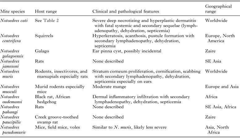
Non-chiropteran Notoedres species feed on rodents, insectivores, other small mammals, procyonids and felids, primarily (Table 1). One lineage includes Notoedres muris and Notoedres musculi, distinguished by eggs having reduced stalks and several setal, anal, or anterior lateral shield changes in males and nymphs (Klompen, Reference Klompen1992). Notoedres muris is somewhat catholic in feeding preference and is geographically widespread, with reports from at least five rat (Rattus) species, hamsters (Cricetus cricetus), voles (Microtus californicus), and other rodents, marsupials including northern brown bandicoot (Isoodon macrourus) and brushtail possum (Trichosurus vulpecula), and the insectivore European hedgehog (Erinaceus europeus) (Heath et al. Reference Heath, Rush-Munro and Rutherford1971; Domrow, Reference Domrow1974; Klompen and Nachman, Reference Klompen and Nachman1990; Klompen, Reference Klompen1992). The related mite, N. musculi, has been found in Europe and Asia on field mice (Apodemus spp.) and mice (Mus musculus) (Klompen, Reference Klompen1992). Notoedres oudemansi, which has prominent dorsal plates, has to date only been reported from Africa, infesting black rats (Rattus rattus) in Rwanda and causing fatal mange in an African hedgehog (Erinaceus albiventris) (Fain, Reference Fain1965; Gregory, Reference Gregory1981; Klompen, Reference Klompen1992). Notoedres pseudomuris also causes skin disease in rodents including mice, field mice and voles (M. transcaspicus) (Klompen, Reference Klompen1992). Other mite species associated with infestation in rodents but for which disease has not been reported include Notoedres pahangi from multiple rat genera and species in southeast Asia and Africa, Notoedres paucipilis from a Creek groove-toothed swamp rat (Pelomys fallax) in Zaire, and Notoedres jamesoni in Rattus spp. in southeast Asia (Klompen, Reference Klompen1992).
Host range, clinical characteristics and geographical distribution of non-chiropteran Notoedres spp. mites.

Reports from the literature of mange caused by Notoedres cati worldwide.

Another rodent specialist is N. centrifera. While less broadly distributed geographically and across host species than some rodent Notoedres species, N. centrifera is notable for its apparent virulence. Notoedres douglasi, which had been considered the squirrel mange mite from North America, was synonymized with Asian N. centrifera in 1964 (Fain, Reference Fain1965; Klompen, Reference Klompen1992). Little has been written about the species in Asia, except that it was collected in all stages from an Indonesian squirrel (Ratufa bicolor), which was held in captivity in the Netherlands (Jansen, Reference Jansen1963). There was no further information about its clinical impact or ecology. In North America, Notoedres centrifera has been reported from the western gray squirrel, the eastern gray squirrel (Sciurus carolinensis), the fox squirrel (Sniger niger), the southern flying squirrel (Glaucomys volans), the eastern chipmunk (Tamias striatus) and porcupine (Lavoipierre, Reference Lavoipierre1964; Carlson et al. Reference Carlson, Daniel and Svend1982; Kazacos et al. Reference Kazacos, Kevin and Herald1983; Evans, Reference Evans1984; Snyder et al. Reference Snyder, Hamir, Hanlon and Rupprecht1991; Klompen, Reference Klompen1992; Burton and Dufour, Reference Burton and Dufour2000; Cornish et al. Reference Cornish, Linders, Little and Vander Haegen2001; Stephenson et al. Reference Stephenson, Swift, Villepique, Clifford, Nyaoke, De la Mora, Moore and Foley2013b ). Aside from the squirrel in captivity in the Netherlands, wild rodents were from California, Oregon, Washington, Illinois, Indiana, Michigan, Kansas, New York, Pennsylvania, Ohio and Massachusetts. Further research on the species in Asia may help clarify whether the Asian and New World mites show any genetic or morphological distinctions.
There are only a few Notoedres species that infest hosts other than bats, rodents and insectivores. Notoedres galagoensis was found on the primate, Demidoff's dwarf galago (Galago demidovii) from Zaire but pathology or disease were not addressed (Fain, Reference Fain1963).
The best-studied member of the genus and the only known zoonotic species is N. cati and while the name suggests it is a cat parasite, it is known to infest at least 18 host species. In 1965, N. cuniculi, a species found in rabbits (Oryctolagus spp.), was synonymized with N. cati (Fain, Reference Fain1965). This species has a distinctive dorsal anus and sucker-like feet on various legs depending on stage and sex of the mite. The name and most veterinary texts suggest that this is a parasite of cats when in fact it infests numerous host species. Among case reports of N. cati infestation that we could find in the literature, one was in an insectivore, two in rodents, four in lagomorphs, 38 in felids, most commonly bobcats and domestic cats, one in a dog, three in procyonids and five in viverrids (Table 1). Several reports described zoonotic disease in humans, including a case report of acariasis by a person from a pet lynx (Lynx sp.)(Sequeira and Dowdeswell, Reference Sequeira and Dowdeswell1942), an outbreak involving dozens of individuals manifesting lesions on hands, legs and face after contact with a large group of infested cats in India (Chakrabarti, Reference Chakrabarti1986), and a description of a rash on arms from exposure to an infested pet cat (Foley, Reference Foley1991).
There appears to be a strong detection and reporting bias associated with feline mange and occasionally human cases, likely because humans tend to notice infestations on cats or themselves more often than on wildlife. In addition, high prevalence or epidemics in a host species may be more likely to be detected than occasional cases. These biases likely affect the apparent geographic distribution. Notoedres cati is distributed worldwide with clinical reports occurring on every continent except Antarctica (Table 1). The greatest concentrations of reported N. cati cases are in Europe, India and North America suggesting observation and reporting biases. North American infestations are mostly reported from the southern USA (especially Southern California, Texas and Florida) and Mexico, but occur as far north as Massachusetts and Michigan while the parasite is reported from South America as well. There are four reports from Africa (Nigeria, Kenya and South Africa), multiple from Europe, and only one from Australia in southern Queensland. There were reports from various parts of southeast and far-east Asia although none in mainland China and none in Russia. The data are highly consistent with spread of this parasite worldwide on introduced housecats, but the origin remains obscure.
CLINICAL CHARACTERISTICS, IMMUNOLOGY AND PATHOLOGY OF NOTOEDRIC MANGE
Life cycle and gross lesions
Clinical mange is a variably severe disease initiated by the burrowing and secretions of a mange mite in the epidermis but strongly modulated by host immune responses as well. Even though mange is often progressive and severe, spontaneous recovery from notoedric mange has been reported (Carlson et al. Reference Carlson, Daniel and Svend1982; Nebraska Game and Parks Commission, 1991; Cornish et al. Reference Cornish, Linders, Little and Vander Haegen2001). Notoedres spp. mites live in all life stages in host skin and, unlike Sarcoptes, do not survive off-host (Paterson, Reference Paterson and Paterson2008) but do crawl on the skin surface at times between moulting, an opportunity for mites to be transferred among host individuals. The entire life cycle of Notoedres spp. mites can be accomplished in about 2 weeks. The larval, nymphal and adult mites create burrows in host skin, with the anatomical location varying depending on host and mite species. In felids, N. cati mites tend to burrow in face and ears (Scott and Horn, Reference Scott and Horn1987; Bowman et al. Reference Bowman, Hendrix, Lindsay, Barr and Bowman2002) and sometimes legs and genital regions (Flynn, Reference Flynn1973). Burrows of adult female mites can extend deeper than the stratum corneum, while nymphal burrows tend to be very shallow (Bowman et al. Reference Bowman, Hendrix, Lindsay, Barr and Bowman2002). Infestation with N. cati in a viverrid was concentrated on the tail (Klompen, Reference Klompen1992). Gray squirrels had N. centrifera lesions primarily on head and pinnae, neck, forelimbs and thorax (Stephenson et al. Reference Stephenson, Swift, Villepique, Clifford, Nyaoke, De la Mora, Moore and Foley2013b ) but the same mite on porcupines primarily affected inguinal regions (Snyder et al. Reference Snyder, Hamir, Hanlon and Rupprecht1991). Anatomic patterns in rodents infested with N. muris varied: they were predominantly on extremities only generalizing to sacral regions in severe cases (Gordon et al. Reference Gordon, Unsworth and Seaton1943) and then later described as predominantly on ear pinnae, nose, eyelids and tail (Watson, Reference Watson1962; Lavoipierre, Reference Lavoipierre1964; Flynn, Reference Flynn1973; Klompen and Nachman, Reference Klompen and Nachman1990). Hedgehogs had lesions and baldness beginning on the back and then generalizing (Gordon et al. Reference Gordon, Unsworth and Seaton1943; Heath et al. Reference Heath, Rush-Munro and Rutherford1971).
Histologic lesions
Clinical outcomes affecting the skin can be alopecia, corneal hyperplasia and skin crusting in squirrels (Carlson et al. Reference Carlson, Daniel and Svend1982; Nebraska Game and Parks Commission, 1991; Stephenson et al. Reference Stephenson, Swift, Villepique, Clifford, Nyaoke, De la Mora, Moore and Foley2013b ), alopecia and corneal hyperplasia in porcupines, pruritus with hyperemic skin and pustular dermatitis in cats (Scott and Horn, Reference Scott and Horn1987), and crusting with lichenification in up to 90% of the skin surface of bobcats (Serieys et al. Reference Serieys, Foley, Owens, Woods, Boydston, Lyren, Poppenga, Clifford, Stephenson, Rudd and Riley2013)(Table 1). Histology shows similarities among severely affected cases. Notoedres muris lesions were restricted to the stratum corneum and were characterized by epidermal cell proliferation, cornification and scab formation (Watson, Reference Watson1962; McKenzie et al. Reference McKenzie, Green and Knox1976). The dermal inflammation of hedgehogs with N. oudemansi featured neutrophils, eosinophils and mast cells (Gregory, Reference Gregory1981). Squirrels with N. centrifera had irregular acanthosis with rete ridge formation, orthokeratotic and parakeratotic hyperkeratosis with serocellular crusting, intracorneal pustules, numerous variably-sized intracorneal and intraepidermal tunnels, superficial bacteria and tunnels contained mites, mite waste products and mite eggs (Stephenson et al. Reference Stephenson, Swift, Villepique, Clifford, Nyaoke, De la Mora, Moore and Foley2013b ). Porcupine lesions included multifocal hyperkeratosis, acanthosis, and variable cellular infiltrates in the dermis (Snyder et al. Reference Snyder, Hamir, Hanlon and Rupprecht1991). In bobcats with N. cati, there was acanthosis and hyperkeratosis, pleocellular necrotizing dermatitis with lichenification and hyperplastic dermatitis (Serieys et al. Reference Serieys, Foley, Owens, Woods, Boydston, Lyren, Poppenga, Clifford, Stephenson, Rudd and Riley2013). Notably in bobcats, cats, squirrels and porcupines, histology reveals numerous mites in skin sections.
Systemic and immunological impacts of mange
Systemic responses to N. cati, N. centrifera, N. muris or N. oudemansi can include anorexia, depression, emaciation, dehydration, secondary bacterial infection, neutrophilia and eosinophilia, and enlarged lymph nodes (Gregory, Reference Gregory1981; Carlson et al. Reference Carlson, Daniel and Svend1982; Foley, Reference Foley1991; Nebraska Game and Parks Commission, 1991; Bowman et al. Reference Bowman, Hendrix, Lindsay, Barr and Bowman2002; Paterson, Reference Paterson and Paterson2008; Serieys et al. Reference Serieys, Foley, Owens, Woods, Boydston, Lyren, Poppenga, Clifford, Stephenson, Rudd and Riley2013; Stephenson et al. Reference Stephenson, Swift, Villepique, Clifford, Nyaoke, De la Mora, Moore and Foley2013b ). Bobcats with severe mange had evidence of chronic disease with anaemia, low serum creatinine, hyperphosphataemia, hypoglycaemia, hypernatremia and hyperchloraemia (although many affected individuals also had evidence of anticoagulant rodenticide toxicosis, complicating the clinical picture) (Serieys et al. Reference Serieys, Foley, Owens, Woods, Boydston, Lyren, Poppenga, Clifford, Stephenson, Rudd and Riley2013). They also tended to have low levels of cholesterol, albumin and globulin, likely due to protein loss through the severely inflamed skin. Some bobcats may lose protein or blood through the intestines as well, based on the findings of elevated blood urea nitrogen, anaemia and colitis (Serieys et al. Reference Serieys, Foley, Owens, Woods, Boydston, Lyren, Poppenga, Clifford, Stephenson, Rudd and Riley2013) although the pathology of this is not well-understood.
The immunopathology is better described for sarcoptic mange in canids and people, attributable to Sarcoptes scabiei of several different strains (Pence and Ueckermann, Reference Pence and Ueckermann2002). In canids, severe mange is more likely with immaturity, malnourishment and exposure to harsh weather conditions. Experimental infestation in young (4-month old) red foxes (Vulpes vulpes) was associated with Type I hypersensitivity primarily manifest as mast cell hyperplasia and eosinophilic infiltration, with associated eosinophilia and neutrophilia (Little et al. Reference Little, Davidson, Rakich, Nixon, Bounous and Nettles1998). Crusted scabies in people is an extreme form of sarcoptic mange characterized by large numbers of mites, high IgG and IgE levels, and the development of skin crusts over extensive regions of the body (Walton et al. Reference Walton, Holt, Currie and Kemp2004). Skin lesions often become secondarily infected and regional enlarged lymph nodes are common. Risk factors for crusted scabies include inability to effectively scratch and groom as with cognitive deficiency or neuropathy, or immunosuppression.
Animals that are treated for notoedric mange do not become immune: for example, several treated, recovered bobcats from southern California were reinfested at later dates after re-release into the wild (Serieys and Riley, personal communication). Such susceptibility to reinfestation is also seen in red foxes with sarcoptic mange (Little et al. Reference Little, Davidson, Rakich, Nixon, Bounous and Nettles1998). Immunogenetic studies of bobcats showed that, during and after a severe mange epidemic, a relatively isolated population of bobcats underwent a population bottleneck, which was manifest in genetic signatures of drift among neutral alleles, and there was increasing evidence of inbreeding, with the strength of both of these even more pronounced than the genetic isolation imposed by local freeways with heavy traffic (Serieys et al. Reference Serieys, Lea, Pollinger, Riley and Wayne2014). Despite the population crash, immune genes maintained diversity by balancing selection. This was noticed in genes for major histocompatibility complex class I associated with adaptive immune stimulation and toll-like receptors that help in stimulation of helper and cytotoxic T-cells.
Mange-associated fatality
Notoedric mange can be fatal but specific causes of death attributable to notoedric mange are not well understood. Fatality as an outcome appears to be associated with mite species, host species, host demographic characteristics and other comorbidities such as coinfection and toxicoses. For example, while mange due to N. oudemansi was a contributor to fatalities in hedgehogs, those individuals also were exposed to two other mite species and blow-fly maggots (Gregory, Reference Gregory1981). Notoedres muris and the mite Caparinia tripilis may co-infest hedgehogs and exacerbate disease (Heath et al. Reference Heath, Rush-Munro and Rutherford1971). Experimental reduction in metabolic rate of rats increased N. muris mange severity while increased metabolism contributed to recovery (Leech and Spence, Reference Leech and Spence1951), suggesting that in hedgehogs, N. muris infestation would be more severe during hibernation (Heath et al. Reference Heath, Rush-Munro and Rutherford1971). Severe mange may cause death in squirrels by inhibiting the animal's ability to forage leading to severe malnutrition and dehydration, by secondary bacterial infection and septicaemia, or by hindering the animal's ability to avoid predation (Bryant, Reference Bryant1921, Reference Bryant1926; Cornish et al. Reference Cornish, Linders, Little and Vander Haegen2001; Stephenson et al. Reference Stephenson, Swift, Villepique, Clifford, Nyaoke, De la Mora, Moore and Foley2013b ). For example, lesions around the eyes could interfere with sight and foraging behaviour. The cause of death for a Notoedres-infested squirrel from California was a fractured skull resulting from blunt trauma, possibly from a fall from a tree, which is a common finding during notoedric mange outbreaks and may be the result of incoordination or weakness secondary to mange (Bryant, Reference Bryant1921; Cornish et al. Reference Cornish, Linders, Little and Vander Haegen2001).
In cats suffering from notoedric mange, death is reportedly more common among kittens and immunosuppressed individuals (Foley, Reference Foley1991; Bowman et al. Reference Bowman, Hendrix, Lindsay, Barr and Bowman2002; Paterson, Reference Paterson and Paterson2008). Many cases of mange in bobcats end with fatality and even those animals for which treatment is attempted often succumb (Riley et al. Reference Riley, Bromley, Poppenga, Uzal, Whited and Sauvajot2007; Serieys et al. Reference Serieys, Foley, Owens, Woods, Boydston, Lyren, Poppenga, Clifford, Stephenson, Rudd and Riley2013). Of 22 individual bobcats treated for severe mange and emaciation, only eight (36%) survived (Serieys, unpublished observation), although there is likely a bias towards discovering and capturing severely ill animals. Two retroviruses of felids that can contribute to immunosuppression and could exacerbate mange are feline leukaemia virus (FeLV) and feline immunodeficiency virus (FIV). Although puma lentivirus prevalence in bobcats in southern California may be as high as 22% (Bevins et al. Reference Bevins, Carver, Boydston, Lyren, Alldredge, Logan, Riley, Fisher, Vickers, Boyce, Salman, Lappin, Crooks and VandeWoude2012), the clinical significance of puma lentivirus infection is not known and to date no bobcats have been identified with severe mange and concurrent retrovirus infection (Serieys and Riley, unpublished observation) (Serieys et al. Reference Serieys, Epeldegui, Lea, Armenta, Riley, VandeWoude, Carver, Schlottman, Wayne, Foley and UittenbogaartSubmitted). An additional stressor on bobcats is exposure to anticoagulant rodenticides (Riley et al. Reference Riley, Bromley, Poppenga, Uzal, Whited and Sauvajot2007), suggested to be related to immune changes associated with the rodenticides (Serieys et al. Reference Serieys, Epeldegui, Lea, Armenta, Riley, VandeWoude, Carver, Schlottman, Wayne, Foley and UittenbogaartSubmitted). Sublethal effects of anticoagulant rodenticides, including sublethal haemorrhage, have been documented in other mammals (Oliver Wheeler, Reference Oliver and Wheeler1978; Littin et al. Reference Littin, O'Connor, Gregory, Mellor and Eason2002) and in raptors (Mendenhall and Pank, Reference Mendenhall and Pank1980). Bobcats from two studies with severe or fatal mange had a 100% prevalence of anticoagulant rodenticide residues in liver tissue, although bobcats and mountain lions without mange show such exposure as well (Riley et al. Reference Riley, Bromley, Poppenga, Uzal, Whited and Sauvajot2007; Serieys et al. Reference Serieys, Foley, Owens, Woods, Boydston, Lyren, Poppenga, Clifford, Stephenson, Rudd and Riley2013). Exposure to higher doses of anticoagulants or more agents was associated with greater likelihood of dying due to severe mange (Serieys et al. Reference Serieys, Armenta, Moriarty, Boydston, Lyren, Poppenga, Crooks, Wayne and Riley2015). Subtle gastrointestinal disease (associated with rodenticide ingestion and intestinal parasitism) could induce the high blood urea nitrogen, anaemia and low blood protein observed in bobcats with mange, and could contribute to malnourishment and immunosuppression. While anticoagulants typically cause acute blood loss in most species, which would manifest with both anaemia and thrombocytopenia, most bobcats with mange had normal platelet counts regardless of anticoagulant exposure status. Interestingly, bobcats with rodenticide exposure (regardless of mange status) had evidence of generalized systemic inflammation based on leukocytes counts and phenotypes, cytokines and clinical chemistry, leading authors to propose that chronic immune activation leads to immune dysfunction, and inhibits bobcats from mounting a mange mite-specific immune response (Serieys et al. Reference Serieys, Epeldegui, Lea, Armenta, Riley, VandeWoude, Carver, Schlottman, Wayne, Foley and UittenbogaartSubmitted). More information on clinical and pathological responses to notoedric mange will be important to understand management and possible treatment.
DIAGNOSIS AND TREATMENT OF MANGE
With an infested animal in hand, diagnosing notoedric mange is straight-forward as long as there is an index of suspicion and any underlying contributing factors can be addressed. For domestic cats, other syndromes that induce skin thickening and related mange signs are more common including flea allergy dermatitis and endocrinopathies. For all other species and for cats outside the well-known hot spots such as in coastal southern California and parts of the tropics, clinicians must remember to assess for mange. Diagnosis is most easily done with a skin scraping and examination under the microscope for mites. Scraping should be done at the periphery of the lesions just deeply enough that tiny pools of blood are seen, and then mites can be examined either in mineral oil on a slide or saline solution. In the majority of cases, mites are reported to be abundant, facilitating diagnosis. If the animal is presented as a carcass, a piece of skin may be digested and the mites floated on a high-density medium to concentrate mites.
When skin cannot be sampled, as is often the case for wildlife including squirrels and bobcats, two techniques for acquiring information efficiently include deploying remote-triggered cameras and using citizen science. Wildlife cameras were used extensively during studies of bobcats in the San Francisco Bay Area, Los Angeles area and near San Diego. An example from San Diego County is shown in Fig. 1. Quality photographs reveal clinical signs consistent with mange but have the drawbacks that the diagnosis cannot be confirmed and assessing the number of affected individuals can be difficult (if the same animal is repeatedly photographed, unless a camera-visible marker or coat pattern can identify individuals). Nevertheless, the observation of a mange-affected bobcat on a camera helps raise concern that infestation is affecting a geographic area. We used citizen science based on an internet-deployed questionnaire to obtain information about cases in western grey squirrels in the Big Bear area of southern California (Rudd et al. Reference Rudd, Stephenson, Clifford and Foley2016). That community featured high proportions of visitors as well as retired individuals, many of whom showed concern for squirrels. When local newspapers reported a mange outbreak, a large number of people were willing to visit an internet site and report their observations retrospectively of squirrels with mange and declines in squirrel numbers, and prospectively as healthy or mangy squirrels began to return to the area after the die-off. As for bobcats however, cases were not confirmed unless dead or very sick individuals were given to researchers, local animal control, veterinarians, or California Department of Fish and Wildlife personnel.
Bobcat with moderate mange captured on remote camera near culvert in San Diego County, California. Photo courtesy: Megan Jennings.

Molecular diagnostic tools can contribute to study of mange in at least three ways: to improve sensitivity in cases where there are few mites in a skin section, as part of a remote sensing study for use in fecal specimens, and to differentiate closely related mite species. Even if mites cannot be visualized from a skin scrape, polymerase chain reaction (PCR) technology can improve sensitivity and confirm a diagnosis. DNA extraction can be done ether from the scraped material in saline or from a digested piece of skin (from a dead animal). There is a published assay for the PCR amplification of an approximately 450 base-pair (bp) fragment of the internal transcribed spacer 2 (ITS-2) region of sarcoptid mites (Zahler et al. Reference Zahler, Essig, Gothe and Rinder1999). ITS-2 has a relatively rapid evolution rate (Schlotterer et al. Reference Schlotterer, Hauser, Vonhaeseler and Tautz1994), while the primers are located in conserved regions, allowing for differentiation of an array of closely related mites. There is good coverage in this region in a large public database (GenBank, http://www.ncbi.nlm.nih.gov, accessed 6/15/2016) for mites and we had good results using the assay for rapid diagnosis applied to squirrel carcasses (Stephenson et al. Reference Stephenson, Swift, Villepique, Clifford, Nyaoke, De la Mora, Moore and Foley2013b ). We also developed an N. cati-specific PCR targeting the ITS-2 to amplify a 241 base pair fragment with a forward primer, which is universal across the subclass Acari and a reverse primer specific for N. cati (Stephenson et al. Reference Stephenson, Clifford, Worth, Serieys and Foley2013a ). The assay was used to amplify mite DNA from bobcat and raccoon skin with an analytic sensitivity of 1·88 mites in 200 mg of sample. The assay does not amplify DNA from S. scabiei, Psoroptes cuniculi or N. centrifera. With minor modification to the Qiagen (Valencia, CA) fecal DNA extraction kit, the N. cati PCR proved useful in bobcat feces as well. Occasionally sarcoptiform mites are detected on fecal flotation of infested felids (in diverse clinical or commercial laboratories using their standard protocols) indicating that the animals ingest and pass the mites in feces after grooming. Fecal surveys were conducted in the San Francisco Bay Area both to determine the presence of infested bobcats and obtain a rough estimate of prevalence (Stephenson et al. Reference Stephenson, Clifford, Worth, Serieys and Foley2013a ). Fecal samples can often be identified morphologically as being from felids, or a PCR assay can be applied to differentiate host species, as was done in the Stephenson study (Stephenson et al. Reference Stephenson, Clifford, Worth, Serieys and Foley2013a ). By choosing samples that are at some distance from each other, the chances of repeatedly sampling the same individual bobcat can be minimized but not eliminated. In principle, individual hosts could be identified using microsatellites or other molecular assays.
In order to use molecular methods to differentiate among Notoedres spp. mites and understand relationships, such as for example the unresolved question as to whether N. cati is identical from cats, rabbits and bobcats (Lavoipierre, Reference Lavoipierre1964; Fain, Reference Fain1965), a reference database is needed. As of June, 2016, the GenBank database contained only two N. cati nucleotide accessions, one from a stray domestic cat in Switzerland and one from a Japanese raccoon dog, although our group has subsequently deposited data from N. centrifera from squirrels. Sequence homology between N. centrifera and N. cati is only 69% and thus it is probable that sequencing of the ITS-2 region from other Notoedres species could yield a highly informative phylogenetic tree. For very closely related mites, a technique such as microsatellites could inform whether there is ecological structure in the overall mite population. For example, microsatellites have revealed distinct S. scabiei subpopulations among cheetahs, gazelles, wildebeest and African lions, with patterns suggestive of transmission from favoured prey to some predators, e.g. from wildebeest to lion (Gakuya et al. Reference Gakuya, Rossi, Ombui, Maingi, Muchemi, Ogara, Soriguer and Alasaad2011). Such a study has not been performed for Notoedres mites.
Treatment of notoedric mange is straightforward for domestic animals and much more problematic for wildlife. Older literature reports treatment with lime sulphur, oral or injectable ivermectin, or malathion, augmented as needed with broad-spectrum antibiotics for secondary bacterial infection and steroids to manage self-mutilation and hypersensitivity (Evans, Reference Evans1984; Klompen and Nachman, Reference Klompen and Nachman1990; Bowman et al. Reference Bowman, Hendrix, Lindsay, Barr and Bowman2002). Modern acaricides such as selamectin or ivermectin (macrocytic lactones) used according to labelling instructions are safe, fast-acting, provide protection for weeks and very effective. A recent report described success in treating Notoedres of laboratory rats and guinea pigs with ivermectin, enrofloxacin and vitamins (Chand et al. Reference Chand, Singh and Singh2014). Some animals (especially wildlife) present for treatment in extreme debilitation and aggressive supportive care is required to manage refeeding syndrome after starvation, slow reversal of dehydration, reversal of electrolyte imbalances and other systemic problems associated with mange. Given the suggestion of anticoagulant toxicosis as a cofactor in bobcat mange, treatment with Vitamin K may be warranted. When attempting to manage an affected wild population, options include trap and treat or development of some delivery device that can apply long-acting acaricides without handling the animals. A promising long-acting product is flumethrin and imidacloprid on a collar (Seresto, Bayer, Shawnee Mission, KS), which has efficacy against some parasites up to 8 months. A collar is available for domestic cats and dogs and off-label uses for wild animals could be evaluated, although the collar scaffold is plastic and must be modified to attach to a sturdier collar that wild animals could not remove. A bait-station with a spot applicator of selamectin or other appropriate drug could be engineered but concerns include whether an individual might be re-dosed and suffer consequences of overdosing and whether this might have desired population impacts.
NOTOEDRIC MANGE EPIDEMIOLOGY AND ECOLOGY
Overview
While the majority of reports of notoedric mange are individual case reports, some epidemics have been described with severe implications for the populations. The two most serious obstacles to understanding such epidemics are defining the specific population at risk and being able to monitor the population effectively to obtain basic epidemiological quantities such as prevalence, incidence, case fatality rates, risk factors, etc. The true impact of mange on wildlife populations is difficult to quantify as populations are also impacted by resource limitation, predation and other factors. Serious epidemics may be recognized but not described in scientific literature. As a result, our descriptions of epidemics here rely in part on unpublished or fragmentary data, but they serve to highlight both data gaps that can be addressed in future work as well as important commonalities and differences across mite and host species in mange epidemics. The vast majority of epidemiological observations have been for N. cati: here we attempt to present the few data available for other mite species as well.
Miscellaneous small mammal Notoedres
A study in Kenya reported prevalence of N. oudemansi in hedgehogs to be 9 of 140 (6·4%) animals examined, with a strong bias towards males (Gregory, Reference Gregory1981). Authors reported that they believed this high prevalence to reflect an observation bias because ‘affected animals [are] more active by days than others’. One clinically unaffected hedgehog was found to have a mite in its facial skin and interestingly, a hedgehog co-housed with an infested one developed mange after 8 days. Other in-contact animals did not develop mange.
A mite with similarities to N. musculi was detected on white-footed mice (Peromyscus leucopus) on Nantucket Island (Telford, Reference Telford1998). This population had been under close scrutiny for years prior, thus the epidemic appeared to have begun abruptly from an unknown source. Sex was not a risk factor for infestation although older mice were at greater risk. The author speculated that other rodent species could be a source but suggested that the lack of overlap between mice and vole or rat burrows would have kept the mites from crossing species.
Squirrel mange
Epidemics of N. centrifera mange have been documented for almost a century. Because almost all cases described before 1990 were in tree squirrels, it is not known if there is another reservoir host species which has escaped detection. A report described 16 porcupines with notoedric mange lesions in their inguinal region from Pennsylvania collected over 8 years (Snyder et al. Reference Snyder, Hamir, Hanlon and Rupprecht1991)- the animals were euthanized and it is not clear if there was a population impact, if these cases were from spillover from squirrels, or if the ecology of mange in squirrels and porcupines overlapped. Where tree squirrels are the only host, then either the mite persists in populations at low prevalence as was stated (Asserson, Reference Asserson1974) or is occasionally reintroduced from nearby populations. Because of the severity and chronicity of squirrel mange in Washington, it was suggested that mange may represent a form of top-down population regulation of western gray squirrels (Cornish et al. Reference Cornish, Linders, Little and Vander Haegen2001; Linders and Stinson, Reference Linders and Stinson2006; Vander Haegen et al. Reference Vander Haegen, Gregory and Linders2007). Since the earliest report in 1917, there have been at least four documented notoedric mange die-offs of western gray squirrels in the Sierra Nevada Mountains of California, Oregon and Washington (Bryant, Reference Bryant1921, Reference Bryant1926; Payne, Reference Payne1940; Cornish et al. Reference Cornish, Linders, Little and Vander Haegen2001). Notoedric mange caused the near extinction of a population in the Yosemite Valley, taking over 20 years to recover (Bryant, Reference Bryant1926; Michael, Reference Michael1940; Payne, Reference Payne1940). Notoedric mange contributes to population declines of western gray squirrels in Washington State, where the declines and range reductions have promoted the State to list the species threatened (Cornish et al. Reference Cornish, Linders, Little and Vander Haegen2001; Linders and Stinson, Reference Linders and Stinson2006).
In 2009, residents and wildlife rehabilitators in the isolated San Bernardino Mountains of California reported a dramatic die-off of western gray squirrels with reports of dead squirrels at the base of trees and live squirrels with hair loss and wounds exhibiting erratic behaviour (Rudd et al. Reference Rudd, Foley, Villepique and Clifford2013). In our previously described citizen science survey, online reporting indicated that cases started at lower elevation, moving up in elevation over approximately a year to Lake Arrowhead and Big Bear Lake. This is consistent with a point source introduction across the desert floor into a highly vulnerable population. These towns historically had a very high density of western gray squirrels with residents reporting multiple western gray squirrels in their yards every day (J. Villepique, CDFW, unpublished observation). After the outbreak, most residents reported seeing a western gray squirrel far less often than previously, at most every several weeks (Stephenson, Foley and Clifford, unpublished observation).
Modern epidemics in Washington and southern California may be fundamentally different from early epidemics in the Sierra Nevada Mountains because of anthropogenic impacts. Squirrels in Washington are confronted with degradation and fragmentation of habitat due to increased urbanization, competition with the non-native eastern gray and fox squirrels, predation and possibly other diseases (Cornish et al. Reference Cornish, Linders, Little and Vander Haegen2001; Linders and Stinson, Reference Linders and Stinson2006; Vander Haegen et al. Reference Vander Haegen, Gregory and Linders2007). It has been suggested that large die-offs might follow mast crop failure leading to nutritional stress, which could cause both increased transmission and increased mortality (Carlson et al. Reference Carlson, Daniel and Svend1982; Cornish et al. Reference Cornish, Linders, Little and Vander Haegen2001). Density was considered a factor in epidemics of eastern squirrel mange (Burton and Dufour, Reference Burton and Dufour2000) as well as in southern California, where western gray squirrel density was very high in part because of feeding squirrels and birds, promoting aggregation around the abundant food resources and disease transmission (Rudd et al. Reference Rudd, Foley, Villepique and Clifford2013). In Washington State, spread of mange mites by biologists trapping and studying squirrels was considered a possible factor in transmission (Cornish et al. Reference Cornish, Linders, Little and Vander Haegen2001). In light of such severe impacts of mange on western gray squirrels, the lack of epidemics in eastern grey squirrels is interesting suggesting inherently lower susceptibility or ecological factors that reduce risk.
Notoedres cati
Understanding the epidemiology of N. cati infestation is a challenge because the true host range of the mite is poorly understood and there is strong bias against identification of mild or subclinical cases, those from not recognized endemic areas, and cases in wildlife. Traditional epidemiological studies are rare for mange outbreaks in cats (Wilson-Hanson and Prescott, Reference Wilson-Hanson and Prescott1982; Foley, Reference Foley1991). Notoedres cati epidemics have been described in white-nosed coatis (Nasua narica), domestic cats, humans, bobcats and European rabbits (Oryctolagus cuniculus). Sixty-six coatis were found with mange in Mexico over 2 years, representing 46% of all captured individuals. Interestingly, very few of other species shared similar hair loss, not confirmed in those animals to be mange, including one gray squirrel (S. colliaei) and six feral cats. Disease was more common at higher host density. Risk factors were evaluated in one study of domestic cats, where older cats had more severe disease during an epidemic on a Florida key which began in a feral cat colony and spread to ‘most neighbourhoods and entire cat households, eventually comprising more than 500 cases (Foley, Reference Foley1991). Among Greek cats, being long-haired was a significant risk factor (Lefkaditis et al. Reference Lefkaditis, Sossidou, Panorias, Koukeri, Paştiu and Athanasiou2015).
There are a few supposed hot-spots of N. cati, such as central coastal areas in California to as far south as San Diego County and parts of the Florida Keys (Foley, Reference Foley1991; Thompson, Reference Thompson2012). Using the data from Table 2, we evaluated whether N. cati was disproportionately reported from warmer areas, tropical areas, high or low latitude, Old or New world, or from countries with higher socioeconomic status (and possibly spending on pet care). We used chi-square goodness-of-fit tests to compare the observed number of cases that would be expected if cases were distributed randomly for each of these risk factors, using median per capita GDP (World Bank, 2013) as an index of socio-economic status and whether or not reporting sites were in tropical locations from (FAO, 2007). None of these were significant, although Australia stood out for its very low prevalence.
The earliest confirmed report of notoedric mange in a bobcat appears to be from the 1950s (Lavoipierre, Reference Lavoipierre1964), although the paper did not specify the location of the case. Certainly most cases in wildlife remain undiagnosed. We examined older literature on bobcats and queried wildlife biologists who have worked with bobcats in California for decades in order to document the absence of mange, recognizing that such an analysis lacks sensitivity. Status reports on bobcat fur harvest sustainability for the California Department of Fish and Game from the 1970s and 1980s revealed no suggestion that mange was a problem for the trade (Grippi, Reference Grippi1975; Zezulak, Reference Zezulak1978; Gould, Reference Gould1988a , Reference Gould b ; ). Workers in far northern Humboldt County and the southern Sierra Nevada mountains confirmed that there was no detectable mange from 2009 to 2015 based on bobcats detected on remote cameras and caught in traps (G. Wengert, IERC, personal communication). Most epidemiologic risk factors have not been examined per se for bobcats with the exception of exposure to anticoagulant rodenticides and use of developed areas, and the early finding that males may be at greater risk (Riley et al. Reference Riley, Bromley, Poppenga, Uzal, Whited and Sauvajot2007; Serieys et al. Reference Serieys, Armenta, Moriarty, Boydston, Lyren, Poppenga, Crooks, Wayne and Riley2015).
In contrast, numerous cases, at times associated with documented population declines, have been noted in the San Francisco Bay area and San Diego, Orange, Santa Barbara, Fresno and Contra Costa Counties (Riley et al. Reference Riley, Boydston, Crooks, Lyren, Gehrt, Riley and Cypher2010; Serieys et al. Reference Serieys, Foley, Owens, Woods, Boydston, Lyren, Poppenga, Clifford, Stephenson, Rudd and Riley2013; Stephenson et al. Reference Stephenson, Clifford, Worth, Serieys and Foley2013a ) (Serieys, Clifford, Boydston, Vickers, Jennings, unpublished observation). Across all the studies, relevant epidemiological findings included that affected bobcats tended to be active during daytime, which is unusual for the species (Serieys et al. Reference Serieys, Armenta, Moriarty, Boydston, Lyren, Poppenga, Crooks, Wayne and Riley2015). The majority (67% of 110 moderate to severe bobcat mange cases from 2002 to 2012) occurred during the dry season (Serieys et al. Reference Serieys, Armenta, Moriarty, Boydston, Lyren, Poppenga, Crooks, Wayne and Riley2015), possibly due to changes in contact patterns among the bobcats, other seasonal factors including concurrent diseases, or some environmental trigger experienced by the mite to increase transmission or burrowing (Riley et al. Reference Riley, Boydston, Crooks, Lyren, Gehrt, Riley and Cypher2010). Interestingly, during the dry season bobcats were 2·8 times more likely to be exposed to anticoagulant rodenticides (Serieys et al. Reference Serieys, Armenta, Moriarty, Boydston, Lyren, Poppenga, Crooks, Wayne and Riley2015).
In the early 2000s, the bobcat population of Santa Monica Mountains National Recreation Area (SMMNRA) in Los Angeles and Ventura counties was dense, with a minimum (based on radio-collared animals) of 60 and 68 bobcats 100 km−2 (Riley et al. Reference Riley, Boydston, Crooks, Lyren, Gehrt, Riley and Cypher2010), which is at least twice as large as other bobcat population estimates (4–28 animals 100 km−2) (Anderson and Lovallo, Reference Anderson, Lovallo, Feldhamer, Thompson and Chapman2003). SMMNRA and the surrounding area is a mix of natural and human-altered areas, including relatively remote natural areas, intensely developed areas that include commercial development and high-density residential development, and altered but relatively open and vegetated areas such as golf courses, landscaped areas in parks and adjacent to office buildings, and low-density residential areas. In 1999, 85% of radiocollared bobcats survived for at least 1 year and a baseline scat index yielded approximately 60 bobcat scat samples per month (Riley et al. Reference Riley, Bromley, Poppenga, Uzal, Whited and Sauvajot2007). In 2007, notoedric mange was implicated as a cause of death in at least 19 bobcats in the area (Riley et al. Reference Riley, Bromley, Poppenga, Uzal, Whited and Sauvajot2007). Associated with this epidemic, survival declined to only 28% and scat found declined to only 20 per month over the following 2 years. Survival gradually improved to 58% as the epidemic waned (Riley et al. Reference Riley, Boydston, Crooks, Lyren, Gehrt, Riley and Cypher2010).
Orange County, also in southern California, has the highest human population density in the State and would appear to provide significant anthropogenic stressors to bobcats. Of 74 bobcats trapped during research studies in western and central Orange County during 2002–2008, only one had mange and concurrent camera-trapping efforts in the study areas did not indicate cases of mange in bobcats (Lyren et al. Reference Lyren, Turschak, Ambat, Haas, Tracey, Boydston, Hathaway, Fisher and Crooks2006; Lyren et al. Reference Lyren, Alonso, Crooks and Boydston2008a , Reference Lyren, Alonso, Crooks and Boydston b ). There were no cases of mange in 18 bobcats captured at a site near the intersection of Orange, Riverside and San Bernardino Counties during 2008–2009, and also no mange seen on images of bobcats by remotely-triggered cameras during this study (Boydston and Crooks, Reference Boydston and Crooks2013). However, in another study, of 89 bobcats opportunistically found dead or near death in Orange County during 2005–2011, 21 (24%) had mange. The cases of mange in Orange County disproportionately occurred on the eastern, inland side where there is lower human development and possibly increased bobcat density (Riley et al. Reference Riley, Boydston, Crooks, Lyren, Gehrt, Riley and Cypher2010).
The epidemiology of bobcat mange in San Diego County has not yet been the subject of publication but mirrors cases and reporting in other counties. There were no cases of mange among 13 captured bobcats, bobcats examined after being killed by cars, or those detected on trail cameras intermittently present in Cuyamaca Rancho State Park, Anza Borrego State Park and California Department of Fish and Wildlife lands in the San Felipe Valley between 2001 and 2008 (W. Vickers and W. Boyce, UC Davis, unpublished observation). No mange was detected on over 100 bobcat images captured on approximately 40 cameras from mid-2012 to early 2013 in the south-central part of the county east of Interstate 15 and south of Hwy 67 and Hwy 78 (W. Vickers and W. Boyce, UC Davis, unpublished observation) nor on trail cameras monitored by California State Parks personnel in Anza Borrego and Cuyamaca State Parks for 2011–2013 (M. Puzzo, personal communication). However, since 2010, 21 bobcats with confirmed or suspected mange were found in riparian, grassland and chaparral habitats in the region of Highway 67 between the towns of Ramona and Lakeside (M. Jennings, San Diego State University, unpublished observation). These included two in 2010, five in 2011, and 14 in 2012 and were collected after vehicular collision, citizen and animal control reports, and sightings on wildlife camera traps. Often the bobcats were in culverts, freeway underpasses, or near golf courses or homes, all in inland areas with low housing density. Between 2011 and 2012, bobcats were monitored using remote cameras, which included 8322 camera nights, 1433 bobcat photographs and 40 (2·8%) bobcats with mange. An animal that was treated at a rehabilitation facility for severe mange was recaptured several years later with no signs of mange.
A mange epidemic also occurred in the San Francisco Bay Area, much farther north than most other cases to date. There were 10 documented cases in Santa Clara County from 2002 to 2005 (Riley et al. Reference Riley, Boydston, Crooks, Lyren, Gehrt, Riley and Cypher2010). In the Mid-Peninsula Regional Open Space District in Santa Clara County, which comprises chaparral, old and disturbed coast live oak grassland and mixed conifer in the northern coast range mountains, prior to 2010, preserve staff and visitors frequently observed bobcats and never reported mange. In 2010 to early 2011, four bobcats with severe skin disease and emaciation were reported dead or moribund (Stephenson et al. Reference Stephenson, Clifford, Worth, Serieys and Foley2013a ). Concurrently, rangers stated that bobcats were no longer seen by rangers or reported by the public (C. Koopman, Mid-Peninsula Open Space District, personal communication). From December 2010 through July 2011, 65 opportunistically-acquired bobcat fecal samples from that area were assessed using N. cati-specific PCR and 8 of these were PCR-positive (Stephenson et al. Reference Stephenson, Clifford, Worth, Serieys and Foley2013a ). Although some samples may have been repeats from the same bobcat individuals, the PCR-prevalence among fecal samples was 12% and when adjusted for the calculated test sensitivity of 53%, the true fecal prevalence was 23%. An additional three bobcats with severe mange were found in relatively urban areas of Santa Clara County and referred for treatment, although unsuccessfully (A. Kinney, Wildlife Care of Silicon Valley, personal communication).
A serious concern is that epidemics of notoedric mange in multiple species could become more frequent, have greater population impacts, or result in eradication of some isolated populations of vulnerable hosts. In addition to changes in contact rates and transmission of mange mites with increased urbanization, habitat fragmentation and anthropogenic stressors, spillover from domestic animals to vulnerable wildlife may be exacerbated by human development and increased domestic animal-wildlife contact. Almost certainly bobcats could transmit N. cati to and from domestic cats depending on contact rates between species. In general, pathogens that require close contact for transmission, such as notoedric mites, may have frequency-dependent transmission, a form of disease transmission where the rate is a function of the number of interactions between susceptible and infected hosts, not the density of available hosts. Frequency-dependent transmission of disease allows a pathogen to continue to spread even as host population size declines, sometimes to the point of local or global extinction of the host, as for example in Tasmanian devil facial tumour (McCallum et al. Reference McCallum, Jones, Hawkins, Hamede, Lachish, Sinn, Beeton and Lazenby2009) where close contact and subsequent transmission may be expected to result in complete eradication of the species across large swaths of Tasmania. The problem may be compounded in mange because of other effects of mange on host demography. For example, in the related sarcoptic mange, fox reproduction declines when mange is prevalent (Rolls, Reference Rolls1969; Morner, Reference Morner1992). Mite infestation in young domestic pigs and red foxes causes lower growth rates, higher nutritional requirements and higher mortality (Arends et al. Reference Arends, Stanislaw and Gerdon1990; Soulsbury et al. Reference Soulsbury, Iossa, Baker, Cole, Funk and Harris2007). Female coyotes with severe sarcoptic mange had lower ovulation and pregnancy rates than uninfested coyotes (Pence and Windberg, Reference Pence and Windberg1994). Pregnancy per se appeared to be a risk factor for sarcoptic mange (Coop and Kyriazakis, Reference Coop and Kyriazakis1999; Fthenakis et al. Reference Fthenakis, Karagiannidis, Alexopoulos, Brozos and Papadopoulos2001) although notoedric mange cases were not more common in pregnant bobcats. Rather, cases were found more commonly during dry seasons in southern California, which is not denning season for these animals (Riley et al. Reference Riley, Boydston, Crooks, Lyren, Gehrt, Riley and Cypher2010; Serieys et al. Reference Serieys, Lea, Pollinger, Riley and Wayne2014). Understanding impacts of notoedric mange on reproduction rates and juvenile mortality is important for future research because these outcomes could suppress host populations even further than widespread adult mortality.
Concluding remarks
Sparse data inform our understanding of notoedric mange. Yet combining and expanding our knowledge of risk factors, natural disease progression, expected trajectories of epidemics and response of both individual cases and epidemics to control attempts could allow us to make progress in understanding and managing notoedric mange. Mange has the capacity to contribute to population regulation and local host extinction. Overly dense populations could be particularly vulnerable to mange, suggesting that managers could discourage practices that allow for intense crowding. It is generally not known whether epidemic or fatal mange is an attribute of highly virulent mite species or highly susceptible host species. Spillover from non-target hosts, particularly non-native or domestic species, constitutes an important threat to vulnerable native species. Where possible, such contact could be minimized through removal or other actions. Although extremely difficult in wildlife, mange is treatable, particularly if caught early. For rodents, baited selamectin or ivermectin stations could be developed. Bobcats with severe mange may be easier to see or capture, and some type of device such as carbon-dioxide or pressured air-powered marker (e.g. paintball gun) could be modified to deliver topical acaricide to some individuals. Because severely debilitated individuals may require supportive care such as fluid and electrolyte support, treatment and rehabilitation protocols should be developed for each species when intensive care is possible and desired. Any predisposing or debilitating factors could be easier management targets than the mange itself, such as, if indicated, reduction in anticoagulant rodenticide deployment, culling to reduce FeLV-infected felids, etc. Ongoing surveillance is important, taking into consideration pre-, inter- and post-epidemic status of the host population and whether other species help reservoir the disease. Prospective studies of mange ecology and epidemiology are essential prerequisites for development of effective strategies to ensure robust populations of affected host species.
ACKNOWLEDGEMENTS
We thank C. Koopman, C. Roessler and the rangers at Rancho San Antonio for helping to facilitate this project and staff at collaborating rehabilitation and veterinary facilities including A. Kinney at Wildlife Center of Silicon Valley, C. Borwick, C. Lassen and VCA Lakeside Animal Hospital, Drs. S. Weldy and K. Krause at Serrano Animal and Bird Hospital, Lake Forest, CA., and N. Thomas and Big Bear Alpine Zoo. Translation of some literature and interpretations were contributed by purpledawn, a user at reddit.com. Blain Glover helped with artwork. Helpful discussion was generated by N. Pedersen, S. Marks, R. Poppenga, R. Wayne and S. Owens. Any use of trade, firm, or product names is for descriptive purposes only and does not imply endorsement by the US Government.
FINANCIAL SUPPORT
Funding was provided by the California Department of Fish and Wildlife, the UC Davis Wildlife Health Center (both to J. F.), Fund for Animals, the Summerlee Foundation, Santa Monica Mountains Fund, US Geological Survey, National Science Foundation Graduate Research Fellowship and Panthera (all to L. E. K. S.).