Implications
The growth period of quail life is an important phase in realizing the long-term great performance. The present study contributes with additional knowledge to the usage of sodium butyrate (SB) in growing quail diets. Addition of SB improved growth performance, blood biochemistry, haematology and histomorphometry of intestine and immune organs of Japanese quail. Feeding quail on diets containing SB throughout the growth periods show its positive impacts.
Introduction
The general trend in the poultry industry is to find safety for the bird, whether in the feed or the environmental conditions. Subsequently, the great conditions lead to improvement in physiological and productive status. Nutritional regulation of those statuses through feed additives as SB may have useful effects and supply a simple way for the amelioration of poultry health and growth. In several studies on poultry, SB was used as a source of butyric acid which is known for its beneficial effects in the gut, since it is solid, stable and much less odorous (Jiang et al., Reference Jiang, Zhang, Gao and Zhou2015). Once SB reaches the stomach (proventriculus and gizzard) of the bird, it quickly releases the sodium and, due to the low pH, butyrate is rapidly converted to the undissociated form, also termed butyric acid. Sodium butyrate can benefit in improving food safety (Cerisuelo et al., Reference Cerisuelo, Marín, Sánchez-Vizcaíno, Gómez, de la Fuente, Durán and Fernández2014), have positive impacts on growth performance (Hu and Guo, Reference Hu and Guo2007; Zhang et al., Reference Zhang, Jiang, Zhu, Gao, Dai, Chen and Zhou2011) and plays an expanded role in intestinal homeostasis (Hodin, Reference Hodin2000). In additiony, butyrate seems to play a role in the intestinal epithelium development of bird through stimulating the duodenal mucosa growth (Hu and Guo, Reference Hu and Guo2007). Supplementation of SB has been related to improving immunity and may modify inflammatory and immune responses (Zhou et al., Reference Zhou, Packialakshmi, Makkar, Dridi and Rath2014). Guilloteau et al. (Reference Guilloteau, Martin, Eeckhaut, Ducatelle, Zabielski and Van Immerseel2010) summarized the physiological effects of SB as follows: (1) control of the intestinal barrier, (2) pathogen reduction, (3) increase of mucin synthesis, (4) regulation of the immune response and (5) positive effects on the intestinal epithelium, such as supplying energy to the colonocytes and enterocytes, and augmenting the intestinal cells differentiation, proliferation and maturation.
Sodium butyrate supplementation in poultry diets enhances the performance, nutrient utilization, feed efficiency and the immune response of birds, and provide energy to the epithelial cells and promote absorption of water and electrolytes as sodium (Dibner and Buttin, Reference Dibner and Buttin2002; El-Sawy et al., Reference El-Sawy, El-Maddawy, Ibrahiem and Bo-Ghazel2015). Likewise, butyrate is considered as a stimulator of broiler performance and intestinal epithelium growth as well as a modulator for the gastrointestinal microbiota composition and activity (Czerwiński et al., Reference Czerwiński, Højberg, Smulikowska, Engberg and Mieczkowska2012). Thus, this study was conducted to investigate the impact of dietary SB supplementation during the different growth periods on the growth performance, blood biochemistry, haematology, immune organs and intestinal histomorphometry of Japanese quail.
Material and methods
All animal care and use procedures were approved by the Department of Poultry Production, Faculty of Agriculture, Fayoum University, Fayoum, Egypt.
Characteristics of the product (sodium butyrate)
The product has some characteristics including its name (Gustor BP-70), the composition (protected with fat and per cent of SB is 70%), the handling (no dust and almost no smell), and the efficacy (gradual release along the digestive tract). Gustor BP-70 was purchased from NOREL MISR Company (Cairo, Egypt).
Experimental design, birds, management and diets
Birds were obtained from the farm of Faculty of Agriculture, Fayoum University (Fayoum, Egypt). Two hundred and forty, unsexed 1-day-old Japanese quail chicks with an average weight of 8.64±0.04 g were kept for 42 days on a floor litter system in separate pens. The study consisted of four experimental groups (three replicates per group of 20 chicks in each). The first group fed basal diet without SB (control, T1), the 2nd group fed 1 g SB/kg basal diet during the first period from 1 to 21 days then basal diet until day 42 (SB 1 to 21, T2), the 3rd group fed 1 g SB/kg basal diet during whole period from 1 to 42 days (SB 1 to 42, T3) and the 4th group fed basal diet from 1 to 21 days then fed 1 g SB/kg basal diet during the second period from 22 to 42 days (SB 22 to 42, T4). Experimental diets were formulated to cover the recommended requirements of growing Japanese quail according to National Research Council (NRC) (1994). Ingredients and chemical composition of the basal diet are listed in Table 1. Chemical analysis of basal diet was performed as recommended by Association of Official Analytical Chemists (2006) for determining moisture (Method 934.01), CP (Method 984.13), crude fibre (Method 978.10), ether extract (Method 920.39) and ash (Method 942.05). The conditions of management of quail were the same in all groups. A continuous lighting program was applied through the experimental period, which lasted for 42 days. Birds were allowed to access ad libitum to feed and water.
Ingredients and chemical composition of the basal diet fed to Japanese quail

1 Chemical constituents for plant concentrate meal: CP, 450 g/kg; ether extract, 15.4 g/kg; crude fibre, 18.1 g/kg; calcium, 40 g/kg; available phosphorus, 25 g/kg; lysine 27 g/kg; methionine, 13.5 g/kg; ME, 11.12 MJ/kg.
2 Each 3 kg of premix supplies one ton of the diet with: vitamin A, 12 000 000 IU; vitamin D3, 2 000 000 IU; vitamin E, 40 g; vitamin K3, 4 g; vitamin B1, 3 g; vitamin B2, 6 g; vitamin B6, 4 g; vitamin B12, 30 mg; niacin, 30 gm; biotin, 80 mg; folic acid, 1.5 g; pantothinic acid, 12 g; Zn, 70 g; Mn, 70 g; Fe, 40 g; Cu, 10 g; I, 1.5 g; Co, 250 mg; Se, 200 mg; Choline, 350 g and complete to 3.0 kg by calcium carbonate.
3 Calculated according to NRC (1994).
Growth performance parameters
Live BW (LBW) was recorded at 1, 21 and 42 days of age. Feed consumption (FC) was recorded on a replicate basis through the test periods (at the first period (1 to 21 days of age), the second period (22 to 42 days of age) and the total period (1 to 42 days of age)) to determine average FC per bird per replicate per period by subtracting the residual feed from the offered one per replicate and divided by the number of birds in the same replicate and period. Body weight gain (BWG) and feed conversion ratio (FCR) were calculated at the same these periods. The mortality rate was recorded daily, and the weight of dead birds was used to adjust the BWG in the period, FC and FCR.
Blood sampling and laboratory analyses
On days 21 and 42 of age, six quails from each treatment were chosen around the average of treatment and slaughtered by severing the jugular vein. The blood sample/bird was collected in two tubes, the first contained ethylenediamine tetraacetic acid to determine haematological parameters as red blood cells (RBCs), packed cell volume (PCV) and haemoglobin (Hb). The second tube (without anticoagulant) was centrifuged at 3000 rpm for 10 min to separate serum which was stored in sterile tubes at −20°C until the time of chemical determinations. Levels of blood components were determined using colorimetrical methods and GENESYS 10 UV spectrophotometer (Thermo Spectronic, Rochester, NY, USA) according to the provider´s instructions. Kits for estimation of blood albumin (Bromocresol green method) and total protein (by Biuret reagent) were purchased from DIAMOND Diagnostics, while cholesterol (cholesterol oxidase and peroxidase method) and triglycerides (glycerol-3-phosphate oxidase–peroxidase method) were purchased from SPINREACT. Moreover, kits for estimation of blood Hb, glucose (glucose oxidase and peroxidase method), creatinine (alkaline-picrate method), uric acid (Uricase and peroxidase method), urea (Urease–Berthelot method), aspartate aminotransferase (AST) and alanine aminotransferase (ALT) were provided from BIODIAGNOSTIC. 2,4-dinitrophenyl hydrazine was used as a colour reagent for both of AST and ALT reactions.
The immune organ index and histological technique and histomorphometry
Six quails per group slaughtered on days 21 and 42. Thymus, bursa of Fabricius and spleen were removed and weighed to calculate the immune organ index. Representative tissue samples (6 birds/treatment) from each intestinal segments (duodenum, jejunum and ileum), thymus gland, spleen and bursa of Fabricius were immersed directly in Bouin’s fixatives, dehydrated in ascending grades of ethyl alcohol and processed using paraffin technique. Then cut using rotatory microtome at 4 to 6 μm and mounted on clean, dry glass slides. The obtained sections were stained with Harris Haematoxylin and Eosin (Bancroft and Marilyn, Reference Bancroft and Marilyn2008). The sections collected serially from each intestinal segment. The depth of ten crypts, the length and diameter of ten villi were measured from each section per segment. The mean was obtained from these values. With regard to immune organs, cortex and medulla diameter and the ratios of cortex to medulla for thymus and Bursa were measured in three well-oriented lobules per section. In spleen, germinal centres/field and germinal centres area/field area were measured. All histomorphometry measurements were obtained according to Sikandar et al. (Reference Sikandar, Zaneb, Younus, Masood, Aslam, Khattak, Ashraf, Yousaf and Rehman2017). The measurements were obtained by the aid of Image J analysis software program, Microsoft Company using LEICA (DFC290 HD system digital camera, Heerbrugg, Switzerland) linked to the light microscope using a 10× objective lens.
Statistical analysis
The obtained data were subjected to one-way ANOVA using IBM SPSS Statistics for Windows (IBM SPSS 22; IBM Corp., Armonk, NY, USA). The research unit was the pen for parameters of growth performance, but it was the individual bird for all of the other parameters. Differences among means were tested by post hoc Duncan’s multiple range tests. A level of P<0.05 was used as the criterion for statistical significance. The model of analysis was as follows:
 $$Y_{{ij}} {\equals}\mu {\plus}S_{i} {\plus}e_{{ij}} $$
where Y ij signifies the observed value, μ the overall mean, S i the treatment effect ith and e ij the random error. The statistical analysis results were shown in tables as mean values and standard error of the mean.
Results
Growth performance
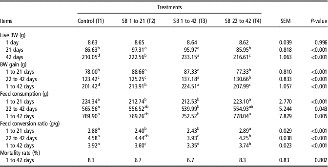
Effect of supplementation of SB in growing quail diets during different periods of age on LBW, BWG, FC and FCR are listed in Table 2. At 21 days of age, quail fed diet containing SB during the first phase (T2 and T3) reflected highly significant (P<0.001) LBW values compared to those fed diets without SB (T1 and T4). At 42 days of age, the group fed diet containing SB during total phase (T3) had significantly heavier LBW than the other groups. With regard to BWG, the groups T2 and T3 gained significantly (P<0.001) higher weight at the first period (1 to 21 days) than T1 and T4. During the period from 22 to 42 and 1 to 42 days of age, the group T3 had significantly heavier BWG than the other groups. The group fed control diet (T1) recorded the lowest (P<0.001) BWG value, whereas T2 and T4 treatments had the moderate values.
Effects of dietary sodium butyrate (SB) supplementation on growth performance of Japanese quail

a,b,c,dMeans having different superscripts within each effect in the same row are significantly different at accompanied probability.
The lowest FC was recorded for groups T2 and T3, but the groups fed diet without SB during the first phase consumed the highest (P<0.001) through 1 to 21 days of age. During the period from 22 to 42 days of age, the group T3 had lower (P=0.043) figures in FC than the control (T1). With regard to FC during 1 to 42 days of age, the group T3 had lower (P=0.005) figures in FC than the control group (T1) which showed the highest figures. The group fed diet supplemented with SB during the first phase (T2 and T3) recorded the best (P<0.001) FCR during 1 to 21 days of age compared with the groups T1 and T4. During 22 to 42 days and 1 to 42 days of age, the best values (P<0.001) in FCR recorded for the group T3 compared with those of the other treated groups (T2 and T4) and the control (T1) which was the worst. No significant differences among groups were detected on the mortality rate percentage.
Blood haematology and biochemistry
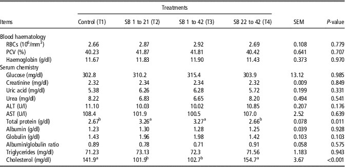
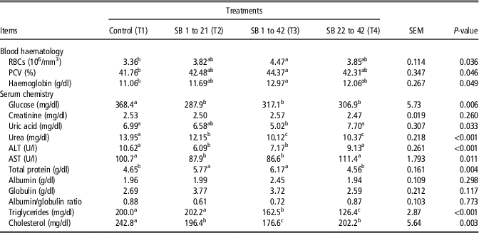
Changes in values of RBCs, PCV and Hb after treatment with dietary SB of groups T2, T3 and T4 compared to group T1 at 21 and 42 d are shown in Tables 3 and 4, respectively. No significant alterations were found after 21 days. However, samples of 42-day-old displayed significant changes, where group T3 showed the highest levels of RBCs (P=0.036), PCV (P=0.046) and Hb (P=0.049). As shown in Table 3, changes in the serum biochemical profiles after 21 days had no significance except for total protein, and cholesterol. Groups T2 and T3 displayed the higher (P=0.011) total protein values compared to the control group T1. Moreover, groups T2 and T3 had close cholesterol levels, which were significantly lower (P<0.001) than the values of group T4 and the control T1.
Effects of dietary sodium butyrate (SB) supplementation on blood haematology and biochemistry of Japanese quail at 21 days

RBCs=red blood cells; PCV=packed cell volume; ALT=alanine transaminase; AST=aspartate transaminase.
a,bMeans having different superscripts within each effect in the same row are significantly different at accompanied probability.
Effects of dietary sodium butyrate (SB) supplementation on blood haematology and biochemistry of Japanese quail at 42 days

RBCs=red blood cells; PCV=packed cell volume; ALT=alanine transaminase; AST=aspartate transaminase.
a,b,cMeans having different superscripts within each effect in the same row are significantly different at accompanied probability.
Significant changes in levels of many blood parameters were noticed after 42 days among all groups (Table 4). Glucose levels in T2, T3 and T4 groups were lower (P=0.006) than T1. Uric acid levels were lower (P=0.033) in group T3 than the close levels in groups T1 and T4. Urea levels were lower (P<0.001) in groups T3 and T4 compared with the groups T1 and T2. Analysis of AST and ALT indicated that, the groups T2 and T3 had lower levels of AST (P=0.011) and ALT (P<0.001) compared to group T1. Levels of total protein in group T3 were higher than group T2 levels, and both were higher (P=0.004) than the close values of groups T1 and T4. Serum levels of cholesterol were low in groups T2 and T4, and more lower in group T3 when compared with group T1 (P=0.003). In addition, triglycerides levels in groups T3 and T4 were significantly (P<0.001) lower than groups T1 and T2 levels.
Histological and histomorphometrical findings of intestinal segments
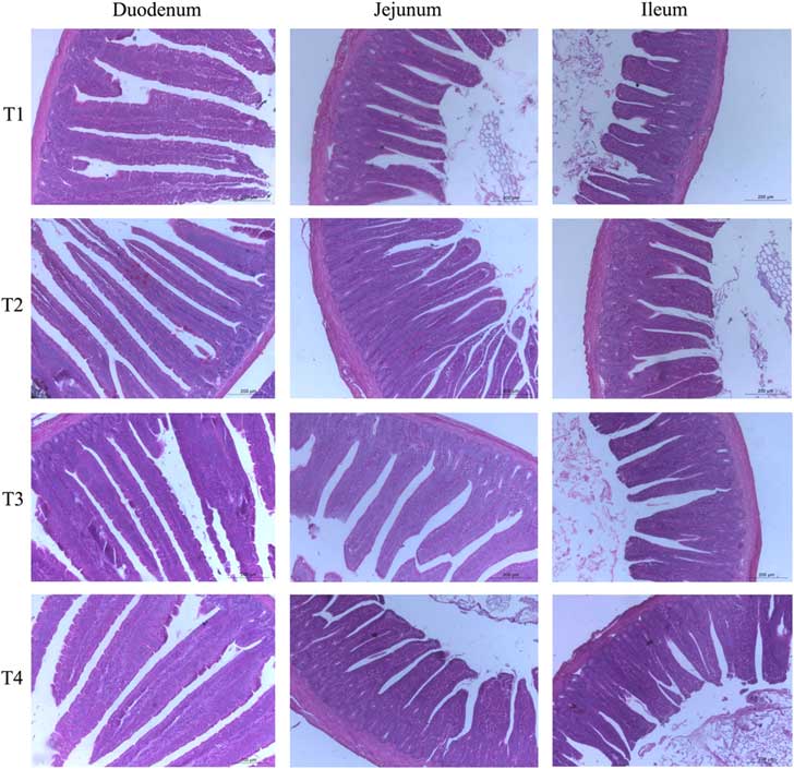
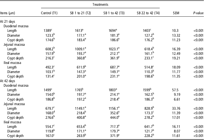
Histomorphometry parameters of the intestinal segments (duodenum, jejunum and ileum) are presented in Table 5. The intestinal mucosa of quail chick possessed normal histological structure. The intestinal mucosa threw into intestinal villi which appeared very long and thin in the duodenum, broad and long in the jejunum as well as short and broader in the ileum. The normal histological structures of the intestine of all groups were observed (Figure 1). The changes in the intestinal mucosa obtained histomorphometrically. At 21 days, the groups T2 and T3 had better (P<0.001) villus length and diameter compared to the T1 and T4. However, at 42 days, the most villus length and diameter occurred (P<0.001) in group T3, while the lowest one occurred in the control group. The intestinal crypts in all intestinal segments of all groups normally appeared with the same structures. The crypt appeared very deep in group T3, while it appeared shallow in the control group.
Photomicrograph of duodenal, jejunal, ileal mucosa of Japanese quail showing normal villi and crypts. Scale bar=200 μm. Haematoxylin and Eosin stain. Magnification 100×. Treatments: T1=control; T2= sodium butyrate (SB) 1 to 21; T3=SB 1 to 42; T4=SB 22 to 42.

Effects of dietary sodium butyrate (SB) supplementation on histomorphometry of intestinal segments of Japanese quail at 21 and 42 days

a,b,cMeans having different superscripts within each effect in the same row are significantly different at accompanied probability.
The immune organs index and histomorphometry
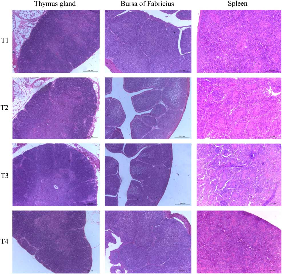
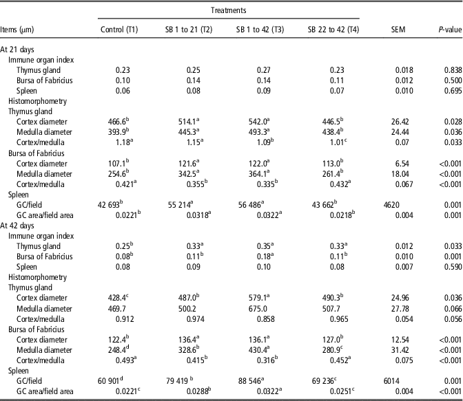
Histomorphometry parameters and index of the immune organs (thymus gland, bursa of Fabricius and spleen) are summarized in Table 6. No significant differences among groups were detected on the immune organ index at 21 days of age. At 42 days of age, the results indicated that the immune organ index was significantly affected by supplementation of SB expect spleen index. The groups fed diet supplemented with SB during any period (T2, T3 and T4) recorded the higher values (P=0.033) of thymus index compared to the control group. Bursa of Fabricius index was significantly increased (P=0.001) in group T3 compared to the other groups (T1, T2 and T4). At 21 days, the groups fed diet containing SB (T2 and T3) had significantly better histomorphometry parameters of thymus gland (cortex diameter (P=0.028), medulla diameter (P=0.036) and cortex/medulla (P=0.033)), bursa of Fabricius (P<0.001), spleen (P=0.001) compared to the T1 and T4. However, at 42 days, group T3 had significantly best values in histomorphometry parameters of the thymus gland (cortex diameter (P=0.036)), bursa of Fabricius (P<0.001), spleen (P=0.001) compared to the other groups and the control had the worst values in these parameters. The immune organs for all quail groups possessed normal histological picture. The thymus gland appeared to consist of a centrally located uniform medulla surrounded by a lobulated cortex overcrowded with lymphocytes. Histomorphometrically, the thicker thymic cortex was presented in group T3, on the same way the thicker thymic medulla was presented in group T3 (Figure 2). Bursa of Fabricius appeared highly folded containing longitudinal folds emerge into the bursal lumen, inside each fold the follicles were organized into two layers separated by connective tissues. The thicker follicular cortex was presented in group T3, on the same way the thicker follicular medulla was presented in group T3 (Figure 2). Spleen appeared enclosed by a thin fibrous capsule around splenic parenchyma which consisted of the periarterial lymphatic sheath, periellipsoidal lymphatic sheath and germinal centres. The higher germinal centre area/splenic field area was in group T3 (Figure 2).
Photomicrograph of thymus gland, bursa of Fabricius and spleen of Japanese quail. Scale bar=200 μm. Haematoxylin and Eosin stain. Magnification 100×. Treatments: T1=control; T2= sodium butyrate (SB) 1 to 21; T3=SB 1 to 42; T4=SB 22 to 42.

Effects of dietary sodium butyrate (SB) supplementation on the immune organs index and histomorphometry of Japanese quail at 21 and 42 days

GC=germinal centres.
a,b,c,dMeans having different superscripts within each effect in the same row are significantly different at accompanied probability.
Discussion
Through the results of growth performance, it could say that supplementation of SB in any period of growth (T2, T3 or T4) presented improvement in parameters of growth performance (LBW, BWG, FC and FCR) compared to the control (T1, without SB throughout the experiment). Moreover, the addition of SB during the whole period (T3, SB 1 to 42 days) gave the best results in parameters of productive performance compared with the addition in the first period only (T2, SB 1 to 21 days) or the last period only (T4, SB 22 to 42 days). The obtained data were in consistency with those of Sikandar et al. (Reference Sikandar, Zaneb, Younus, Masood, Aslam, Khattak, Ashraf, Yousaf and Rehman2017) who noticed that body gain increased and that of FCR improved (P<0.05) in the groups fed SB compared to the control. In another agreement, the growth performance of group fed SB was numerically better compared to the control group (Chamba et al., Reference Chamba, Puyalto, Ortiz, Torrealba, Mallo and Riboty2014). Likewise, supplementation of SB in broiler diet significantly augmented weight gain and better FCR as compared to the control during finisher and grower phases (Mallo et al., Reference Mallo, Puyalto and Rama Rao2012; Chamba et al., Reference Chamba, Puyalto, Ortiz, Torrealba, Mallo and Riboty2014). The improvement of performance is due to several functions for SB. Moquet et al. (Reference Moquet, Onrust, Van Immerseel, Ducatelle, Hendriks and Kwakkel2016) assumed that improved performance may be due to the ability of SB to make the gut environment acidic. Addition SB in the diets may improve the intra-luminal digestibility of proteins and mineral which may give rise to weight gain as cited by Zhang et al. (Reference Zhang, Jiang, Zhu, Gao, Dai, Chen and Zhou2011). On the contrary, Leeson et al. (Reference Leeson, Namkung, Antongiovanni and Lee2005) declared that several levels of dietary SB did not improve BWG or FCR of broilers. Moreover, Zhang et al. (Reference Zhang, Jiang, Zhu, Gao, Dai, Chen and Zhou2011) clarified that no significant difference in BWG, FC and FCR of broilers fed various levels of SB or without SB. As in the intestines functionality of day-old birds and the digestive enzymes activity is not sufficiently developed, SB is not released wholly. As a result, the feed digestion and nutrients absorption are not completely carried out. These causes depressed BWG and worst FCR during the starter phase of growth in response to SB which affects the general performance of birds as mentioned by Ahsan et al. (Reference Ahsan, Cengiz, Raza, Kuter, Chacher, Iqbal, Umar and Çakir2016). Therefore, it is recommended feeding on SB for longer periods to show its positive effect on the growth. In addition to the above, it can be stated that the reasons of positive influence of SB supplementation are various, and most of which revolve around the role of SB in improving the immunity and intestinal health. This is may be due to that SB stimulates the functional development of the gastrointestinal tract in terms of digestion and absorption of nutrients. Moreover, SB prevents harmful effects on kidney and liver function, where have the protective impact over hepatocytes. Therefore, to improve the growth performance and physiological status of quail, feeding SB through growth periods may be the most welcome approach.
Blood haematology and biochemistry analysis indicated that dietary treatment over a full period of 42 days (group T3) was the best choice during raising Japanese quail as there was an increase in RBCs, PCV and Hb, excluding occurrence of anaemia. Likely, El-Sawy et al. (Reference El-Sawy, El-Maddawy, Ibrahiem and Bo-Ghazel2015) reported that there was a significant rise in the values of PCV, Hb and RBCs in the SB-treated chicks. This rise may be caused by the increase in the number of F-reticulocytes, erythroid progenitors and F-programmed progenitors induced by SB inclusion (Constantoulakis et al., Reference Constantoulakis, Papayannopoulou and Stamatoyannopoulos1988). The group T3 displayed lower levels of serum triglycerides and cholesterol, and higher values of total protein compared to the other groups. Similar effects were reported and indicated that the metabolism of cholesterol, lipids and glucose could be affected by the action of short-chain fatty acid (SCFAs), which include, propionate, butyrate and acetate (den Besten et al., Reference den Besten, van Eunen, Groen, Venema, Reijngoud and Bakker2013; Khatibjoo et al., Reference Khatibjoo, Mahmoodi, Fattahnia, Akbari-Gharaei, Shokri and Soltani2018). Likewise, Gao et al. (Reference Gao, Yin, Zhang, Ward, Martin, Lefevre, Cefalu and Ye2009) illustrated that butyrate reduced blood levels of fatty acids (FA), cholesterol and triglycerides. Likely, it was recently reported that treatment of broiler chickens with SCFAs lowered the cholesterol and glucose levels in serum (Khatibjoo et al., Reference Khatibjoo, Mahmoodi, Fattahnia, Akbari-Gharaei, Shokri and Soltani2018). Likewise, supplementation of butyric acid led to a reduction in serum triglyceride and total cholesterol level (Zhang et al., Reference Zhang, Jiang, Zhu, Gao, Dai, Chen and Zhou2011; Kamal and Ragaa, Reference Kamal and Ragaa2014). Moreover, SCFAs decline the activity of 3-hydroxy-3-methylglutaryl-CoA reductase (HMGCR) and synthase (HMGCS), depressing synthesis of cholesterol (den Besten et al., Reference den Besten, van Eunen, Groen, Venema, Reijngoud and Bakker2013). In addition, SCFAs reduce levels of plasma glucose by activating free FA receptors (FFAR-2 and FFAR-3) and rise hormones activities of the gut as glucagon-like peptide-1 and peptide YY, which act to raise pancreatic insulin secretion and decline glucagon secretion (Le Poul et al., Reference Le Poul, Loison, Struyf, Springael, Lannoy, Decobecq, Brezillon, Dupriez, Vassart, Van Damme and Parmentier2003; den Besten et al., Reference den Besten, van Eunen, Groen, Venema, Reijngoud and Bakker2013). The current results displayed that treatment with SB as SCFAs reduced the levels of triglycerides at 42 days which could be explained with the finding of Ge et al. (Reference Ge, Li, Weiszmann, Wang, Baribault, Chen, Tian and Li2008) who reported that SCFAs enhanced FA oxidation and conversely inhibited lipolysis and de novo synthesis. Likely, it was described that SCFAs reduced serum cholesterol and triglycerides (Fushimi et al., Reference Fushimi, Suruga, Oshima, Fukiharu, Tsukamoto and Goda2006). Cholesterol and triglycerides values changed differently at 21 and 42 days by SB supplementation may be due to the long-term ability of SCFAs on reducing the enzyme activity of hepatic HMGCS and HMGCR. Moreover, it was found that SB-induced oxidation of FA and production of ketone bodies by fibroblast growth factor-21 activation in serum (Li et al., Reference Li, Gao, Zhang, Ye, Xu, Ye and Jia2012). Fibroblast growth factor-21 which plays a main role in lipid metabolism, and then butyrate may be a potential regulation of lipid homeostasis.
Analysis of biochemical indices displayed that there were significant increases in serum total protein levels in SB-treated chicks for a long period (T3). Similarly, El-Sawy et al. (Reference El-Sawy, El-Maddawy, Ibrahiem and Bo-Ghazel2015) stated that SB-treated birds had significantly higher in the serum total protein compared to that of the control. This may be attributed to the increase in activity of proteolytic enzymes, the improvement in pancreatic secretions, stimulating the digestive enzymes activity, the increase of nutrient digestibility and the higher absorption rate caused by increased intestinal villus length. A reference for biochemical analysis of some serum parameters in mature Japanese quail was described by Scholtz et al. (Reference Scholtz, Halle, Flachowsky and Sauerwein2009) who stated that results could be affected by species, sex, age and diet. Moreover, the biochemical analysis may help in the clinical diagnosis of diseases; for example, ALT and AST are two enzymes produced by the liver for handling transamination reactions and used as tests for liver function. However, AST is not considered a specific sign of hepatocellular disease as it occurs in skeletal and cardiac muscles and RBCs (Harr, Reference Harr2002; Scholtz et al., Reference Scholtz, Halle, Flachowsky and Sauerwein2009). It was stated that hepatocellular disease could be diagnosed by high values of serum AST and ALT (Harr, Reference Harr2002). Moreover, as one of the best kidney function tests, analysis of serum creatinine indicated the normal state of kidney function after SB treatment. It was found that there was a significant reduction in serum AST and ALT levels after treatment with SB which resulted in lowering nitrogenous output in the form of urea and uric acid formation, where uric acid is the main product nitrogenous waste in birds (Harr, Reference Harr2002). However, high values of uric acid and decreased elimination of urate by the kidneys due to renal disease results, which is characterized by uric acid or urate deposition in joints and tissues (Maiuolo et al., Reference Maiuolo, Oppedisano, Gratteri, Muscoli and Mollace2016). Urea and uric acid values changed differently at 21 and 42 days by SB supplementation may be due to the long-term ability of SB in decreasing urease and uricase activities (Wang et al., Reference Wang, Wang, Liao, Wu, Liang, Laudadio and Tufarelli2016). This effect of SB treatment seemed to protect the quails against the toxic effects of increased uric acid levels. Some studies showed that using of butyric acid various forms as dietary supplementation have not changed levels of ALT and AST and creatinine in serum (Kamal and Ragaa, Reference Kamal and Ragaa2014).
Similar to current observation in histomorphometry for intestine, Leeson et al. (Reference Leeson, Namkung, Antongiovanni and Lee2005) and Shahir et al. (Reference Shahir, Moradi, Afsarian and Esmaeilipour2013) found significantly improve in crypt depth and villus length by SB addition in broiler diet. Moreover, Mallo et al. (Reference Mallo, Puyalto and Rama Rao2012) demonstrated that the lengths and widths of the intestinal villi can be significantly improved by the addition of SB. El-Sawy et al. (Reference El-Sawy, El-Maddawy, Ibrahiem and Bo-Ghazel2015) indicated that the intestines of SB-treated groups presented a significant improvement in villi length compared to the control group. Newly, Abdelqader and Al-Fataftah (Reference Abdelqader and Al-Fataftah2016) concluded that butyric acid addition in broilers diet can decrease intestinal epithelia damage then accelerate and recovery of intestinal histological characteristics and growth performance. Sikandar et al. (Reference Sikandar, Zaneb, Younus, Masood, Aslam, Khattak, Ashraf, Yousaf and Rehman2017) found improved histomorphometrics in SB-offered groups in jejunum and duodenum compared to the control. The addition of SB to poultry diets may promote restitution of intestinal epithelial cell and stimulate the growth of the intestinal mucosa (Hu and Guo, Reference Hu and Guo2007). Moreover, butyrate acts as a direct source of energy to stimulate the proliferation and differentiation of epithelial cells, improve intestinal barrier function and benefits to maintain the villus structure (Kinoshita et al., Reference Kinoshita, Suzuki and Saito2002). The action of SB in the improvement of intestine status may be due to that butyrate stimulates intestinal blood flow and the gastrointestinal hormones synthesis. Butyric acid leads to increase the secretion of peptides which lead to enhanced proliferation of enterocytes for improved repair of damaged mucosa and increased height of villi. In healthy avian the increase in immune organs index is correlated with enhanced immune responses. Bursa and thymus index in the current study increased at 42 days of age in the group fed SB from 1 to 42 days. As well, Sikandar et al. (Reference Sikandar, Zaneb, Younus, Masood, Aslam, Khattak, Ashraf, Yousaf and Rehman2017) stated that thymus and bursa weighed more in the group fed SB compared to the control. It is clear that, addition SB in the diet may modify the function of T and B cells in the later phases of the age and can regulate the immunity (Ahsan et al., Reference Ahsan, Cengiz, Raza, Kuter, Chacher, Iqbal, Umar and Çakir2016). Spleen, thymus and bursa are the key actors of the immune system and their histomorphometry parameters in the current study significantly improved in the quail fed SB in any period during the growing phase. These findings similar to results of Sikandar et al. (Reference Sikandar, Zaneb, Younus, Masood, Aslam, Khattak, Ashraf, Yousaf and Rehman2017) who stated that the greater germinal centre area and sized thymus medulla in the spleen of SB-treated chickens, and an increasing trend of bursal parameters detected in the group fed SB indicated that SB may activate the immune system in chicken. From current results, it was thought that the improvement of growth performance fed SB supplementation may be due to the improvement in morphometry of intestine and immune organs is correlated with enhanced the growth and health of birds.
Conclusions
The results of current study declared that dietary supplementation of SB in any period from growth phase of quail improved growth performance, blood biochemistry, haematology and histomorphometry of intestine and immune organs. Supplementation of SB throughout the growth period 1 to 42 days of age showed the best results. Overall the results indicate that, it is recommended feeding quail on diets containing SB through the whole growth period to show its affirmative impact on the growth and physiological indices.
Acknowledgements
The authors thank the administration of poultry farm, Faculty of Agriculture, Fayoum University, Egypt.
Declaration of interest
The authors declare that there is no conflict of interest.
Ethics statement
Animal care and maintenance were performed in accordance with guidelines of Egyptian Research Ethics Committee and the guidelines contained in the Guide for the Care and Use of Laboratory Animals (2011).
Software and data repository resources
None of the data were deposited in an official repository.