Introduction
Cnidarian jellyfish are major oceanic plankton and neuston (Banse Reference Banse1975; Nishikawa Reference Nishikawa2004). Earlier, it was believed that only a few fish species consumed jellyfish as their food (Arai Reference Arai1988) which is because the soft tissues of jellyfish are rapidly digested in fish stomachs (Arai Reference Arai2005), making it difficult to detect them in conventional gut content analyses. In contrast to the previous conception, recent DNA analysis of fish gut contents revealed the presence of cnidarians in their stomachs and emphasised their importance as a food source for marine fish (Dischereit et al. Reference Dischereit, Throm, Werner, Neuhaus and Havermans2024). In addition to the predator–prey relationships between fish and cnidarian jellyfish species, a complex food web in the ocean is created by many organisms including those traditionally recognised as fish food, such as small fish and arrow worms (Belgrano Reference Belgrano2005; Sommer et al. Reference Sommer, Charalampous, Scotti and Moustaka-Gouni2018).
Trematodes are a group of parasites that inhabit more than two hosts in their life cycles (Olsen Reference Olsen1967). Many of the trematode species have larval (miracidia, cercaria, and metacercaria), parthenital (sporocysts and rediae), and adult (sexual adults or maritae) stages (Littlewood and Bray Reference Littlewood and Bray2014). In the life cycles, molluscs are the first intermediate hosts for sporocysts or rediae that generate cercariae by asexual proliferation. The cercariae are released from the molluscs to develop into metacercariae in the second intermediate host, such as cnidarian jellyfish (Browne et al. Reference Browne, Pitt and Cribb2020; Cribb et al. Reference Cribb, Cutmore, Wee, Browne, Morales and Pitt2024; Diaz Briz et al. Reference Diaz Briz, Martorelli, Genzano and Mianzan2012; Køie Reference Køie1991; Louvard et al. Reference Louvard, Yong, Cutmore and Cribb2024; Motta et al. Reference Motta, Caffara, Fioravanti, Bottaro, Avian, Terlizzi and Tedesco2023; Pitt et al. Reference Pitt, Browne, Cutmore, Diaz, Wee and Cribb2025). The metacercariae are then ingested by the definitive vertebrate hosts and develop into adults, which release eggs into the environment through host faeces. Furthermore, predators of the second intermediate host harbouring metacercariae might become paratenic hosts, which are potential sources of infection for the definitive host. Nevertheless, owing to the difficulty of morphological identification, there is still no clear information on the species and hosts of stages other than adults, including metacercariae, for most trematode species.
To address the abovementioned issues, molecular methods have been used for the species identification of metacercaria in cnidarian jellyfish and other host groups (Ansai et al. Reference Ansai, Sekine, Munakata, Sase, Sasaki, Nitta, Suzuki, Abe, Takano, Fukumori, Nakatsubata, Suzuki and Waki2025; Cribb et al. Reference Cribb, Cutmore, Wee, Browne, Morales and Pitt2024; Louvard et al. Reference Louvard, Yong, Cutmore and Cribb2024; Nakao et al. Reference Nakao, Waki, Sasaki, Anders, Koga and Asakawa2017). In previous studies, suitable barcoding fragments for species identification can be obtained from the nuclear 28S ribosomal RNA (rDNA), the second internal transcribed spacer region (ITS2 rDNA), and mitochondrial cytochrome c oxidase subunit 1 (COI) regions, and some combinations of these regions were often used (Cribb et al. Reference Cribb, Cutmore, Wee, Browne, Morales and Pitt2024; Seo et al. Reference Seo, Ansai, Sase, Saito, Takano, Kojima and Waki2024). Among the regions, COI is especially suitable for the species identification of wide groups of trematodes (Ansai et al. Reference Ansai, Sekine, Munakata, Sase, Sasaki, Nitta, Suzuki, Abe, Takano, Fukumori, Nakatsubata, Suzuki and Waki2025; Cribb et al. Reference Cribb, Cutmore, Wee, Browne, Morales and Pitt2024; Martin et al. Reference Martin, Downie and Cribb2020; Seo et al. Reference Seo, Ansai, Sase, Saito, Takano, Kojima and Waki2024). However, the sequence information for adults, which can be identified on the basis of morphological analyses, of many species is currently unavailable. Therefore, to identify the species of those metacercariae using molecular approaches, it is essential to obtain the sequence of their adults for data comparisons.
Metacercariae have been detected in cnidarian jellyfish frequently around the world (Browne et al. Reference Browne, Pitt and Cribb2020; Cribb et al. Reference Cribb, Cutmore, Wee, Browne, Morales and Pitt2024; Køie Reference Køie1991; Louvard et al. Reference Louvard, Yong, Cutmore and Cribb2024; Motta et al. Reference Motta, Caffara, Fioravanti, Bottaro, Avian, Terlizzi and Tedesco2023; Pitt et al. Reference Pitt, Browne, Cutmore, Diaz, Wee and Cribb2025). Because cnidarian jellyfish are beginning to be considered major food for marine fish, the metacercariae in jellyfish should infect host fish via predation. Indeed, adult trematodes, from which metacercariae were found in cnidarian, have been detected in marine fish (Cribb et al. Reference Cribb, Cutmore, Wee, Browne, Morales and Pitt2024; Køie Reference Køie1991; Louvard et al. Reference Louvard, Yong, Cutmore and Cribb2024), which suggests the importance of the jellyfish as a source of infection to their definitive host fish. However, many of the metacercariae from jellyfish were not identified to their species levels (Pitt et al. Reference Pitt, Browne, Cutmore, Diaz, Wee and Cribb2025). Moreover, recent findings of metacercaria in cnidarians are focused on the Australian coast, the Mediterranean Sea, and the Atlantic Ocean (Browne et al. Reference Browne, Pitt and Cribb2020; Cribb et al. Reference Cribb, Cutmore, Wee, Browne, Morales and Pitt2024; Diaz Briz et al. Reference Diaz Briz, Martorelli, Genzano and Mianzan2012; Louvard et al. Reference Louvard, Yong, Cutmore and Cribb2024; Motta et al. Reference Motta, Caffara, Fioravanti, Bottaro, Avian, Terlizzi and Tedesco2023; Pitt et al. Reference Pitt, Browne, Cutmore, Diaz, Wee and Cribb2025), with no clear understanding of the species of metacercaria and host jellyfish in other waters. We focused on the metacercariae of cnidarian jellyfish around Japan. We examined trematodes obtained from cnidarian jellyfish in Japan and used DNA barcoding. Our study underscores the importance of jellyfish as the second intermediate or paratenic host of fish trematodes around Japan.
Material and methods
Sample collection
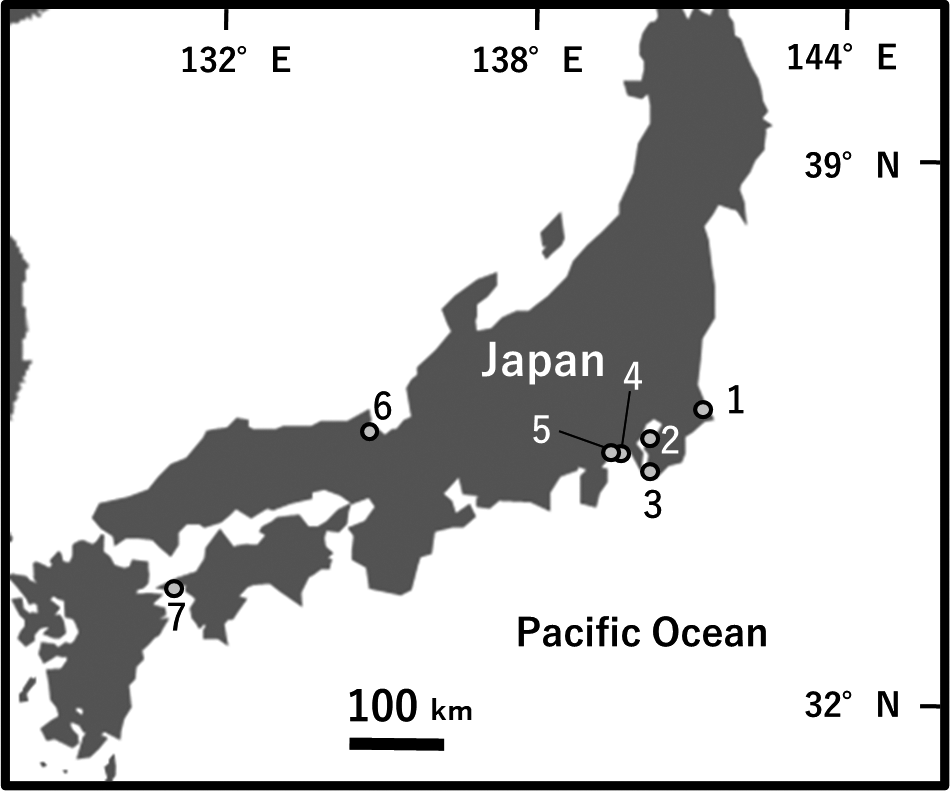
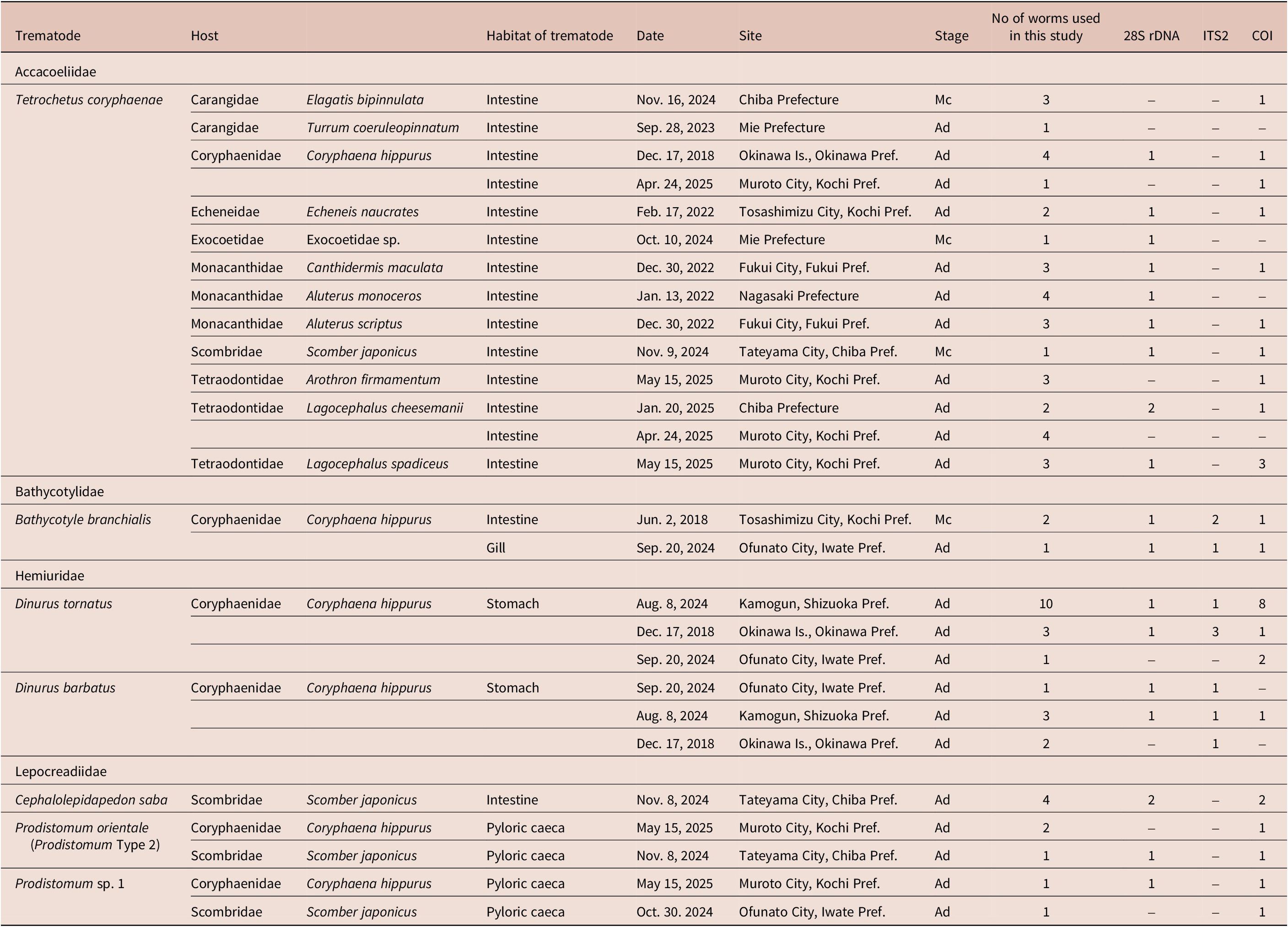
Cnidarian jellyfish were sampled from seven localities in the coastal regions of Japan between 2023 and 2025 (Table 1, Fig 1). The hosts were subsequently transported to the laboratory and identified based on their morphological characteristics, as described by Minemizu et al. (Reference Minemizu, Kubota, Hirano and Lindsay2015) and Church et al. (Reference Church, Abedon, Ahuja, Anthony, Destanović, Ramirez, Rojas, Albinsson, Trasobares, Bergemann, Bogdanovic, Burdick, Cunha, Damian-Serrano, D’Elía, Dion, Doyle, Gonçalves, Rajal, Haddock, Helm, Gouvello, Lewis, Magalhães, Mańko, Mayorga-Adame, de Mendoza, Moura, Munro, Nel, Oguchi, Perelman, Prieto, Pitt, Roughan, Schaeffer, Schmidt, Sellanes, Wilson, Yamamoto, Lazo-Wasem, Simon, Decker, Coughlan and Dunn2025). Host tissues were then dissected under a stereomicroscope. When metacercariae were detected, they were observed in live conditions, and photographs of some selected worms were taken under a light microscope (BX53, Olympus). The detected metacercariae were subsequently placed between a cover slip and glass slide, fixed in 70% or 99% ethanol, or transferred directly into 70% ethanol for fixation. These samples were preserved in 70% or 99% ethanol. Marine fish were also obtained from fishery markets or recreational anglers in eight Prefectures in Japan between 2018 and 2025 (Table 2), when there was an opportunity, and their trematodes were used for molecular comparisons with metacercariae in jellyfish. Moreover, a metacercaria from a stranded arrow worm was used in this study. Adults and metacercariae detected in these hosts were fixed in 70% or 99% ethanol or 5% formalin between glass slides and subsequently stored in ethanol or formalin. These trematode specimens were used for morphological and molecular analyses.
Metacercariae detected from cnidarian and chaetognathan hosts. Site numbers correspond to those given in Fig 1

I: infection intensity.
Sampling sites of cnidarian and chaetognathan hosts in Japan. The locality numbers correspond to those provided in Table 1.

Adults and metacercariae of trematodes from fish hosts sampled in this study.

Pref., Prefecture; Stage, developmental stage in the host (Ad, adults; Mc, metacercariae); 28S rDNA, ITS 2, and COI: numbers of specimens of which sequences were obtained.
Morphological observations
The trematode samples fixed between glass slides were stained with acetocarmine or Heidenhein’s iron–haematoxylin, dehydrated in an ethanol series, cleared in xylene or creosote, and mounted on slides with Canada balsam, followed by observation under a light microscope. Permanent mounts of selected adults and metacercaria, fixed in ethanol, were also prepared after cutting a piece of tissue from each specimen for molecular analyses. These specimens were observed under a light microscope (BX53, Olympus). A camera lucida (U-DA, Olympus) attached to the microscope was used for making line drawings. Permanent mounts of metacercariae and adults were deposited in the Ibaraki Nature Museum (INM-1-131100–131180).
DNA barcoding and phylogenetic analyses
Alkaline lysates of small tissue pieces of selected metacercariae and adults were individually prepared as templates for polymerase chain reaction (PCR), as described by Nakao et al. (Reference Nakao, Waki, Sasaki, Anders, Koga and Asakawa2017) (Table 3). DNA templates for some tiny lepocreadiid and hemiurid metacercariae were obtained using their whole bodies. Partial fragments of 28S rRNA and COI regions were amplified using the following primer sets. 28S rRNA: dig12 and 1500R (Tkach et al. Reference Tkach, Kudlai and Kostadinova2016) or LSU5 (Littlewood et al. Reference Littlewood, Curini-Galletti and Herniou2000) and 1600R (Williams et al. Reference Williams, Reid and Littlewood2003); COI: JB3 (Bowles et al. Reference Bowles, Hope, Tiu, Liu and McManus1993), CO1-R trema (Miura et al. Reference Miura, Kuris, Torchin, Hechinger, Dunham and Chiba2005), and JB3 and another reverse primer JB4.5 (Bowles et al. Reference Bowles, Blair and McManus1992). Moreover, for the species identification of metacercariae of two Prodistomum species (Prodistomum orientale and Prodistomum Type 4), the following primer sets of COI of the other region were used for data comparisons with those in a previous study (Cribb et al. Reference Cribb, Cutmore, Wee, Browne, Morales and Pitt2024): Dig_cox1Fa and Dig_cox1R (Wee et al. Reference Wee, Cribb, Bray and Cutmore2017). For the species identification of the metacercariae of Dinurus and Bathycotyle species, the partial fragments of ITS2 rDNA were also amplified using the primer sets as described by Louvard et al. (Reference Louvard, Yong, Cutmore and Cribb2024): 3S (Bowles et al. Reference Bowles, Hope, Tiu, Liu and McManus1993) and ITS2.2 (Cribb et al. Reference Cribb, Anderson, Adlard and Bray1998). These ITS2 rDNA primers were selected based on availability of comparative datasets in the online databases. These primers were also used for sequencing. Whenever required, the following internal sequencing primers were used for 28S rDNA: ECD2S (reverse, Williams and Ozawa, Reference Williams and Ozawa2006), D3m (reverse, Takano and Kano Reference Takano and Kano2014), and 900F (forward, Lockyer et al. Reference Lockyer, Olson, Østergaard, Rollinson, Johnston, Attwood, Southgate, Horak, Snyder, Le, Agatsuma, McManus, Carmichael, Naem and Littlewood2003). A commercial service (Eurofins Genomics Inc., Tokyo, Japan) was used to perform the Sanger sequencing of PCR amplicons. The BioEdit sequence alignment editor (Hall Reference Hall1999) was used to prepare the alignment datasets of 28S rDNA and COI.
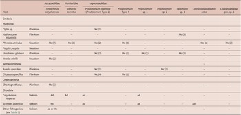
Summary of infections of trematodes in sampled host species.

Developmental stage in the host: Ad, adults; Mc, metacercariae. The numbers in parentheses indicate the number of metacercariae identified by molecular analyses.
The systematic positions of metacercariae at the species or higher taxonomic levels were predicted by comparing their 28S rDNA, ITS2, and COI sequences with published data using the Basic Local Alignment Search Tool (BLAST) against the DDBJ/EMBL/GenBank databases in accordance with the method described by Ansai et al. (Reference Ansai, Sekine, Munakata, Sase, Sasaki, Nitta, Suzuki, Abe, Takano, Fukumori, Nakatsubata, Suzuki and Waki2025). We identified a set of most similar sequences and the related species/genera to each of our sequences using BLAST. We also compared our sequences of metacercariae with those of our adult trematodes. Regarding the COI, if the closely related sequences (>85% similarity) of our metacercariae were not found in our adults and in the databases, the COI sequences were not used for species identification. The sequences of our samples and the closely related sequences found in the databases were used to construct an alignment data set using BioEdit. Then, intraspecific divergence values for their overlapping regions (858–1191 sites for 28S rDNA: 495 and 536 sites for ITS; 348–802 sites for COI) were computed based on the p-distance using the genetic software MEGA X (Kumar et al. Reference Kumar, Stecher, Li, Knyaz and Tamura2018). The divergence values of related species were used as thresholds to decide whether it was reasonable to assume the conspecificity of metacercariae versus those from the online databases and our adults. If the divergence values between our metacercariae and species-identified trematodes were less than or subequal to the threshold intraspecific distance value of the related species/genera, they were deemed conspecific; otherwise, their higher taxonomic group (genus or family) was examined by phylogenetic analyses using 28S rDNA sequences in MEGA X. The maximum likelihood method was used for reconstructing the phylogenetic trees under the General Time Reversible + G models selected by MEGA X (Nei and Kumar, Reference Nei and Kumar2000). Nodal support was determined with 1000 bootstrap replicates. Conspecificity of adults was also confirmed based on the sequence comparisons when species identifications were difficult only by morphological analyses.
Through comparisons of our 28S rDNA and ITS2 sequences with published data using BLAST against the DDBJ/EMBL/GenBank databases, we found that our adults of Dinurus barbatus, and an adult and metacercariae of Bathycotyle branchialis detected in Coryphaena hippurus, were molecularly closely related to unidentified metacercariae in jellyfish reported by Louvard et al. (Reference Louvard, Yong, Cutmore and Cribb2024). We also conducted morphological and molecular analyses to determine the conspecificity of the unidentified metacercariae in the previous study and our study worms using the same above-described methods. A phylogenetic tree based on 28S rDNA (645 site-overlapping region) was built for Bathycotyle branchialis and its related species to determine relationships between our worm and unidentified metacercariae in a past study by the same method as described above.
Results
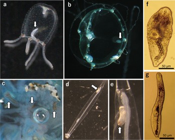
We sampled eight cnidarian species (245 individuals) from seven localities (Fig 1, Table 1), among which infection with metacercariae was detected in seven host species (Fig 2, Table 1 and 3). Based on morphological observations and molecular analyses of 28S rDNA (1343–1607 bp), ITS2 (521–662 bp), and COI (376–890 bp), the metacercariae obtained from the jellyfish were classified into nine species belonging to three families. Metacercariae of Tetrochetus coryphaenae (see below) were also found in an arrow worm and marine fish. We also report adults in fish hosts which were morphologically and/or molecularly identified as follows. In addition, unidentified metacercariae in the online databases, which were identified at their species levels in our analyses, were included in this section.
Metacercariae (arrows) in cnidarian (a–c) and chaetognathan (d and e) hosts (non-scale), and isolated from hosts (f and g). (a) Opechona sp. 1 in Hydrocoryne miurensis. (b) Prodistomum orientale in Clytia sp. (c) Tetrochetus coryphaenae in Physalia utriculus. (d, e) Tetrochetus coryphaenae in Chaetognatha sp. (f) Prodistomum sp. 1 from Urashimea globosa. (g) Lepocreadiidae sp. 1 from Physalia utriculus.

All measurements in the descriptions are expressed as mean values, with ranges in parentheses, and are in micrometres unless otherwise stated.
Family Accacoeliidae Odhner, 1911
Genus Tetrochetus Looss, 1912
Adults of Tetrochetus species were sampled from nine fish species (Table 2). These adults exhibited morphological characteristics of the genus Tetrochetus – oesophagus long and slender, ventral sucker rounded, and sinus-sac absent (Gibson et al. Reference Gibson, Jones and Bray2002). On the basis of our morphological and molecular analyses, all adults were identified as a single species, Tetrochetus coryphaenae Yamaguti, Reference Yamaguti1934, as follows. Metacercariae of this species were detected from two cnidarian species, Physalia utriculus and Velella velella. In addition to these jellyfish hosts, metacercariae were also detected in an arrow worm and three fish species.
Tetrochetus coryphaenae Yamaguti, Reference Yamaguti1934
Of the adults found in the nine host fish species, the morphological characteristics of adults obtained from two tetraodontid species, Lagocephalus cheesemanii (Clarke 1897) and Lagocephalus spadiceus (Richardson 1845), were different from those of adults obtained from the other fish species (e.g. Coryphaena hippurus Linnaeus, 1758). These two forms of adults were morphologically identified as Tetrochetus hamadai and Tetrochetus coryphaenae, respectively (see below). Adults morphologically identified as Tetrochetus hamadai were found only in the two tetraodontid hosts, while those identified as Tetrochetus coryphaenae were found only in the other host fish species. However, our molecular analysis revealed that these two forms belong to a single species. We then defined the former as the ‘T. hamadai’ type, which was defined as a morphological variation of Tetrochetus coryphaenae, and the latter as the ‘T. coryphaenae’ type.
Morphological descriptions
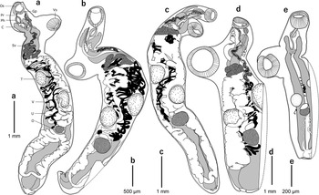
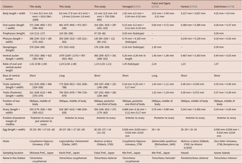
Adult (‘T. corypahenae’ type) (Fig 3a–c, measurements shown in Table 4). Body elongated and narrow. Oral sucker rounded, subterminal. Prepharynx and pharynx muscular. Oesophagus long, slender, dividing at level of anterior margin of ventral sucker, with small out-pocketings. Caeca long and wide, extending from level of pharynx to posterior body terminal. Ventral sucker subequal to or larger than oral sucker, rounded, situating in anterior one-fourth to one-sixth of body. Testes oval, situated obliquely at middle to posterior one-third of body. Anterior testis subequal to posterior testis, smaller than half of body width. Sinus-sac absent. Seminal vesicle long, posterior margin reaching cluster of uterus. Genital pore on ventral margin of oral sucker. Ovary oval. Vitellaria tubular. Posterior margin of vitellaria reaching middle of ovary or extending to posterior margin of ovary. Eggs numerous, elongate oval, not less than 25 μm in length. Excretory vesicle connecting posterior margin of caecum. Excretory pore at posterior margin of body.
Adults and metacercaria of Tetrochetus coryphaenae. The dotted lines show the outline of the tissue cut out for PCR. (a–c) ‘T. coryphaenae type’ adult. (d) ‘T. hamadai type’ adult. (a) Adult from Coryphaena hippurus. (b) Adult from Aluterus scriptus. (c) Adult from Aluterus monoceros. (d) Adult from Lagocephalus cheesemanii. (e) Metacercaria from Physalia utriculus. Abbreviations: C, caecum; Gp, gonad pore; O, ovary; Os, oral sucker; Ph, pharynx; Pr, prepharynx; Sv, seminal vesicle; T, testis; U, uterus; V, vitellarium; Vs, ventral sucker.

Comparison of morphological measurements of Tetrochetus coryphaenae. All measurements are in μm unless indicated otherwise

* Size of two testes.
** This species was divided into two species, Lagocephalus cheesemanii (Clarke 1897) and Lagocephalus spadiceus (Richardson 1845) (Abe and Tabeta Reference Abe and Tabeta1983; Abe et al. Reference Abe, Tabeta and Kitahama1984).
Remarks. This genus includes 13 species (WoRMS Editorial Board 2025) among which Tetrochetus coryphaenae can be distinguished from 10 of the other 12 species based on the following morphological characteristics: narrow body, testes whose widths are greater than half the width of the body, ventral sucker subequal to or larger than oral sucker and at anterior one-fourth to one-sixth of body and eggs not less than 25 μm in length. The remaining two species, Tetrochetus aluterae (Hanson, Reference Hanson1955) and Tetrochetus mitenevi Zubchenko, Reference Zubchenko1978, exhibit the same morphological characteristics as those of Tetrochetus coryphaenae. We concluded that Tetrochetus aluterae and Tetrochetus mitenevi were junior synonyms of Tetrochetus coryphaenae because we could not find any distinguishing characteristics between the three species. Madhavi and Bray (Reference Madhavi and Bray2018) stated that Tetrochetus coryphaenae and Tetrochetus aluterae can be distinguished by their egg size (25–29 μm in length in Tetrochetus aluterae in the original description (Hanson Reference Hanson1955)); however, the sizes are within the range of the egg size of our specimen (25–39 μm). Furthermore, we detected adults with the distinct morphological characteristics of Tetrochetus coryphaenae from the type host species of Tetrochetus aluterae, scrawled filefish Aluterus scriptus (Osbeck, 1765) (Monacanthidae) (Hanson Reference Hanson1955). In addition, Tetrochetus aluterae was suspected as a synonym of Tetrochetus coryphaenae in previous studies (Madhavi and Bray Reference Madhavi and Bray2018; Siddiqi and Cable Reference Siddiqi and Cable1960), thereby supporting our morphological analyses. Tetrochetus mitenevi has been detected in John dory Zeus faber (Zeidae) in the Atlantic Ocean (Zubchenko Reference Zubchenko1978). Zubchenko (Reference Zubchenko1978) reported that Tetrochetus mitenevi and Tetrochetus coryphaenae could be distinguished from each other based on the body and sucker size. Nonetheless, the morphological characteristics of Tetrochetus mitenevi are similar to those of our specimens and to the original description of Tetrochetus coryphaenae (Yamaguti, Reference Yamaguti1934), based on body size (4.25 mm × 0.9 mm in Tetrochetus mitenev vs 2.8–4.8 mm × 538–895 μm in our adults of Tetrochetus coryphaenae from Aluterus scriptus) and oral and ventral suckers (oral and ventral sucker length 0.28 mm and 0.44 mm in Tetrochetus mitenevi vs 281–353 and 290–427 μm in our specimens, respectively) (Table 4).
Adult (‘T. hamadai’ type) (Fig 3d, measurements shown in Table 4). Body elongated and wide. Oral sucker rounded, subterminal. Prepharynx and pharynx muscular. Oesophagus long, slender, dividing at level of anterior margin of ventral sucker, with small out-pocketings. Caeca long and wide, extending from level of pharynx to posterior body terminal. Ventral sucker clearly larger than oral sucker, rounded, situating at anterior one-fourth of body. Testes oval, at middle of body, situated obliquely. Anterior testis subequal to posterior testis, with the width smaller than a half of body width. Sinus-sac absent. Seminal vesicle long, posterior margin reaching anterior margin of anterior testis. Genital pore on ventral margin of oral sucker. Ovary oval. Vitellaria tubular. Posterior margin of vitellaria extending to posterior margin of ovary. Eggs numerous, elongate oval. Excretory vesicle connecting posterior margin of caecum. Excretory pore at posterior margin of body.
Remarks. Morphological characteristics of our specimens resembled those of Tetrochetus hamadai (Fukui and Ogata Reference Fukui and Ogata1935), such as the following: wide body, large ventral sucker, and vitellaria whose posterior margin extends posterior margin of ovary. Among species in this genus, Tetrochetus macrorchis Yamaguti, Reference Yamaguti1970, and Tetrochetus scomberesoxis Nikolaeva, 1966, resemble Tetrochetus hamadai because they have a wide body. Nevertheless, these two species can be distinguished from Tetrochetus hamadai based on their testes that are larger than half the width of the body (vs smaller in Tetrochetus hamadai) and the posterior margin of vitellaria that does not extend beyond the posterior margin of ovary (vs extending in Tetrochetus hamadai). Although our specimens were identified as Tetrochetus hamadai as above, molecular analyses using the 28S rDNA and COI sequences of these specimens (= ‘T. hamadai’ type) and ‘T. coryphaenae’ type specimens in our study indicated that they were a single species as follows.
28S rDNA sequences (891–1450 bp) were determined from eight adults, including three ‘T. hamadai’ and five ‘T. coryphaenae’ types. All sequences were identical including two sequences of Tetrochetus coryphaenae which were deposited in online databases in their overlapping region (858 sites, Supplementary Table S1). This result suggests that the ‘T. hamadai’ type and ‘T. coryphaenae’ type specimens, and Tetrochetus coryphaenae yield sequences in the databases that can be identified as the same species – Tetrochetus coryphaenae. Furthermore, 379- to 900-bp-long COI sequences were obtained from six adults of the ‘T. coryphaenae’ type, and their intraspecific divergence values in the overlapping region (364 sites) ranged from 0 to 0.0274 (Supplementary Table S2). Here, the divergence values between the ‘T. coryphaenae’- and ‘T. hamadai’-type adults ranged from 0.0110 to 0.0302. Although this maximum value was slightly higher than those calculated in ‘T. coryphaenae’-type adults, these differences were interpreted as within the intraspecific variation. Therefore, we identified these two types as a single species. The morphological variations between these types are probably owing to differences in host species, as co-infection of these two types was not found in the same host species.
In addition, T. hamadai was originally described by Fukui and Ogata (Reference Fukui and Ogata1935) based on adults in the type host puffer Sphoeroides spadiceus (Tetraodontidae). However, this host species was later categorised into two species, Lagocephalus cheesemanii and Lagocephalus spadiceus (Abe and Tabeta Reference Abe and Tabeta1983; Abe et al. Reference Abe, Tabeta and Kitahama1984). As the original description of Tetrochetus hamadai did not provide morphological characteristics of the type host specimen, it remains unclear as to which of the two fish species is the type host. However, ‘T. hamadai’-type adults were detected in both candidates for the type host species in our study and judged as Tetrochetus coryphaenae.
Metacercaria (Fig 3e). Body hue light brown in live condition. Body elongated and wide, 2.4 mm (1.1–4.0 mm) in length and 721 (541–902) in width. Oral sucker rounded, subterminal, 216 (174–247) in length and 247 (201–303) in width. Prepharynx and pharynx muscular. Prepharynx 21 (15–28) in length. Pharynx 71 (51–81) in length and 68 (54–86) in width. Oesophagus long, slender, dividing at level of anterior margin of ventral sucker, with small out-pocketings. Caeca long and wide, extending from level of pharynx to posterior body terminal. Ventral sucker well-developed, rounded, situated in anterior one-third of body, 416 (264–549) in length and 383 (265–499) in width. Testes oval, at middle of body. Anterior testis 298 (242–355) in length and 200 (190–209) in width. Posterior testis 235 (141–299) in length and 214 (158–285) in width. Ovary oval, 134 (90–177) in length and 160 (121–198) in width. Excretory vesicle connecting posterior margin of caecum. Excretory pore at posterior margin of body.
Remarks. Based on the comparison of 28S rDNA and COI sequences, our metacercariae and Tetrochetus coryphaenae adults were identified as a single species (Supplementary Tables S1, S2). Our 28S rDNA sequences were identical to those of Tetrochetus coryphaenae sequences deposited in the DDBJ/EMBL/GenBank databases (OR814155 and OR814156, Louvard et al. Reference Louvard, Yong, Cutmore and Cribb2024), supporting our species identifications.
Hosts of adults in the present study. ‘ T. coryphaenae’ type: Aluterus monoceros (Linnaeus, 1758), Aluterus scriptus (Osbeck, 1765), Arothron firmamentum (Temminck and Schlegel, 1850), Canthidermis maculata (Bloch, 1786), Coryphaena hippurus Linnaeus, 1758, Echeneis naucrates Linnaeus, 1758, Turrum coeruleopinnatum (Rüppell, 1830). ‘ T. hamadai’ type: Lagocephalus cheesemanii (Clarke, 1897), Lagocephalus spadiceus (Richardson, 1845).
Host jellyfish of metacercariae in the present study: Physalia utriculus (Gmelin, 1788); Velella velella (Linnaeus, 1758)
Host chaetognathan of metacercaria in the present study: Chaetognatha sp.
Host fish of metacercariae in the present study: Elagatis bipinnulata (Quoy and Gaimard, 1825); Exocoetidae sp.; Scomber japonicus Houttuyn, 1782
New Japanese name: Kurage-biidoro-kyuchu (‘Kurage’ is ‘jellyfish’ in Japanese. ‘Biidoro’ is ‘a Japanese traditional glass trinket’. ‘Kyuchu’ is ‘trematode’ in Japanese).
Synonyms: Tetrochetus aluterae (Hanson, Reference Hanson1955); Tetrochetus hamadai Fukui and Ogata, Reference Fukui and Ogata1935; Tetrochetus mitenevi Zubchenko, Reference Zubchenko1978.
Depositions: INM-1-131100–131127 (adults), INM-1-131128–131146 (metacercariae)
DNA markers: 28S rDNA, LC889164–889168 (adults), LC889169–889172 (metacercariae); COI, LC889205–889214 (adults), LC889215–889221 (metacercariae)
Bathycotylidae Dollfus, 1932
Bathycotyle Darr, 1902
We detected one adult specimen of this genus from a gill of Coryphaena hippurus. This specimen was identified as belonging to this genus based on the following morphological characteristics: body large, ovary between testes, and absence of sinus-sac and sinus-organ (Gibson et al. Reference Gibson, Jones and Bray2002). Our molecular analyses revealed that our study samples of adult and the unidentified metacercariae in the jellyfish, namely Sclerodistomidae sp. A in Louvard et al. (Reference Louvard, Yong, Cutmore and Cribb2024), were the same species. We also report two metacercariae of Bathycotyle branchialis detected from the intestine of Coryphaena hippurus (Fig 4b).
Adult and metacercaria of Bathycotyle branchialis form Coryphaena hippurus, and its maximum likelihood tree. In (a) and (b), the dotted lines show the outline of the tissue cut out for PCR. (a) Adult. (b) Metacercaria. (c) Maximum likelihood tree of Bathycotyle branchialis and its related trematodes based on partial sequences of nuclear 28S ribosomal DNA (645 sites) with Dinurus spp. (LC889175 and LC889178) as outgroup taxa. Bootstrap values above 50% are shown. Database accession numbers are indicated after species names. The bold name represents newly obtained sequences of Bathycotyle branchialis. Scale bar indicates number of substitutions per site.

Bathycotyle branchialis Darr, 1902
Morphological descriptions
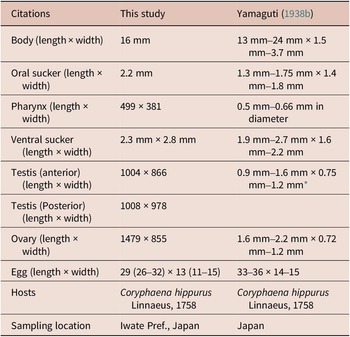
Adult (Fig 4a, measurements shown in Table 5). Body large, elongated. Oral sucker muscular, opening anteroventrally. Pharynx muscular. Oesophagus indistinct. Ventral sucker muscular, surmounted by body wall. Testes oval, separated from each other by ovary. Ovary oval, between two testes. Vitellaria narrow and twisted, from anterior testis to posterior body margin. Eggs elliptical. Excretory pore opening at posterior terminal of body.
Comparison of morphological measurements of Bathycotyle branchialis. All measurements are in μm unless indicated otherwise

* Size of two testes.
Metacercaria (from fish host, Fig 4b). Body subcylindrical and wide, 2.9 mm (2.7 mm–3.1 mm) in length and 610 in width. Oral sucker muscular, rounded, opening anteroventrally. Pharynx muscular, 174 (115–232) in length and 173 (153–193) in width. Ventral sucker muscular, rounded, 628 (562–694) in length and 623 (568–678) in width, situating anterior two-third of body. Testes oval, separated from each other by ovary. Anterior testis 54 (41–68) in length and 43 (41–45) in width. Posterior testis 62 in length and 31 (26–36) in width. Ovary oval, between two testes, 69 (67–72) in length and 64 (61–67) in width. Excretory pore opening at posterior terminal of body.
Remarks. This genus includes only one species, Bathycotyle branchialis. Moreover, the morphological characteristics of our adult specimen were thought to be similar to those of Bathycotyle branchialis in the past study (Yamaguti Reference Yamaguti1938b) (Table 5); therefore, we identified our specimen as Bathycotyle branchialis.
Based on comparisons of 28S rDNA and COI sequences between our adult, metacercariae, and unidentified metacercariae (Sclerodistomidae sp. A in Louvard et al. Reference Louvard, Yong, Cutmore and Cribb2024) in the jellyfish Velella velella and Porpita porpita in Australia, these samples were identified as a single species – Bathycotyle branchialis (Supplementary Tables S3 and S4). Regarding ITS2, the intraspecific values in the superfamily Hemiuroidea, to which Bathycotylidae belongs, were 0 in the 495-site overlapping region. Here, the ITS2 divergence value between our metacercariae and Sclerodistomidae sp. A in Louvard et al. (Reference Louvard, Yong, Cutmore and Cribb2024) was zero, indicating that they are a single species. Conversely, the divergence values between these metacercariae and our adult was 0.0040, which was slightly higher than the maximum intraspecific value observed within the superfamily. This result suggests the existence of two species within the family Bathycotylidae, despite only one species currently being recognised, although these worms were determined as a single species based on 28S rDNA and COI sequences. Further detailed morphological and molecular analyses based on adult specimens are needed to resolve the inconsistency in the divergence values in this species.
In Louvard et al. (Reference Louvard, Yong, Cutmore and Cribb2024), the metacercaria with a long forebody was identified as Sclerodistomidae sp. A based on molecular analyses, which was supported by morphological evidence because some sclerodistomid adults also have a long forebody (Gibson et al. Reference Gibson, Jones and Bray2002). In the 28S rDNA tree (Fig 4c), Bathycotyle branchialis (our sequence, LC889174), Sclerodistomidae sp. A (OR814167) in Louvard et al. (Reference Louvard, Yong, Cutmore and Cribb2024), and Prosogonotrema bilabiatum (Sclerodistomidae, PV614122) formed a distinct clade (BP = 91). This suggests that the two families, Bathycotylidae and Sclerodistomidae, are closely related, exhibiting similar morphological characteristics in their metacercariae. This may be the reason why the metacercariae of Bathycotyle branchialis were identified as Sclerodistomidae in the previous study. Furthermore, morphological and molecular analyses including other sclerodistomid adults are required to determine whether the two families should be considered synonymous.
Host of adult in the present study: Coryphaena hippurus Linnaeus, 1758
Host fish of metacercariae in the present study: Coryphaena hippurus Linnaeus, 1758
New Japanese name: Shiira-no-kamikazari (‘Shiira’ means ‘Coryphaena hippurus’ in Japanese. ‘Kamikazari’ is ‘a hair accessory’. ‘No’ is used to indicate possession or association).
Synonyms: Bathycotyle coryphaenae Yamaguti, 1938; Sclerodistomidae sp. A (in Louvard et al. Reference Louvard, Yong, Cutmore and Cribb2024).
Depositions: INM-1-131147 (adult), INM-1-131148–131149 (metacercariae)
DNA markers: 28S rDNA, LC889173 (adult), LC889174 (metacercaria); ITS2, LC889194 (adult), LC889195–889196 (metacercariae); COI, LC889223 (adult), LC889222 (metacercaria)
Family Hemiuridae Looss, 1899
Genus Dinurus Looss, 1907
Hemiurid adults were sampled from Coryphaena hippurus. These adults were classified as members of Dinurus based on the following morphological characteristics: body surface with plications, long pars prostatica, seminal vesicle trilobed, and presence of sinus-sac (Gibson et al. Reference Gibson, Jones and Bray2002). These adults were divided into two species based on the following morphological characteristics. Molecular analysis indicated that hemiurid metacercariae detected from the jellyfish Physalia utriculus were identified as Dinurus tornatus. In addition, unidentified metacercaria in Louvard et al. (Reference Louvard, Yong, Cutmore and Cribb2024) was identified as Dinurus barbatus by our molecular analyses.
Dinurus tornatus (Rudolphi, 1819)
Morphological descriptions
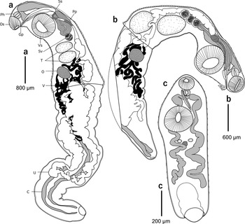
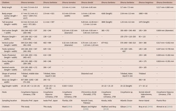
Adult (Fig 5a, measurements shown in Table 6). Body elongate, slender. Body surface with plications. Body proper (soma) equal in length to its tail (ecsoma). Oral sucker rounded, subterminal. Pharynx muscular. Oesophagus indistinct. Ventral sucker rounded, length twice of oral sucker. Testes oval, in tandem, located close together. Pars prostatica long. Seminal vesicle subglobular, trilobed, small and middle lobe large. Sinus-sac long and well-developed. Genital pore on ventral margin of oral sucker. Ovary oval. Vitellaria tubular. Eggs numerous, oval. Excretory pore at posterior margin of body.
Adults and metacercaria of the genus Dinurus. (a) Adult of Dinurus tornatus obtained from Coryphaena hippurus. (b) Adult of Dinurus barbatus obtained from Coryphaena hippurus. The dotted line shows the outline of the tissue cut out for PCR. (c) Metacercaria of Dinurus tornatus obtained from Physalia utriculus. Abbreviations: C, caecum; Gp, gonad pore; O, ovary; Os, oral sucker; Ph, pharynx; Pp, pars prostatica; Ss, sinus-sac; Sv, seminal vesicle; T, testis; U, uterus; V, vitellarium; Vs, ventral sucker.

Comparison of morphological measurements of Dinurus species. All measurements are in μm unless indicated otherwise

* Size of two testes.
Metacercaria (Fig 5c). Body proper (soma) subcylindrical, 1198 in length and 409 in width. Oral sucker rounded, subterminal, 124 in length and 135 in width. Pharynx muscular, 61 in length and 64 in width. Ventral sucker rounded, larger than oral sucker, 239 in length and 229 in width. Testes situated vertically. Left testis 27 in length and 31 in width, and right testis 21 in length and 21 in width. Sinus-sac long, 215 in length and 14 in width. Ovary oval, 25 in length and 31 in width. Tail (ecsoma) shrunken in our specimen.
Remarks. Genus Dinurus includes 13 species (WoRMS Editorial Board 2025), among which Dinurus tornatus can be distinguished from the other species based on the following characteristics: sinus-sac long, seminal vesicle small, trilobed and middle lobe large; ventral sucker larger than oral sucker; vitellaria tubular; eggs smaller than 19 μm in length. Dinurus dorabi and Dinurus tornatus possess a body proper (soma) that is equal in length to its tail (ecsoma) and have a long sinus-sac and ventral sucker twice the length of the oral sucker. Nevertheless, they can be distinguished by the shape of trilobed seminal vesicles (distorted oval in Dinurus dorabi vs subglobular in Dinurus tornatus). Based on the 28S rDNA, ITS2, and COI sequences, the metacercariae obtained from the jellyfish Physalia utriculus and the adults of Dinurus tornatus obtained from Coryphaena hippurus were identified as a single species (Supplementary Tables S6–S8).
Host of adults in the present study: Coryphaena hippurus Linnaeus, 1758
Host jellyfish of metacercariae in the present study: Physalia utriculus (Gmelin, 1788)
New Japanese name: Mahi-mahi-kyuchu (‘Mahi-mahi’ is named after the local name of the host fish Coryphaena hippurus)
Synonym: Distoma tornatum Rudolphi, 1819
Depositions: INM-1-131159–131173 (adults), INM-1-131174 (metacercaria)
DNA markers: 28S rDNA, LC889175–889176 (adults), LC889177 (metacercaria); ITS2, LC889197–889200 (adults), LC889201 (metacercaria); COI, LC889224–889233, LC889236 (adults), LC889234–889235 (metacercariae)
Dinurus barbatus (Cohn, 1902)
Morphological descriptions
Adults (Fig 5b, measurements shown in Table 6). Body elongate, slender. Body surface with plications. Body proper (soma) equal in length to its tail (ecsoma). Oral sucker rounded, subterminal. Tubular tufts present ventrally behind oral sucker. Pharynx muscular. Oesophagus indistinct. Ventral sucker rounded, clearly larger than oral sucker. Testes oval, tandem or oblique, positioned close together. Pars prostatica long. Seminal vesicle trilobed, with lobes equal in size. Sinus-sac short. Genital pore on ventral margin of oral sucker. Ovary oval. Vitellaria digitiform. Eggs numerous, oval. Excretory pore at posterior margin of body.
Remarks. Among the 13 species in the genus (WoRMS Editorial Board 2025), Dinurus barbatus can be distinguished based on the following morphological characteristics: body proper (soma) equal in length to its tail (ecsoma), ventral sucker twice the length of oral sucker, tubular tufts present ventrally behind oral sucker, sinus-sac short and seminal vesicle trilobed with lobes equal in size. Dinurus breviductus Looss, 1907 and Dinurus longisinus Looss, 1907 resemble Dinurus barbatus because of the following morphological characteristics: body proper (soma) equal in length to its tail (ecsoma), ventral sucker twice the length of oral sucker, sinus-sac short. Nevertheless, Dinurus barbatus can be distinguished from these two species based on the presence of tubular tufts (present in Dinurus barbatus vs absent in the other two species). Furthermore, Dinurus longisinus can be distinguished from Dinurus barbatus by the shape of the seminal vesicle (lobes equal in size in Dinurus barbatus vs a large middle lobe in Dinurus longisinus).
Our molecular analyses revealed that our adults obtained from Dinurus barbatus and the unidentified jellyfish metacercariae reported by Louvard et al. (Reference Louvard, Yong, Cutmore and Cribb2024), namely Hemiuridae sp. A, from the jellyfish Porpita porpita were identified as a single species based on the divergence values of 28S rDNA and ITS2 sequences (Supplementary Tables S6 and S8).
Host of adults in the present study: Coryphaena hippurus Linnaeus, 1758
New Japanese name: Hige-ari-mahi-mahi-kyuchu (‘Hige’ implies ‘beard’ and ‘mustache’ in Japanese. ‘Ari’ implies ‘present’)
Synonyms: Lecithocladium barbatum Cohn, 1902; Hemiuridae sp. A (Louvard et al. Reference Louvard, Yong, Cutmore and Cribb2024)
Depositions of adults: INM-1-131175–131180
DNA markers of adults: 28S rDNA, LC889178–889179; ITS, LC889202–889204; COI, LC889237
Family Lepocreadiidae Odhner, 1905
We detected adults of this family from Coryphaena hippurus and Scomber japonicus. The adults were identified to genus or species levels through molecular or morphological analyses.
Lepocreadiid metacercariae were detected from jellyfish and were divided into seven species based on our molecular analyses. Metacercariae of two of the seven species were identified to Cephalolepidapedon saba and Prodistomum orientale based on the comparisons of 28S rDNA and COI sequences of our adults and online databases. Also, metacercariae of two species, Prodistomum sp. 1 and Prodistomum Type 4, were conspecific to adult specimens of ours and online databases, respectively, although their species names were not determined. The species names of the remaining three species could not be determined because comparisons using 28S rDNA and COI sequences did not identify their conspecific species. Genera of two of the three unidentified species were determined using the 28S rDNA tree (Opechona sp. 1 and Prodistomum sp. 2). The taxonomic position of the remaining one species in this family was not determined in this study; therefore, we labelled it as Lepocreadiidae sp. 1.
Genus Cephalolepidapedon Yamaguti, Reference Yamaguti1970
Two adults obtained from Scomber japonicus are considered a member of the genus Cephalolepidapedon based on the following morphological characteristics: oral sucker funnel-shaped, slightly enlarged oral spines, blind caeca, and pseudo-oesophagus absent (Cribb and Bray Reference Cribb and Bray2003). The two adults were morphologically identified as a single species as follows. Morphological descriptions of this metacercaria were not provided because the whole worm was used for DNA extraction, and paragenophore specimens for this species were not found in this study.
Cephalolepidapedon saba Yamaguti, Reference Yamaguti1970
Morphological descriptions
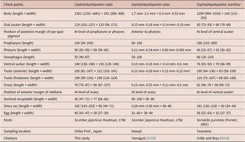
Adults (Fig 6a, b, measurements shown in Table 7). Body elongate. Eye-spot pigment, from level of oral sucker to pharynx. Oral sucker funnel-shaped with slightly enlarged oral spines. Pseudo-oesophagus absent. Caeca blind at posterior margin. Ventral sucker rounded. Testes subglobular, tandem, situated posterior to ovary. Cirrus-pouch claviform, situating at level of ventral sucker. Genital pore just anterior to ventral sucker. Ovary oval. Most of uterus between ventral sucker and ovary. Vitellaria surrounding caeca, from level of ovary to posterior margin of body. Eggs oval.
Adults and metacercariae of the family Lepocreadiidae. (a) Adult of Cephalolepidapedon saba. (b) Terminal genitalia of Cephalolepidapedon saba. (c and d) Adults of Prodistomum sp. 1 obtained from Scomber japonicus (c) and Coryphaena hippurus (d). (e) Metacercaria of Opechona sp. 1 obtained from Urashimea globosa. (f) Metacercaria of Prodistomum sp. obtained from Physalia utriculus. Abbreviations: C, caecum; E, oesophagus; Eg, egg; Ev, excretory vesicle; Gp, gonad pore; O, ovary; Os, oral sucker; Ph, pharynx; Pp, pars prostatica; Ss, sinus-sac; Sv, seminal vesicle; T, testis; V, vitellarium; Vs, ventral sucker.

Comparison of morphological measurements of Cephalolepidapedon species. All measurements are in μm unless indicated otherwise

* Size of two testes.
Remarks. Two species belong to the genus Cephalolepidapedon (Cribb and Bray, Reference Cribb and Bray2003), of which Cephalolepidapedon saba can be distinguished from another species, Cephalolepidapedon warehou Bray and Cribb, Reference Bray and Cribb2003, based on the position of the posterior margin of the eye-spot pigment (at level of prepharynx or pharynx in C. saba vs ventral sucker in C. warehou) and the anterior margin of vitellaria (at level of ovary in C. saba vs ventral sucker in C. warehou). Intraspecific distance values of COI sequences of one metacercaria detected from the jellyfish Physalia utriculus and adults detected from Scomber japonicus indicated that these worms belong to a single species (Supplementary Table S9). 28S rDNA was not amplified for the metacercaria.
Hosts of adults in the present study: Scomber japonicus Houttuyn, 1782
Host jellyfish of metacercaria in the present study: Physalia utriculus (Gmelin, 1788)
New Japanese name: Saba-no-osama-kyuchu (‘Saba’ is ‘Scomber fish’ in Japanese. ‘Osama’ is ‘king’ based on the oral sucker with spines that resembles a crown)
Synonyms: Apocreadium misakiense (Shimazu, 1986); Lepocreadium misakiense Shimazu, 1986; Opechona acanthoris Gaevskaya and Aleshkina, 1985; Stephanostomum scombri Korotaeva, 1974.
Depositions of adults: INM-1-131150–131152
DNA markers: 28S rDNA, LC889180–889181 (adults); COI, LC889265–889266 (adults), LC889267 (metacercaria)
Genus Opechona Looss, 1907
Opechona sp. 1
Metacercariae were obtained only from jellyfish for this species.
Morphological descriptions
Metacercaria (Fig 6e). Body elongated and wide, 817 (676–886) in length and 189 (158–208) in width. Oral sucker funnel-shaped, subterminal, 164 (127–186) in length and 160 (153–173) in width. Eye-spot pigment at level of prepharynx. Prepharynx 52 (41–65) in length. Pharynx muscular, 95 (85–106) in length and 67 (59–79) in width. Oesophagus indistinct. Ventral sucker rounded, situating at middle of body, 84 (67–94) in length and 95 (66–114) in width. Testes oval, tandemly situated posterior to ovary. Anterior testis 52 (38–68) in length and 67 (40–92) in width. Posterior testis 58 (44–69) in length and 67 (40–92) in width. Ovary irregularly shaped, 50 (36–63) in length and 38 (32–44) in width. Excretory pore at posterior margin of body.
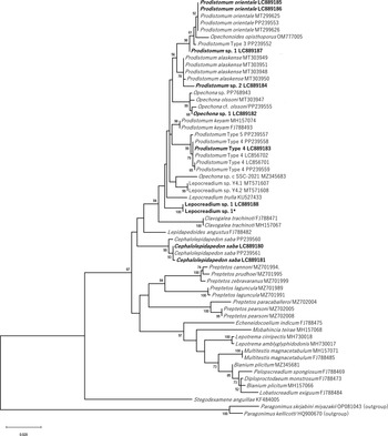
Remarks. No closely related sequence was found for the COI fragments obtained from metacercariae with large oral suckers in the jellyfish Hydrocoryne miurensis and Urashimea globosa (e.g., 78.07% similarity, Stegodexamene anguillae, JN244813). Moreover, the intraspecific distance values of 28S rDNA sequences did not identify the species of these metacercariae (Supplementary Table S10). On the basis of the 28S rDNA tree (919 overlapping sites), our metacercaria formed a clade with other Opechona species, supported by a high bootstrap value (Fig 7, bp = 99), indicating that the study sample can be identified as a member of Opechona.
Maximum likelihood tree of lepocreadiid trematodes inferred from partial sequences of nuclear 28S ribosomal DNA (919 sites) with Paragonimus spp. (HQ900670 and OP081043) as outgroup taxa. Bootstrap values above 50% are shown. Database accession numbers are indicated after species names. Bold names represent newly obtained sequences. Scale bar indicates number of substitutions per site. Asterisk represents a sequence which is not deposited to the online database and identical to LC889188.

Host jellyfish of metacercariae in the present study: Hydrocoryne miurensis Stechow, 1908; Urashimea globosa Kishinouye, 1910
Depositions of metacercariae: INM-1-131153–131154
DNA markers: 28S rDNA, LC889182; COI, LC889268–889269
Genus Prodistomum Linton, 1910
Five adults were sampled from two host fish, Coryphaena hippurus and Scomber japonicus, and identified as the genus Prodistomum Linton, 1910, through morphological and molecular analyses as follows
Metacercariae of four Prodistomum species were detected through molecular analysis in this study. Considering the small size of these metacercariae (from 200 to 350 μm in body length after fixations), we did not attempt preparation of hologenophores that would preserve enough of the body for morphological characteristic analysis. Moreover, more than two species of this genus were detected through molecular analysis from a single host individual. Therefore, we could not prepare the whole paragenophore morphological specimens of this genus. Instead, we provide the following brief descriptions of the unidentified metacercaria of this genus.
Morphological descriptions
Metacercaria of Prodistomum sp. (Fig 6f). Body subcylindrical, 347 in length and 157 in width. Oral sucker oval, 42 in length and 47 in width. Prepharynx 39 in length. Pharynx oval, 21 in length and 16 in width. Ventral sucker at half or anterior two-third of body, 37 in length and 42 in width. Eye-spot pigment, from level of prepharynx to oesophagus. Testes tandemly situated. Anterior testis 14 in length and 20 in width. Posterior testis 14 in length and 19 in width. Ovary 23 in length and 22 in width, anterior to anterior testis. Excretory pore opening posterior terminal of body. Excretory vesicle reaching caecum bifurcation.
Host jellyfish of the metacercaria: Physalia utriculus (Gmelin, 1788)
Deposition of the metacercaria: INM-1-131155
Prodistomum orientale (Layman 1930) (Prodistomum Type 2 in Cribb et al. Reference Cribb, Cutmore, Wee, Browne, Morales and Pitt2024)
Two and one adults from Coryphaena hippurus and Scomber japonicus, respectively, were broken, making morphological identification of their taxonomic position difficult. Morphological characteristics were not provided for the damaged specimens. Metacercariae obtained from Clytia sp., Physalia utriculus, Urashimea globosa, Aurelia coerulea, and Chrysaora pacifica were thought to be conspecific with these damaged adults based on comparisons of 28S rDNA and COI sequences (Supplementary Tables S11 and S12). These metacercariae and adults were identified as Prodistomum orientale (=Prodistomum Type 2 in Cribb et al. Reference Cribb, Cutmore, Wee, Browne, Morales and Pitt2024) based on comparisons of the sequences of our specimens with those reported by Cribb et al. (Reference Cribb, Cutmore, Wee, Browne, Morales and Pitt2024) (Supplementary Tables S11 and S13).
Hosts of adults in the present study: Coryphaena hippurus Linnaeus, 1758, Scomber japonicus Houttuyn, 1782
Host jellyfish of metacercariae in the present study: Physalia utriculus (Gmelin, 1788); Clytia sp.; Urashimea globosa Kishinouye, 1910; Aurelia coerulea von Lendenfeld, 1884; Chrysaora pacifica (Goette, 1886)
New Japanese name: Kurage-hoshikuzu-kyuchu (‘Kurage’ implies ‘jellyfish’ in Japanese. ‘Hoshikuzu’ is ‘stardust’)
Synonyms: Acanthocolpoides guevarai Lopez-Roman, 1979; Acanthocolpoides israelensis Fischthal, 1980; Acanthocolpoides pauloi Travassos and Teixeira de Freitas, 1965; Lepocreadium ghanense Fischthal and Thomas, 1970; Lepocreadium ghanensis Fischthal and Thomas, 1970; Lepocreadium puriense Gupta and Gupta, 1987; Lepocreadium puriensis Gupta and Gupta, 1987; Lepocreadium retrusum Linton, 1940; Lepocreadium scombri (Yamaguti, 1938) Neolepidapedon retrusum (Linton, 1940); Opechona orientalis (Layman, 1930); Opechona scombri Yamaguti, 1938; Pharyngora orientalis Layman, 1930; Prodistomum guevarai (Lopez-Roman, 1979); Prodistomum israelensis (Fischthal, 1980); Prodistomum orientalis (Layman, 1930); Prodistomum pauloi (Travassos and Teixeira de Freitas, 1965); Prodistomum Type 2 (see below, Cribb et al. Reference Cribb, Cutmore, Wee, Browne, Morales and Pitt2024)
Depositions of adult: INM-1-131158
DNA markers: 28S rDNA, LC889185 (adult), LC889186 (metacercaria); COI, LC889192–889193, LC889253–889255 (adults), LC889190, LC889256–889264 (metacercariae)
Prodistomum sp. 1
Although body parts were slightly damaged and cut for PCR templates (Fig 6c, d), the remaining tissues of two adults obtained from Coryphaena hippurus and Scomber japonicus (two adults in total) were identified as the genus Prodistomum based on the following morphological characteristics: body elongate oval, pseudo-oesophagus present, ovary oval and just anterior to anterior testis, uterus anterior to testes, internal seminal vesicle oval, excretory vesicle running right of uterus, excretory pore posterior terminal of body (Jones et al. Reference Jones, Bray and Gibson2005). These two adults were identified as a single species, as described later. Metacercariae obtained from Urashimea globosa were molecularly identified as this species (Fig 2f).
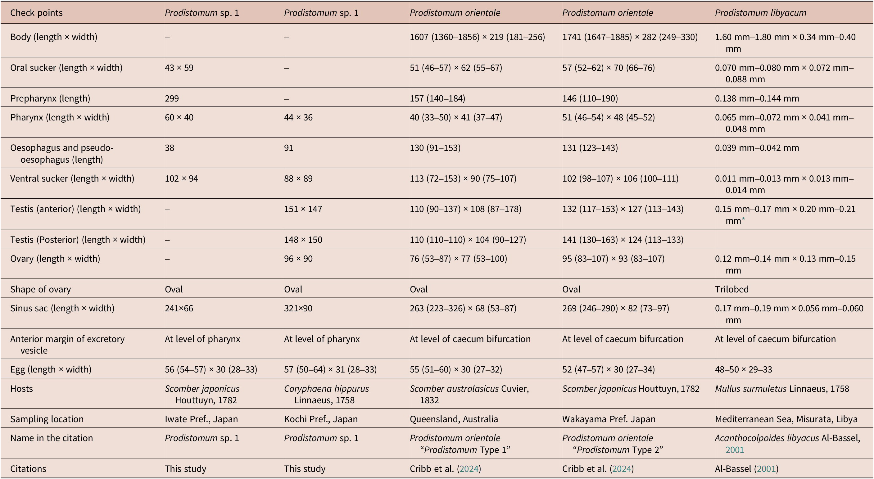
Morphological descriptions (Fig 6c, d, measurements shown in Table 8). Body elongate oval. Eye-spot pigment, from level of oral sucker to pharynx. Oral sucker small. Pseudo-oesophagus present. Prepharynx much longer than oesophagus and pseudo-oesophagus. Ventral sucker rounded. Uterus anterior to testes. Ovary oval, just anterior to anterior testis. Internal seminal vesicle oval. Genital pore anterior to ventral sucker. Anterior margin of vitellaria at level of anterior margin of seminal vesicle. Excretory vesicle running right of uterus and reaching pharynx. Excretory pore at posterior terminal of body.
Comparison of morphological measurements of Prodistomum species. All measurements are in μm unless indicated otherwise

* Size of two testes.
Remarks. This genus includes 19 species (WoRMS Editorial Board 2025). Morphological characteristics of the present specimens were similar to those of two species, Prodistomum orientale and Prodistomum libyacum, owing to the following morphological characteristics: prepharynx much longer than oesophagus and pseudo-oesophagus, anterior margin of vitellaria at level of anterior margin of seminal vesicle (Al-Bassel Reference Al-Bassel2001; Cribb et al. Reference Cribb, Cutmore, Wee, Browne, Morales and Pitt2024). The present specimens differed from these two species based on the anterior margin of the excretory vesicle (reaching pharynx in our specimens vs at level of caecum bifurcation in the other two species), suggesting that our specimens were possibly undescribed species. Because our specimens were cut for the template, we could not record the detailed morphological characteristics based on our specimens. Therefore, we labelled these as Prodistomum sp. 1.
Species of metacercariae obtained from Urashimea globosa were thought to be conspecific with the adults of Prodistomum sp. 1, based on the similarity of 28S rDNA and COI sequences (Supplementary Tables S11 and S12). The species name was unclear based on the divergence values of 28S rDNA and COI sequences (Supplementary Tables S11 and S12). Because the sequences formed a clade with other Prodistomum species in the 28S rDNA tree (e.g., Prodistomum orientale (PP239553), Fig 7, bp = 98), we determined it as as a member of Prodistomum
Hosts of adults in the present study: Coryphaena hippurus Linnaeus, 1758, Scomber japonicus Houttuyn, 1782
Host jellyfish of metacercariae in the present study: Urashimea globosa Kishinouye, 1910
Depositions of adults: INM-1-131156–131157.
DNA markers: 28S rDNA, LC889187 (adult); COI, LC889249–LC889250 (adults), LC889249–889250 (metacercariae)
Prodistomum Type 4
In this study, metacercariae were obtained only from jellyfish for this species. Metacercariae obtained from Chrysaora pacifica, Physalia utriculus, and Urashimea globosa were identified as a single species based on the divergence values of 28S rDNA and COI sequences (Supplementary Tables S11 and S12). Our metacercaria was identified as Prodistomum Type 4 reported as a sporocyst and adults from Purpuradusta gracilis and Scomber japonicus, respectively, from the Pacific coast of Japan (Cribb et al. Reference Cribb, Cutmore, Wee, Browne, Morales and Pitt2024; Ansai et al. Reference Ansai, Sekine, Munakata, Sase, Sasaki, Nitta, Suzuki, Abe, Takano, Fukumori, Nakatsubata, Suzuki and Waki2025) based on COI sequences (Supplementary Tables S11 and S13).
Host jellyfish of metacercariae in the present study: Chrysaora pacifica (Goette, 1886); Physalia utriculus (Gmelin, 1788); Urashimea globosa Kishinouye, 1910
DNA markers: 28S rDNA, LC889183; COI, LC889191, LC889238–889247
Prodistomum sp. 2
A metacercaria species obtained from Aurelia coerulea was not identified based on our analyses (Supplementary Tables S11 and S12). Based on the 28S rDNA tree (919 overlapping sites), this metacercaria formed a clade with Prodistomum alaskense (e.g., MT303949) (Fig 7, bp = 78). Therefore, we judged it as a member of the genus Prodistomum, namely Prodistomum sp. 2. This one metacercaria was obtained for this species.
Host jellyfish of metacercaria in the present study: Aurelia coerulea von Lendenfeld, 1884
DNA markers: 28S rDNA, LC889184; COI, LC889251
Lepocreadiidae sp. 1
For this species, metacercariae were obtained only from jellyfish host Physalia utriculus (Fig 2g). We obtained 28S rDNA and COI sequences of two metacercariae; however, both were insufficient for identification at the species level. Our 28S rDNA sequences resembled those of lepocreadiid trematodes, such as Opechona kahawai (98.38% similarity, FJ788491) and Prodistomum alaskense (98.33% similarity, MT303949), causing difficulty in determining their genus. Moreover, based on the 28S rDNA tree, we did not determine their taxonomic position in this family (Fig 7). Also, the closely related COI sequences of our metacercariae were not detected in our samples and in the databases (e.g., 79.10% similarity, Schistosomatium douthitti, AY157193). Therefore, we labelled them as Lepocreadiidae sp. 1. Morphological characteristics of metacercariae were similar to those of Prodistomum metacercaria in live conditions; however, morphological descriptions and depositions were not provided because the whole worm was used for DNA extraction.
Host jellyfish of metacercariae in the present study: Physalia utriculus (Gmelin, 1788)
DNA markers: 28S rDNA, LC889188; COI, LC889270–889271
Discussion
In the present study, metacercariae belonging to three families (Accacoeliidae, Hemiuridae, and Lepocreadiidae) were detected in jellyfish, as reported in previous studies (Browne et al. Reference Browne, Pitt and Cribb2020; Cribb et al. Reference Cribb, Cutmore, Wee, Browne, Morales and Pitt2024; Diaz Briz et al. Reference Diaz Briz, Martorelli, Genzano and Mianzan2012; Køie Reference Køie1991; Louvard et al. Reference Louvard, Yong, Cutmore and Cribb2024; Motta et al. Reference Motta, Caffara, Fioravanti, Bottaro, Avian, Terlizzi and Tedesco2023). These metacercariae were classified into nine species, of which six were molecularly identified as being the same species as those found in fish hosts in the present and previous studies (Cribb et al. Reference Cribb, Cutmore, Wee, Browne, Morales and Pitt2024), although species names of two of the six were not determined. The remaining three trematode species could not be identified at the species level in this study because the DNA barcodes of identified adults were not yet obtained. Nevertheless, these three species were thought to belong to the family Lepocreadiidae, a major group of fish trematodes (e.g., Madhavi and Bray Reference Madhavi and Bray2018). These findings suggest that cnidarian jellyfish are important second intermediate or paratenic hosts for fish trematodes in Japanese waters, as has been reported in other areas in previous studies (e.g., Cribb et al. Reference Cribb, Cutmore, Wee, Browne, Morales and Pitt2024; Louvard et al. Reference Louvard, Yong, Cutmore and Cribb2024). A comprehensive future study of marine fish would enable detecting of their adult stage conspecific with the cnidarian metacercariae of the three unidentified species.
The cnidarian trematodes were frequently detected in two fish hosts, Coryphaena hippurus and Scomber japonicus. Regarding Coryphaena hippurus, adults of four of the nine cnidarian trematode species were detected. Moreover, Dinurus barbatus and Bathycotyle branchialis, whose adults were detected in Coryphaena hippurus in the present study, were reported to use neuston jellyfish as hosts of metacercariae in Australia (Louvard et al. Reference Louvard, Yong, Cutmore and Cribb2024). Similarly, four of the nine cnidarian trematode species were detected in Scomber japonicus in the present and previous studies (Cribb et al. Reference Cribb, Cutmore, Wee, Browne, Morales and Pitt2024; Shimazu Reference Shimazu1989; Yamaguti Reference Yamaguti1938a). These results indicate that cnidarian jellyfish are consistent food sources for the two fish species and that these trematodes use jellyfish as the major infection source for transporting them to these definitive hosts.
The metacercariae of Tetrochetus coryphaenae were detected from two species of neuston jellyfish in the present study. The first intermediate hosts of trematodes of the family Accacoeliidae have not been found, and the second intermediate hosts of this group have not been determined experimentally. Therefore, it is impossible to determine whether these neuston jellyfish were the second intermediate hosts or the paratenic hosts. Metacercariae of Tetrochetus coryphaenae were also detected in an arrow worm and three fish species. Jellyfish, including the neuston jellyfish Physalia physalis, feed on arrow worms (Bailey and Batty Reference Bailey and Batty1983; Purcell, Reference Purcell1984; Möller, Reference Möller1984; Mutlu, Reference Mutlu2001), and the neuston jellyfish Physalia species feeds on small fish (Munro et al. Reference Munro, Vue, Behringer and Dunn2019). Therefore, the neuston jellyfish, which harboured the metacercariae in the present study, were possibly infected by predating on arrow worms and fish hosts (Fig 8).
Hypothetical routes of trematode transmission related to the cnidarian, chaetognathan, and fish hosts examined in the present study in the coastal regions of Japan. White, blue, and green solid lines indicate the infection route of the metacercariae of Tetrochetus coryphaenae, Prodistomum spp., and Dinurus tornatus to their definitive hosts, respectively. White dotted lines represent the infection route of metacercariae to their paratenic hosts of Tetrochetus coryphaenae. Blue and green dotted lines indicate the infection route of cercariae to the second intermediate host of Prodistomum spp. and D. tornatus, respectively. Alphabets represent the hosts and possible infection routes. a: Neuston cnidarian jellyfish. b: Planktonic cnidarian jellyfish. c: Chaetognatha. d: Coryphaena hippurus, e: Echeneis naucrates. f: Medusivorous fish, such as Carangidae, Monacanthidae, and Tetraodontidae. g: Scomber japonicus. h: Paratenic host fish of Tetrochetus coryphaenae. i: Unknown copepods, the second intermediate host of D. tornatus. j: Infection of metacercariae from copepods to neustonic cnidarian jellyfish (possibly not direct infection). k: Unknown mollusc, the first intermediate host of D. tornatus. l: Unknown mollusc and Purpuradusta gracilis, the first intermediate host of Prodistomum spp. (the host mollusc of Prodistomum Type 4 is Purpuradusta gracilis (Ansai et al. Reference Ansai, Sekine, Munakata, Sase, Sasaki, Nitta, Suzuki, Abe, Takano, Fukumori, Nakatsubata, Suzuki and Waki2025)). m: Unknown mollusc, the first intermediate host of Tetrochetus coryphaenae. n: Unknown infection routes from the mollusc to the cnidarian and chaetognathan hosts of Tetrochetus coryphaenae.

Adults and metacercariae of Tetrochetus coryphaenae were detected in nine and three fish species, respectively. These fish hosts of metacercariae were probably paratenic hosts infected via predation because the metacercariae were detected in the intestines of the host. Cnidarian jellyfish are reported to be fed on fish belonging to the families Carangidae, Coryphaenidae, Monacanthidae, Scombridae, and Tetraodontidae (Arai Reference Arai1988, Reference Arai2005; Denadai et al. Reference Denadai, Santos, Bessa, Bernardes and Turra2012; Kojima Reference Kojima1961; Miyajima-Taga et al. Reference Miyajima-Taga, Masuda, Kurihara, Yamashita and Takeuchi2015, Reference Miyajima-Taga, Masuda, Kurihara, Komi, Yamashita and Takeuchi2018). Therefore, the host fish belonging to these families (Table 2) were probably infected by feeding on infected jellyfish (Fig 8). Among the host fish, because Echeneis naucrates and Coryphaena hippurus are piscivores, they may have also become infected by feeding on fish hosts that harboured metacercariae (Moltó et al. Reference Moltó, Hernández, Sinopoli, Besbes-Benseddik, Besbes, Mariani, Gambin, Alemany, Morales-Nin, Grau, Camiñas, Báez, Vasconcellos, Ceriola and Catalán2020; Smith Reference Smith1997). As mentioned, the metacercaria of Tetrochetus coryphaenae was detected from an arrow worm. Because arrow worms are a common food source for marine fish (Bushula et al. Reference Bushula, Pakhomov, Kaehler, Davis and Kalin2005; Nagasawa and Marumo Reference Nagasawa and Marumo1981), they were also a possible infection source for fish hosts examined in the present study. Because the second intermediate hosts of the family Accacoeliidae were not experimentally determined as described earlier, it is impossible to determine whether arrow worms act as the second intermediate hosts or the paratenic hosts. In a past study, metacercariae of Tetrochetus coryphaenae were detected in the neustonic gastropods Glaucus atlanticus and Janthina janthina (Louvard et al. Reference Louvard, Yong, Cutmore and Cribb2024), which are known to prey on neuston cnidarian jellyfish (Lalli and Gilmer Reference Lalli and Gilmer1989). However, these gastropods may not be an infection source for other hosts because there are no reports of neustonic gastropod species being eaten by other organisms.
The metacercariae of Bathycotyle branchialis were detected in the intestines of the host fish Coryphaena hippurus and also in the neuston jellyfish Velella velella and Porpita porpita in Australia (Louvard et al. Reference Louvard, Yong, Cutmore and Cribb2024). Because these jellyfish were detected in the stomachs of Coryphaena hippurus (Kojima Reference Kojima1961), the metacercariae of Bathycotyle branchialis in the jellyfish probably invaded the host through the oral route. In contrast, adults were detected on the gill of the host fish in the present and previous studies (Yamaguti Reference Yamaguti1938b). Therefore, the metacercariae of Bathycotyle branchialis are probably transported to the intestine and subsequently transmigrated to the gills of the host fish in their life cycle.
The metacercariae and adults of the hemiurid trematode Dinurus tornatus were detected in the neuston jellyfish Physalia utriculus and in the fish Coryphaena hippurus, respectively. Because Coryphaena hippurus has been reported to feed on neuston jellyfish (Kojima, Reference Kojima1961), the fish were probably infected with Dinurus tornatus by feeding on the infected neuston jellyfish (Fig 8). Planktonic copepods commonly act as the second intermediate hosts for hemiurid trematodes (Køie Reference Køie1979, Reference Køie1989, Reference Køie1990, Reference Køie1991; Marcogliese Reference Marcogliese1995). Therefore, cercariae of Dinurus tornatus released from unknown molluscs might first invade copepods. Planktonic copepods have been reported to be preyed upon by planktonic jellyfish (Nagasawa Reference Nagasawa1987; Zenitani et al. Reference Zenitani, Kono, Watari and Tsukamoto2013). However, there is no record of neuston jellyfish preying on copepods. Therefore, neuston jellyfish may become infected by feeding on unknown organisms that have eaten copepods.
Metacercariae of seven species of the family Lepocreadiidae were detected in cnidarian jellyfish. Because lepocreadiid cercariae can invade jellyfish directly and develop into metacercariae (Køie Reference Køie1975; Stunkard Reference Stunkard1969, Reference Stunkard1980a, Reference Stunkardb), the jellyfish hosts of the metacercariae should represent the second intermediate hosts (Fig 8). Because the plankton jellyfish Chrysaora pacifica has been reported to feed on other jellyfish (Sato et al. Reference Sato, Hiromi and Kadota1996), they may become infected orally by predating on cnidarian jellyfish and act as the paratenic host. Of the seven lepocreadiid species, the first intermediate host has been determined only for Prodistomum Type 4, which uses the marine gastropod Purpuradusta gracilis (Ansai et al. Reference Ansai, Sekine, Munakata, Sase, Sasaki, Nitta, Suzuki, Abe, Takano, Fukumori, Nakatsubata, Suzuki and Waki2025). Prodistomum orientale, Prodistomum Type 4, Prodistomum sp. 1, and Cephalolepidapedon saba were found to use Coryphaena hippurus and/or Scomber japonicus as the definitive hosts (Cribb et al. Reference Cribb, Cutmore, Wee, Browne, Morales and Pitt2024; Shimazu Reference Shimazu1989; Yamaguti Reference Yamaguti1938a), both of which feed on gelatinous neuston and zooplankton, respectively (Arai Reference Arai1988, Reference Arai2005; Kojima Reference Kojima1961); thus, they may become infected with those trematodes by feeding on the jellyfish.
Supplementary material
The supplementary material for this article can be found at http://doi.org/10.1017/S0022149X25100989.
Acknowledgements
The present study was kindly helped by Akari Fukutoyama, Takanori Ishikawa, Risa Niida, Kaede Sato, and Nobutaka Taniuchi. Besides, we appreciate the officers and crew of the research vessel Shinyo-maru of the Tokyo University of Marine Science and Technology.
Financial support
This research received no specific grant from any funding agency, commercial, or not-for-profit sectors.
Competing interests
The authors declare none.
Ethical standard
Formal ethical approval was not required for this study because it involved unregulated fish and invertebrate species.