Hyperglycaemia as the main cause of endothelial dysfunction and vascular ageing
The biological role of vascular endothelium
Endothelial cells (ECs) play a key role in the haemostasis and maintenance of vessel functions. ECs maintain a physical barrier between a vessel wall and the lumen. Additionally, the endothelial monolayer is a potent secretory tissue that releases a number of mediators that regulate several physiological processes, such as coagulation (Ref. Reference vanHinsbergh1), platelet aggregation (Refs Reference Palmer2, Reference Montoro-García3), fibrinolysis (Ref. Reference Stegenga4) and vascular permeability (Ref. Reference Dogné5).
Under normal conditions, quiescent ECs maintain vessel homeostasis. The activation of ECs by pro-inflammatory cytokines, hypoxia or shear stress leads to a dramatic alteration in the EC phenotype. Activated ECs express larger amounts of growth factors, inflammatory mediators and adhesion molecules. The characteristic features of endothelial dysfunction are reduced endothelium-mediated vasorelaxation, impaired fibrinolytic ability and increased oxidant stress (Refs Reference Popov6–Reference Hirose10). At the cellular level, the disintegration of intercellular junctions formed by calcium-dependent adhesion molecules, VE-cadherins, is considered to be the most distinguished EC deterioration effect observed during endothelial dysfunction. This in turn leads to an increase in leukocyte adherence, rolling and subsequent extravasation (Refs Reference vanHinsbergh1, Reference Funamoto9–Reference Fraisl12).
Endothelial hyperpermeability and microvessel leakage are thought to be crucial for the development of a variety of pathologies, including enhanced thrombotic processes and haemorrhagic shock, which are observed in diabetes mellitus (DM) (Refs Reference Dogné5, Reference Funamoto9, Reference Mundi13).
Endothelial dysfunction as a preliminary stage of the ageing processes
During the life span of cells, irreversible ageing processes are physiological. In terms of ‘endothelial ageing’, the number of unfavourable changes in endothelium function begins to appear prominently and finally becomes irreversible, whereas the term ‘endothelial dysfunction’ refers to a loss of the physiological properties of the cell in a temporal manner. The intracellular and intercellular interactions that occur during ‘endothelial ageing’ appear to be crucial, and they can influence the microenvironment and alter cell signalling, including altered NO signalling, revasculating factor expression (integrin β2, SDF-1) and release of endothelial progenitor cells (Ref. Reference Avogadro14).
In physiological conditions, when the metabolic control is satisfactory, the blood glucose levels are maintained between 3.6 and 5.8 mmol/l. Human ECs are locally exposed to such glucose concentrations; thus, every recurrent (the short-term hyperglycaemia incubation (60–240 min) and long-term incubation(48 h)) increase in glucose level affects ECs with short- or long-term consequences (Refs Reference Popov6, Reference Brunssen15). Acute local hyperglycaemia (16.6 mmol/l) achieved by brachial intra-arterial infusion of 50% dextrose impairs endothelium-dependent vasodilatation in healthy subjects (Ref. Reference Williams16). In normoglycaemia, the EC phenotype is described as quiescent (Ref. Reference Popov6). The quiescence of these cells is closely controlled by several molecules, such as human Bone Morphogenetic Protein-9 (BMP-9) (Ref. Reference David17), transcription factor E2-2 (Ref. Reference Tanaka18), Angiopoietin 1 (Ang1) (Refs Reference Singh19, Reference Fukuhara20) and very low-density lipoprotein (LDL) receptor (Ref. Reference Jiang21). The expression of the anti-angiogenic R-ras gene is a biomarker of vascular quiescence (Ref. Reference Xu22).
Hyperglycaemia, in the context of ECs, is defined as exposure of the cells to glucose concentrations higher than 10 mmol/l (experiments carried out in vivo or in vitro) (Ref. Reference Popov6). ECs cultured under hyperglycaemic conditions lose their quiescence and become dysfunctional (Ref. Reference Popov6). Studies performed on vascular ECs cultured in hyperglycaemic conditions (30.5 mmol/l D-glucose) indicated that high glucose concentrations induce pro-adhesive and pro-inflammatory phenotypic changes (Refs Reference Avogadro14, Reference Rajapakse23, Reference Liu24). Under these conditions, the expression of interleukin (IL)-1β and adhesion molecules, such as vascular cell adhesion molecule-1 (V-CAM1) or endothelial selectin (P-SEL), was upregulated (Ref. Reference Liu24). Moreover, increased levels of IL-8, IL-6, vascular endothelial growth factor (VEGF), and tumour necrosis factor-α (TNFα) were also observed (Ref. Reference Bhatwadekar25).
Several cellular functions are regulated via interactions between cytoskeletal elements (actin filaments and microtubules) and adhesive molecules. The rearrangements of the EC cytoskeleton and adhesives change cell shape (Ref. Reference Shakhov26). Targosz-Korecka et al. showed that long-lasting hyperglycaemic conditions result in cytoskeletal changes such as stress fibre formation and F-actin polymerisation. After 14 passages, cells cultured under hyperglycaemic conditions (25 mM D-glucose) exhibited a modification in the structure of the cortical actin cytoskeleton, which led to the formation of intercellular gaps and increased the permeability of the endothelium. A decrease in the integrity of the endothelial barrier and cohesiveness of the EC layer was observed after 20 passages (Ref. Reference Targosz-Korecka27).
The nuclear factor Nrf2 is a key regulator in diabetes-induced endothelial dysfunction (Ref. Reference Sharma28). The duration of diabetes increases the expression of pro-inflammatory genes, such as chemokine ligands CCL2 and CCL5, which are controlled by Nrf2. The upregulation of mRNA levels for these chemokines was detected in aortic ECs in short-term diabetes induced in type 1 diabetic mice, whereas in the later stages of diabetes, upregulation was detected in the venous and aortic ECs. These observations suggest that during diabetes, endothelial dysfunction is induced both in arteries and veins to a different extent and in a different manner (Ref. Reference Bucciarelli29).
Under physiological conditions, ECs show a low number of intracellular organelles, which are involved in degradation (lysosomes) or biosynthetic activities (Golgi apparatus and endoplasmic reticulum (ER)) and are produced in the basal lamina. Studies performed using human aortic ECs cultured under hyperglycaemic conditions and in vivo rodent studies have shown a significant enrichment in the number of biosynthetic organelles in these conditions (Refs Reference Popov6, Reference Popov30). The Golgi complex is observed in the capillary and aortic ECs, and interestingly, large volumes of rER have been observed in ECs of the athero-susceptible aortic arch, femoral artery and retinal venules. Direct and prolonged exposure to hyperglycaemia was associated with cytoskeleton reorganisation and mitochondrial fragmentation (Ref. Reference Trudeau31). It is known that diabetes is linked to the thickening of the basal lamina of capillary ECs, resulting in a reduction in the number of metabolic products and nutrients transported between the tissue and circulation leading to endothelial dysfunction (Ref. Reference Dokken32).
Hyperglycaemia as a link to vascular ageing
Hyperglycaemia is one of the principal factors inducing endothelial dysfunction, the basis and hallmark of which are an increased synthesis of extracellular matrix (ECM) proteins, which plays a key role in the development of vascular complications in diabetic patients, such as retinopathy, nephropathy, hypertension and atherosclerosis (Refs Reference Cade8, Reference Avogadro14, Reference Klaassen33, Reference Naka34). Hyperglycaemia is known to modify the mechanical properties of ECs by increasing the stiffness of the cell membrane (Ref. Reference Targosz-Korecka27).
A crucial phenomenon leading to ageing of ECs is the so-called ‘glycaemic memory’, which means the development of vascular complications, such as membrane stiffness or upregulation of cell adhesion molecules (ICAM and VCAM) (Ref. Reference Targosz-Korecka27) after prolonged periods of hyperglycaemia despite a subsequent normalisation of glucose concentration. These phenotypic changes in ECs are controlled via epigenetic mechanisms, among which DNA methylation, histone modification and non-coding RNAs are considered important (Ref. Reference Reddy35).
Another key feature of EC ageing is an endothelial-to-mesenchymal transition (EndoMT), a gradual process of phenotypic changes, from endothelium to mesenchymal cells. This process is observed when cells express lower levels of endothelial markers, such as vascular endothelial cadherin (VE-cad), claudin and cytokeratin-18, and lose their function along with higher expression of mesenchymal markers, such as vimentin, α-smooth muscle actin, and types I and III interstitial collagens, and acquire their function (Ref. Reference Sánchez-Duffhues36). This process is regulated by transforming growth factor-β (TGF-β) family receptors and Wnt signalling pathways (β-catenin-dependent and -independent), which contribute to EndoMT in both vascular embryogenesis and cardiac tissue regeneration (Refs Reference Sánchez-Duffhues36, Reference Van Meeteren37). For example, a population of endocardial ECs that give rise to mesenchymal progenitors tend to migrate into the heart valves or differentiate into vSMCs and pericytes that cover the larger arteries in the heart (Ref. Reference Chen38). A similar process has been observed in human aortic ECs exposed to glucose and in the hearts and kidneys of patients with DM (Refs Reference Li39, Reference Widyantoro40). Hyperglycaemia causes numerous modifications in the vascular tissue, which may accelerate atherosclerosis. Investigations of the vasculature of patients with diabetes and diabetic animals have shown three major mechanisms that are associated with pathological changes observed in these models: protein kinase C (PKC) activation, non-enzymatic glycosylation of proteins and lipids and oxidative stress. PKC is a key player in one of the Wnt signalling pathways, indicating that it could be involved in diabetes-augmented atherosclerosis (Ref. Reference Aronson41). Interestingly, in diabetic nephropathy (DN), exosomes derived from hyperglycaemic glomerular ECs have a significant impact on podocytes. Protecting glomerular ECs from undergoing EndoMT and inhibiting the release of TGF-β1-containing exosomes inhibits podocyte dysfunction and, therefore, renal fibrosis (Ref. Reference Wu42).
Wnt signalling pathways
The Wnt signalling network has been studied for over 30 years, but it is still not completely understood (Refs Reference Chen38, Reference Korinek43). The proteins involved in these pathways are evolutionarily highly conserved and present in a wide spectrum of organisms, ranging from marine anemones to humans, implying that Wnt pathways regulate fundamental cell processes. Indeed, Wnt signalling has been shown to regulate embryonic development through body axis patterning, cell specialisation, migration, polarisation, proliferation and differentiation (Ref. Reference Lowry44). Mutations in proteins of the Wnt network result in abnormalities of the bone (Ref. Reference Fahiminiya45), heart and skeletal muscle structure (Ref. Reference Chen46), and may lead to the development of cancer, DM (Ref. Reference Kanazawa47) or obesity (Ref. Reference Christodoulides48).
The key players that initiate the Wnt cascade include a large family of Wnt proteins, a family of transmembrane receptors, Fz (Frizzled), which is a group of co-receptors, the most important of which are lipoprotein receptor-related protein (LRP)-5/6, receptor tyrosine kinase (Ryk), and tyrosine-protein kinase transmembrane receptor (ROR2) (Figs 1 and 2). A crucial protein for the activation of the Wnt pathway is a cytoplasmic phosphoprotein Dishevelled (Dvl), which acts as a molecular switch to decide which of the Wnt pathways will be activated (Ref. Reference Habas49). It has three domains, namely, DIX (amino-terminal), PDZ (central) and DEP (carboxy-terminal), and different combinations of these domains interact with each of the downstream Wnt pathways (Ref. Reference Habas49). Although the current understanding of the aforementioned signalling is as a large network of mutual protein interactions, the division is still convenient for explanatory reasons.
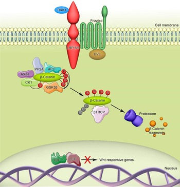
The β-catenin-dependent pathway in the absence of Wnt ligands. In the absence of Wnt ligands, DKK1 (Wnt antagonist) binds to the LRP5/6 receptor. β-catenin interacts with a degradation complex, which consists of APC, PP2A, AXIN, CK1 and GSK-3β. β-catenin is phosphorylated by CK1 and GSK-3β, which leads to the binding of β-TRCP to β-catenin and its ubiquitylation and degradation in proteasomes. Transcription of Wnt-responsive genes is blocked.

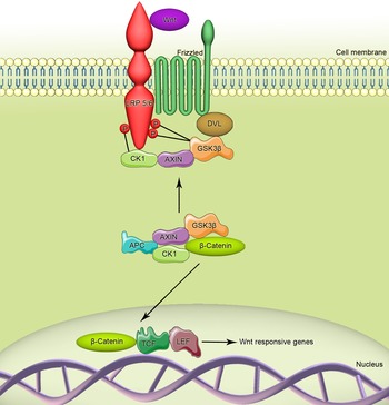
The β-catenin-dependent pathway in the presence of Wnt ligands. Wnt ligands bind to the Frizzled receptor and LRP5/6. GSK-3β and CK1 phosphorylate LRP5/6, which leads to the recruitment of Dvl and Axin to LRP5/6 and Frizzled. β-Catenin translocates to the nucleus, and after binding to LEF1/TCF, transcription factors induce Wnt-responsive gene transcription.

The Wnt pathway is involved in the embryonic development of the pancreas (Ref. Reference Dessimoz50), insulin secretion (Ref. Reference Schinner51) and regulation of proliferation (Ref. Reference Rulifson52) and survival of β-cells (Ref. Reference Liu53). Experiments conducted on LRP receptor knockout mice showed abnormalities in insulin secretion, leading to glucose intolerance (Ref. Reference Fujino54). A reduction in the expression of the TCF7L2 gene in human pancreatic islets resulted in an increase in the rate of apoptosis as well as a reduction in the level of phosphorylated Akt kinase (Refs Reference Liu53, Reference Liu55).
The Wnt pathway is active in human ECs (Ref. Reference Liu55). Various Wnt signalling components, such as Fz receptors, Wnt ligands and effector TCF transcription factors, have been identified in ECs (Refs Reference Cheng56–Reference Mao58).
In vitro, sub-confluent ECs display more angiogenic characteristics than confluent ECs, with a specific TCF activity. In confluent cells, β-catenin was detected in intercellular gap junctions, whereas in sub-confluent cells, β-catenin localised in the nucleus and the cytoplasm. A diverse pattern of Wnt, Fzd, SFRP and Dkk gene expression in human ECs has also been observed, which depends on the differentiation status of the cells (Ref. Reference Goodwin59).
A possible mechanism for the development of endothelial disorders in diabetic conditions is EndoMT regulated by the crosstalk of the TGF-β and Wnt pathways (Ref. Reference Sánchez-Duffhues36). Increased β-catenin expression enhances TGF-β sensitivity in hyperglycaemic conditions (Refs Reference Chen38, Reference Wu42). Unravelling these yet unclear relationships will have a great impact on our understanding of hyperglycaemia-induced endothelial dysfunction and vascular complications.
The β-catenin-independent Wnt pathways
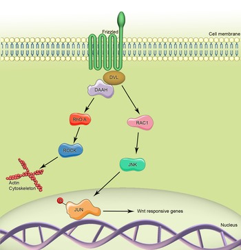
The planar cell polarity pathway is independent of β-catenin (β-cat) and is activated by an interaction between one of the Wnt ligands and Fz. Similar to the β-catenin-dependent pathway, Dvl is recruited, and its PDZ and DEP domains form a complex with Dishevelled-associated activator of morphogenesis 1 (DAAM1) (Fig. 3). This activates the Rho protein, and subsequently, the ROCK kinase – a protein responsible for cytoskeleton arrangement. Dvl also interacts with Rac1, which leads to actin polymerisation (via JNK) and the binding of profilin to actin filaments, which results in actin reorganisation and change in cell polarity (Ref. Reference Anagnostou60).
The PCP pathway in the presence of Wnt ligands. After binding of the Wnt ligand to the Frizzled receptor, Dvl and DAAM are activated. These two proteins activate the small GTPases Rho A and RAC-1, which leads to the activation of ROCK and JNK kinases. JNK phosphorylates JUN, which is translocated to the nucleus to regulate gene expression.

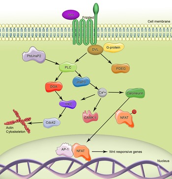
The Wnt/calcium pathway is the second β-cat-independent Wnt pathway. Upon Wnt–Fz interaction, the PDZ and DEP domains of Dvl are directly activated; Dvl also interacts with a G protein, resulting in the activation of either phospholipase C (PLC) or phosphodiesterase (PDE) (Refs Reference Chen38, Reference Komiya61) (Fig. 4). An active PLC causes a rise in the levels of diacylglycerol (DAG) and inositol triphosphate (IP3) and subsequently results in the release of ionic calcium from the ER. This, via the activation of Cdc42, regulates cytoskeleton-dependent processes such as adhesion and migration of the cell. The increase in calcium level also activates CamKII (Ca2+/calmodulin-dependent protein kinase II), which activates kinases such as TAK1 (TGF-β-activated kinase I) and NLK (nemo-like kinase) that inhibit β-cat signalling in the β-catenin-dependent Wnt pathway (Ref. Reference Habas49). On the other hand, the Dvl-G protein interaction can produce the opposite effect if PDE is activated. In this case, calcium release is not increased, and downstream processes do not occur (Ref. Reference Anagnostou60).
The β-catenin-independent pathway in the presence of Wnt ligands. After binding of the Wnt ligand to the Frizzled receptor, the Dvl protein activates PLC, leading to the cleavage of PtdIns(4,5)P into DAG and InsP3. InsP3 increases the cytoplasmic free-calcium level, which subsequently activates CAMKII, PKC and calcineurin. Calcineurin activates NFAT, which is translocated into the nucleus and activates the transcription of Wnt-responsive genes.

The β-catenin-dependent Wnt pathway
The involvement of β-cat distinguishes the β-catenin-dependent Wnt pathway from β-catenin-independent pathways. β-catenin plays two crucial roles within the cell: it is part of the cadherin anchoring system mediating cell–cell interactions and acts as a transcription co-activator in the cell nucleus. In the absence of Wnt ligands, excess β-cat is degraded by a protein complex consisting of Axin, adenomatous polyposis coli (APC), protein phosphatase 2A (PP2A), glycogen synthase kinase 3 (GSK3) and casein kinase 1α (CK1α) (Fig. 1) (Ref. Reference Minde62). Binding of a Wnt ligand to Fz and a simultaneous activation of the LRP 5/6 co-receptor recruits Axin to the plasma membrane, resulting in the disassembly of the degradation complex. Moreover, activated Dvl inactivates GSK-3. Owing to the inactivation of Axin and GSK-3, β-cat is not destroyed and is subsequently transported into the nucleus, where it acts as a co-activator for the TCF/LEF transcription factors and activates the target gene expression (Fig. 2) (Ref. Reference Chandramouli63). Target genes for β-cat-TCF/LEF appear to be specific for the cell type. No universal target genes have been identified thus far. One of the most plausible candidates may be Axin2/conductin (Ref. Reference Jho64), which is involved in the autoregulation of the pathway. For an extensive, updated list of Wnt target genes, the following website ‘the Wntwebpage’ is recommended http://web.stanford.edu/group/nusselab/cgi-bin/wnt/.
Experiments carried out on two macrophage cell lines (RAW264.7 and J774.2) indicate that glucose concentration regulates β-catenin levels by promoting autocrine activation of the β-catenin-dependent pathway. This activation is mediated by changes in N-linked glycosylation of proteins and the hexosamine pathway (Ref. Reference Anagnostou60). The complexity of the catenin-dependent Wnt pathway in cell reprogramming in preadipose cells, where the Wnt antagonist Dickkopf1 (Dkk1) and its receptors (Krm1, LRP5 and LRP6), is coordinately regulated during the early stages of human adipogenesis in vitro (Ref. Reference Christodoulides65). Crosstalk between cytokine and Wnt signalling networks has also been reported; for example, TNF-α inhibits adipogenesis via a β-catenin/TCF4 (TCF7L2)-dependent pathway, which suggests a possible mechanism in endothelial dysfunction (Ref. Reference Cawthorn66).
The effects of β-catenin-dependent Wnt signalling
High glucose concentrations disrupt endothelial adherent junctions (AJs) by dissociating the VE-cad-β-cat complex via activation of the Wnt/β-cat pathway. This process is mediated by tyrosine phosphorylation of VE-cad through PKC-β and MLC phosphorylation (Ref. Reference Haidari67). Blocking the E1E2 domain of Wnt co-receptor LRP6 attenuates the accumulation of β-cat and overexpression of VEGF, intercellular adhesion molecule-1 (ICAM-1), and TNF-α induced by high glucose concentrations in retinal ECs (Ref. Reference Lee68). This suggests that antibodies blocking β-catenin-dependent Wnt signalling can protect against vascular leakage and inflammation in the retina of DR models, which may be used as therapeutic agents in combination with other anti-angiogenic compounds. The possible molecular mechanism underlying this is the regulation of the β-cat and TCF/LEF target genes, including myc and cycD1, which promote vascular cell survival, proliferation and migration (Refs Reference Holnthoner69–Reference Hou71).
Another important molecule linking Wnt signalling to cell longevity is a protein component of the telomerase complex, telomerase reverse transcriptase (TERT), which provides the reverse transcriptase activity of telomerase and is responsible for DNA synthesis at the ends of chromosomes. TERT is essential for maintaining genome stability and cell immortality. High telomerase activity is detected in many human cancers, whereas it is maintained at a low level in somatic cells (Ref. Reference Cesare72). Since uncontrolled cell viability underlies a number of diabetes-related conditions, such as atherosclerosis, DR and DN, regulating telomerase activity is an attractive approach in the development of novel therapeutic strategies for these diseases.
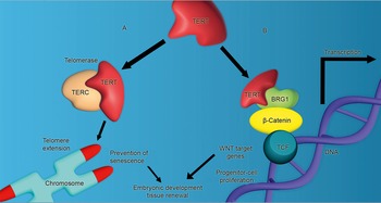
c-Myc, Sp1 (specificity protein 1), AP2 (activator protein 2) and HIF-1 (hypoxia-inducible factor 1) are known regulators of human TERT in normal and cancer cells (Refs Reference Bilsland73, Reference Deng74). Wnt pathways have been found to regulate TERT (Fig. 5). FH535, a β-cat/TCF complex inhibitor, significantly inhibits telomerase activity in many cell lines, such as HCT116, MCF7 and MCF10A (Ref. Reference Zhang75). The results of the same study showed that activation of the Wnt pathway stimulates hTERT expression and telomerase activity. In this context, an interesting novel molecule is BRG1, which is a subunit of a protein complex responsible for alterations in chromatin conformation, facilitating transcription. BRG1 has been shown to be a component of TERT protein complexes. Binding of β-cat to BRG1 results in stimulation of β-cat target gene expression. Therefore, the BRG1–TERT complex might be a molecular link between β-cat and TERT. Experiments carried out on cells from the murine small intestine showed that a complex composed of β-cat, TERT and TCF protein bound to β-cat target genes, whereas the deletion of TERT decreased the expression of Wnt target genes in mouse embryonic stem cells (Ref. Reference Park76).
TERT and β-catenin interaction. (A) During cell division, the telomerase complex repairs the chromosome ends in progenitor cells. TERT ensures reverse transcriptase activity to this complex and uses TERC as a template. (B) TERT increases the transcriptional activity of the β-catenin/TCF complex via interaction with BRG1. Both functions of TERT might increase the proliferation of progenitor cells and prevent cellular senescence at the same time.

Hyperglycaemia and Wnt signalling in diabetes-associated neoplasms
Some types of cancers are known to be more abundant in diabetic and obese individuals (Ref. Reference Khandekar77). This phenomenon has been partially attributed to the growth factor activity of insulin, which is present in higher concentrations in obese patients with insulin resistance or early stages of diabetes. However, in 2013, Chocarro-Calvo et al. (Ref. Reference Chocarro-Calvo78) showed that glucose is essential for β-catenin accumulation in the nucleus. Following Wnt activation, β-catenin accumulates in the cytoplasm, but in the absence of glucose, it is not transported to the nucleus. Upon the addition of glucose, β-catenin is rapidly translocated to the nucleus, where it forms a complex with LEF1, which functions as a p300 acetylase complex and simultaneously reduces the activity of SIRT1 deacetylase. This leads to acetylation of β-catenin, its accumulation in the nucleus, and subsequent activation of target genes. It needs to be emphasised that glucose alone, without a Wnt ligand, did not inactivate GSK-3β. Moreover, hyperglycaemia has been shown to be a potent amplifier of β-cat/Wnt activity in cancer cell lines associated with diabetes and hyperglycaemic conditions. In hepatocellular carcinoma, high glucose concentrations activate β-catenin-dependent Wnt signalling, which is mediated by DKK4 suppression and enhanced the β-cat activity, leading to loss of check at G0/G1/S phases (Ref. Reference Chouhan79). This may partly explain the relationship between hyperglycaemia and tumour progression.
A relevant role of DM has been observed in the development of solid organ malignancies, for example, pancreatic (Refs Reference Coughlin80, Reference Jee81), breast (Refs Reference Baron82, Reference Lipscombe83), liver (Refs Reference Lipscombe83, Reference Wideroff84), endometrial (Refs Reference Friberg85–Reference Vigneri87), colorectal (Refs Reference Coughlin80, Reference Yun88) and bladder cancers (Refs Reference Tseng89, Reference Lewis90). The strongest association with liver and pancreatic cancer was observed in patients with DM2.
Bearing in mind that hyperinsulinaemia is often associated with hyperglycaemia, some experiments were performed on cancer cell lines cultured in high glucose (11 mM) and insulin (100 ng/ml) concentrations. Both conditions caused increased proliferation of cancer cell lines such as SW480 (human colorectal carcinoma), MCF-7 (human breast adenocarcinoma), HT29 (human colon carcinoma), MDA MB468 (human breast adenocarcinoma), PC3 (human prostate cancer) and T24 (human bladder carcinoma) (Ref. Reference Masur91).
Large population studies show that there is a different pattern for the risk of incidence and mortality of a number of cancer types in T1DM and T2DM individuals. T1DM is associated with a higher incidence of pancreas, liver, oesophagus, colon and rectum. Moreover, for women with T1DM, a higher risk of stomach, thyroid, brain, lung, endometrium and ovary cancers has been reported. In T2DM, almost all site-specific cancers are more likely to develop, with the highest risk recorded for liver and pancreas. Given the different pathogenesis of T1DM and T2DM, with hyperinsulinaemia present only in the latter, it seems that hyperglycaemia might be the dominant trigger eventually leading to alterations in epigenetic regulation (Refs Reference Chocarro-Calvo78, Reference Chouhan79). The pro-inflammatory role of insulin signalling has been observed in the development and progression of different types of cancers in both types of DM (Refs Reference Coughlin80, Reference Lipscombe83–Reference Friberg85). Interestingly, there seems to be a protective role of both types of DM with regard to prostate cancer incidence. For T2DM, when co-existing obesity is very frequent, this has been justified by obesity-related lower testosterone levels; however, this does not explain the relationship with T1DM (Ref. Reference Harding92).
Wnt signalling and atherogenesis in hyperglycaemic conditions
Atherosclerosis is common among patients with DM. It is marked by the appearance of plaques consisting of LDL cholesterol, leukocytes, smooth muscle cells and lipids in the arterial walls (Ref. Reference Yan93). The formation of an atherosclerotic plaque is a complex process, and the plaques are more frequently found at branching points within the vasculature, coinciding with turbulent blood flow, rather than the straight sections where the luminal ECs undergo lower shear stress. As described before, the activated and inflamed endothelium loses its protective abilities as it undergoes EndoMT and acts as a source of osteogenic progenitors, leading to vascular calcification and allowing enhanced transendothelial migration of leukocytes (Ref. Reference Sánchez-Duffhues36). There is growing evidence that the Wnt pathway is also an important part of the pathophysiology of these processes. Wnt5A acts via the Ca2+-dependent pathway and has been shown to induce the expression of pro-inflammatory cyclooxygenase-2 in ECs (Ref. Reference Kim94). Wnt5A has also been observed to be abundant in macrophages within atherosclerotic plaques (Refs Reference Holnthoner69, Reference Christman95). The expression of Wnt5A mRNA is activated by exposure to ox-LDL in human macrophages (Ref. Reference Bhatt96). Serum concentrations of Wnt-5A were higher in patients with advanced atherosclerosis than in healthy individuals (Ref. Reference Malgor97). Moreover, Wnt signalling appears to be an important part of the pathophysiology of myocardial infarction (MI) and its complications. The inhibition of Fz receptors by analogues of either Wnt3a/Wnt5A or sfrp2 leads to a reduction in the extent of the MI and development of subsequent heart insufficiency (Ref. Reference Laeremans98).
Furthermore, Wnt signalling is crucial for the loosening of AJs in an endothelial monolayer, facilitating the infiltration of leukocytes into the vessel wall. AJs are formed by interactions of transmembrane proteins, namely, VE-cad, of neighbouring cells. The cytosolic domains of these proteins are anchored to the actin cytoskeleton by a number of proteins, the most important of which are p120, plakoglobin, α-catenin and β-cat (Ref. Reference Dejana99). Tyrosine phosphorylation within the cytoplasmic domain of VE-cadherin causes dissociation of catenins and destabilisation of AJs with a subsequent rise in endothelial permeability (Ref. Reference Schulte100). A side effect of such dissociation is a higher accumulation of β-cat in the cytosol available for translocation to the nucleus and activation of target genes.
Haidari et al. demonstrated that exposure of ECs to hyperglycaemic conditions results in tyrosine phosphorylation of VE-cadherin, dissociation of β-cat from AJs, and enhanced transendothelial migration (Ref. Reference Haidari67). Notably, this process was mediated by PKC-β, a downstream effector in the β-catenin-independent Wnt signalling pathway. Moreover, a high concentration of glucose led to PKC-mediated GSK3β phosphorylation with a subsequent rise in the cytosolic pool of β-cat and activation of the β-cat/TCF/Lef-1 responsive promoter. Hyperglycaemia significantly increased the expression of β-catenin target genes such as cycD1 (cyclin D1) and u-PA (urokinase), indicating that exposure to high glucose levels activates the β-catenin-dependent Wnt pathway in ECs. These findings may provide an important link between diabetes and accelerated atherogenesis (Ref. Reference Park76).
Another study reported that VEGF stimulates uPA expression by inducing EC hyperpermeability through β-cat-dependent urokinase-type plasminogen activator receptor (uPA/uPAR) activation (Refs Reference Behzadian101, Reference Esser102). These results suggest that the crosstalk of the TGF-β and Wnt pathways is important in endothelial dysfunction and that the u-PA/uPAR and Wnt pathways might provide a molecular bridge linking the inflammation and progression of atherosclerosis in patients with diabetes. Additionally, hyperglycaemia promotes a pro-coagulant phenotype in ECs by upregulating the expression of plasminogen activator inhibitor (PAI-1) and P selectin (P-Sel) as well as inducing the secretion of fibrinogen (Ref. Reference Goldberg103).
TUG1 is a long non-coding RNA (lncRNA) that is highly expressed in ECs (Ref. Reference Chen104). TUG1 overexpression was detected in CAD tissues compared with normal arterial tissues. Upregulation of TUG1 was observed in high-dose glucose-induced HUVECs. This overexpression of TUG1 stimulated a number of genes in HUVECs: cell cycle-related, proliferation-related and Wnt pathway-related (e.g., β-catenin, c-myc). The Wnt pathway might be associated with TUG1-promoted migration and proliferation of ECs. This could be a possible mechanism of TUG1 involvement in the regulation of atherosclerosis in DM (Ref. Reference Lee68).
DKK-1, an inhibitor of β-catenin-dependent Wnt signalling, has been observed at higher concentrations in clinical and experimental atherosclerosis compared with normal tissue. The main sources of DKK-1 are ECs and platelets (Ref. Reference Ueland105). The plasma concentration of DKK-1 was higher in patients with T2DM compared with healthy controls, which was associated with platelet activation markers and increased levels of endothelial dysfunction. Additionally, improved glycaemic control downregulates DKK-1 expression in T2DM (Ref. Reference Lattanzio106).
Wnt in diabetic retinopathy (DR)
The key factors underlying the development of DR are inflammation (mediated by VEGF, ICAM and TNFα), microvascular damage and oxidative stress, which lead to a disruption of the blood–retina barrier and retinal ischaemia. Subsequent retinal vascular leakage and neovascularisation result in loss of sight (Refs Reference Klein107, Reference van Hecke108). Since VEGF, TNFα and ICAM are target genes of the Wnt signalling pathway, this pathway has become an important field of research in the pathophysiology of DR (Ref. Reference Chen109). The β-catenin-dependent Wnt pathway has been shown to be upregulated in both human and rodent models of DR. Using a murine model, Zhou et al. (Ref. Reference Zhou110) showed that under ischaemic conditions, as well as in induced diabetes, β-catenin knockout in Muller retinal cells reduces neovascularisation, vascular leakage and inflammation in comparison with those in wild-type mice. At the molecular level, the lack of β-cat reduced the production of VEGF and TNFα. These results further support the detrimental effect of the Wnt pathway in the pathogenesis of DR. Additionally, pericyte loss, which is an important hallmark of DR, was significantly lower in β-cat knockout mice than in wild-type mice exposed to the same hyperglycaemic conditions (Ref. Reference Vigneri87). Furthermore, Lee et al. (Ref. Reference Lee68) demonstrated that application of an anti-LRP6 monoclonal antibody on retinal ECs led to reduced β-cat accumulation, lower expression of VEGF5 and TNFα induced by hyperglycaemia in vitro. Furthermore, an intravitreal administration of the same antibody decreased vascular leakage and neovascularisation in a rat DR model (Ref. Reference Lee68). Similarly, DKK1, a natural inhibitor of the Wnt signalling pathway, has been shown to alleviate retinal inflammation and vascular leakage in DR (Refs Reference Holnthoner69, Reference Yun88). Another negative regulator of Wnt/β-cat signalling, Adenomatosis Polyposis Coli Downregulated 1 Protein (Apcdd1), is expressed in retinal ECs during angiogenesis and barrier formation. Apcdd1-deficient mice exhibit a transient increase in vessel density. Moreover, mice that overexpress Apcdd1 in retina ECs have reduced vessel density but increased barrier permeability (Ref. Reference Mazzoni111).
Another study focusing on fibrosis in DR has shown that introducing SERPINA3 K, a serine proteinase inhibitor, reduces fibrosis in a rat DR model by antagonizing connective tissue growth factor (CTGF), a potent fibrogenic factor, and minimizing the production of ECM proteins. Thus, the effect of SERPINA3 K is likely to be achieved via the inhibition of the Wnt signalling pathway (Ref. Reference Zhang112).
Adherens junctions and the β-catenin-dependent Wnt pathway
In ECs, β-catenin performs several functions owing to its ability to bind different molecular partners (Fig. 2). One of them is the formation and stabilisation of tight AJs in association with the extracellular membrane protein E-cadherin (E-cad). The central part of the β-cat amino acid chain contains binding sites for E-cad (a crucial protein for cell–cell interactions), LEF-1 (a transcription factor) and APC (a part of the β-catenin degrading complex). As all these binding sites overlap, β-cat can interact only with one of these partners at a given time (Refs Reference Hülsken113, Reference Orsulic114). Thus, only upon dissociation from E-cad does β-cat become available for the other two proteins and can function as the Wnt pathway component. This phenomenon has already been demonstrated in Xenopus (Ref. Reference McMahon115) and Drosophila (Ref. Reference Cox116). Moreover, β-cat released from AJs upon their dissociation forms a pool from which it is transported to the nucleus upon activation of the Wnt pathway (Ref. Reference Kam117). Loss of E-cad expression has been reported to result in β-cat accumulation in the cytoplasm, with its subsequent nuclear translocation and interaction with LEF (Ref. Reference Eger118). These observations confirm the involvement of the β-catenin-dependent Wnt pathway in the disintegration of the EC monolayer, leading to the loss of one of its basic roles in maintaining vessel homeostasis.
Research in progress and outstanding research questions
Chronic hyperglycaemia extensively influences the vasculature, leading to multiple organ complications in patients suffering from DM. As DM is a growing problem worldwide, there is and will be more demand for new therapies to treat these complications. Previous studies indicate the possible involvement of Wnt signalling in the development of hyperglycaemia-related complications such as atherosclerosis, DR and certain types of cancers.
Given the importance of hyperglycaemia in disrupting physiological cellular processes and leading to multiple complications, it is crucial to remember that normalizing blood glucose concentration remains the basic course of therapy in diabetes-related health issues. One of the most commonly prescribed anti-hyperglycaemia drugs, metformin, has been shown to reduce the risk and slow the progression of several types of cancer. Many mechanisms have been proposed as an explanation of this phenomenon, the most simple of which may be reducing hyperglycaemia and its cellular consequences (Ref. Reference Aljada119). However, there are elements of the pathogenesis of DM complications that are irreversible, such as metabolic memory, which requires a more complex approach and further investigation into potential therapies.
The current pharmacotherapy for slowing the process of vascular complications in patients with diabetes is rather narrow and is mostly focused on lowering LDL cholesterol levels. Most likely, the most widely used pharmacological agents are statins, which in addition to lowering LDL, have been shown to stabilise the atherosclerotic plaque, preventing thrombus formation. These, however, are not always effective and have a number of well-characterised adverse effects, such as liver dysfunction or rhabdomyolysis, which may lead to acute kidney injury. Similar reservations apply to fibrates, which also reduce lipid levels and predispose them to nearly the same adverse effects. An emerging new player in the field is the anti-PCSK-9 antibody, evolocumab, which targets and inactivates LDL cholesterol receptors. In light of the presented data, the effect of evolocumab on Wnt signalling in diabetic vascular complications appears to be a very interesting alternative to the current use of statins.
One of the most important vascular complications in diabetes is fibrosis, which is directly related to EndoMT. ECs are progenitors of cardiac pericytes and vascular smooth muscle cells, both of which are involved in tissue regeneration and vascular remodelling (Refs Reference Widyantoro40–Reference Wu42). Blocking Wnt signalling with a homologous peptide fragment of wnt3a/wnt5a has been reported to reduce infarct expansion and prevent heart failure after MI (Ref. Reference Laeremans98). Additionally, the occurrence of infarction-related fibrosis can be reduced by a small molecule allosteric activator of aldehyde dehydrogenase-2 (ALDH2), which downregulates β-cat, phosphorylated GSK-3β and Wnt-1 (Ref. Reference Zhao120). Based on the aforementioned studies, inhibition of Wnt signalling can be considered to reduce vascular complications, both in patients with diabetes and non-diabetic individuals.
Untreated ocular complications of chronic hyperglycaemia lead to proliferative DR, diabetic macular oedema and eventually loss of sight. Several sight-saving therapies, such as laser photocoagulation or vitrectomy, are available; however, none of them is fully satisfactory, and there is a need for the development of new therapies in this area (Ref. Reference Stegenga4). Among the new therapeutic candidates are anti-VEGF agents (Refs Reference Baron82, Reference Lipscombe83). Although some promising results have been presented for these candidates, this therapy is not effective for all patients. As described above, the β-catenin-dependent Wnt pathway appears to be involved in many pathological processes, such as neoangiogenesis or inflammation, resulting in VEGF, TNFα and ICAM expression and eventually leading to DR. As such, this signalling system could be a potential target for alleviating inflammation and neoangiogenesis. Thus far, the anti-LRP6 monoclonal antibody is the only potential therapeutic approach targeting the β-cat-dependent system proposed to treat DR (Ref. Reference Lee68). Another potential therapeutic target could be Apcdd1 or ALDH2, overexpression or over-activity of which downregulates Wnt-dependent genes (Refs Reference Mazzoni111, Reference Zhao120).
Finally, disrupting the Wnt pathway could be considered a potential approach to limit the development of certain types of cancers that are more frequent in patients with chronic hyperglycaemia.
OMP-18R5, a monoclonal antibody interacting with five Fzd receptors, inhibited different human tumour growth in xenografts, diminished tumour-initiating cell frequency and exhibited synergistic activity with standard chemotherapeutic agents (Ref. Reference Gurney121). NSC654259, another small molecule that targets the cysteine-rich domain of Frizzled, has shown attenuating properties in multiple types of tumour cell lines (Ref. Reference Lee122).
An eye-specific miRNA-184 expressed in the lens, cornea and retina (Ref. Reference Ryan123) was significantly downregulated in a murine model of oxygen-induced retinopathy (Ref. Reference Takahashi124). Bioinformatics analysis showed that it targets several components of Wnt signalling such as Wnt ligands (Wnt-9, Wnt-16) and Wnt receptors (Frizzled-3, Frizzled-7) (Refs Reference Chen109, Reference Shen125), showing therapeutic potential in proliferative retinopathy.
Increased levels of LRP6 have been observed in the retina of DR animal models (Ref. Reference Chen109). Mab2F1, a monoclonal antibody against the E1E2 domain of LRP6, inhibited Wnt signalling upregulation caused by high glucose levels and suppressed the expression of inflammatory factors (VEGF, ICAM-1 and TNF-a) (Ref. Reference Lee68).
In summary, there is growing evidence that abnormalities at different stages of Wnt signalling result in inflammation, neoangiogenesis and abnormal gene expression. All these phenomena underlie a series of complications in diabetes. Regulating these processes, for example, by inhibiting the Fz or LRP receptors or downregulating the Wnt β-catenin-dependent pathway, could lead to the development of a multipotent therapy, improving the condition of patients suffering from diabetic complications.
Summary
Many diseases that are associated with EC dysfunction are related to an impaired Wnt pathway. Accumulating evidence confirms the idea that in hyperglycaemic conditions, the endothelium loses its protective abilities, becoming activated (inflamed) and undergoing EndoMT, enhancing the expression of Wnt-dependent genes involved in cell proliferation and permeability. In these processes, β-cat signalling appears to be the most important, showing the need for further investigation to establish whether Wnt pathway inhibitors or antagonists may have clinical relevance in the treatment of vascular diabetic complications such as accelerated atherosclerosis and its consequences, DR or DN.
Acknowledgements
This work was founded by the National Science Center, grant PRELUDIUM 8 number UMO-2014/15/N/NZ5/01606. The present publication contains a portion of Martyna Durak-Kozica's doctoral dissertation.
Highlights
• Diabetes favours oncogenesis and accelerates cardiovascular complications.
• Hyperglycaemia affects endothelial functions and induces inflammatory processes leading to atherosclerosis and impaired angiogenesis.
• Wnt signalling is activated during hyperglycaemia and causes endothelial dysfunction and EndoMT.
• Targeting Wnt pathways improves hyperglycaemia-related endothelial complications.