Introduction
The spread of tumours through nerves is known as peri-neural invasion (PNI).Reference Amit, Na’ara and Gil1 The prevailing definition of PNI is a ‘tumour in close proximity to a nerve and involving at least 33% of its circumference or tumour cells within any of the three layers of the nerve sheath.’Reference Liebig, Ayala, Wilks, Berger and Albo2 Peri-neural invasion is an important pathological feature of many malignant neoplasms, including head and neck cancer, gastrointestinal tumour, breast cancer, pancreatic cancer, prostate cancer and bile duct cancer. Positive PNI often indicates poor prognosis and decreased survival rate.Reference Liebig, Ayala, Wilks, Berger and Albo2
Hypopharyngeal cancer (HPC) is the least common type of cancer of the head and neck but has the worst prognosis. Morbidity and mortality rates for HPC have continued to increase over the past decade.Reference Lin, Lin and Lin3 Peri-neural invasion in hypopharyngeal squamous cell carcinoma (HPSCC), a subtype of HPC, is also associated with poor prognosis, namely decreased survival and increased risk of metastasis.Reference Joo, Cho, Lee, Kim and Kim4 However, the correlation between PNI and the clinicopathological parameters of head and neck squamous cell carcinoma (HNSCC) remains unclear.Reference Joo, Cho, Lee, Kim and Kim4–Reference Schmitd, Scanlon and D’Silva7 This study aimed to investigate the positivity rate of PNI in HPSCC and determine its association with the clinicopathological parameters of HPSCC. We anticipate that our findings will potentially provide new insights about PNI and further guide clinical decisions.
Materials and methods
Patients
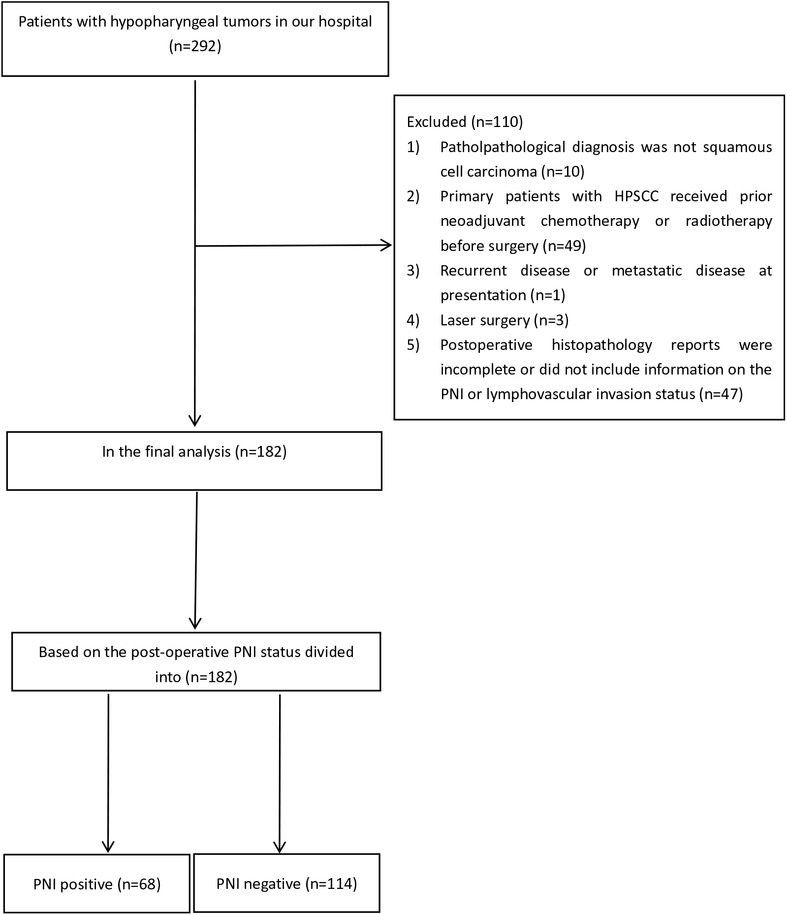
Complying with Good Clinical Practice guidelines in China and the Declaration of Helsinki (2013), the Institutional Review Board of Shandong Provincial ENT Hospital approved this retrospective study (Approval Number 2024-18-01) and waived the requirement for informed consent. We enrolled patients with hypopharyngeal tumours between September 2019 and March 2024. The criteria for exclusion were: (1) non-squamous cell cancer (SCC) post-operative pathology; (2) patients with primary HPSCC who received radiotherapy or neoadjuvant chemotherapy prior to surgery; (3) recurrent or metastatic disease; (4) laser surgery; and (5) incomplete post-operative histopathology reports or no report of PNI or lymphovascular invasion (LVI) status. Figure 1 shows the detailed process for patient recruitment. Ultimately, 182 consecutive patients were identified and included in the final analysis. The clinical and histopathological parameters of all participants were retrospectively analysed, including sex, age, primary tumour sites, tumour thickness (TT, the total thickness of the tumour along its invasive axis), tumour diameter (TD, the longest diameter in the resected specimen), histologic differentiation, tumour–node–metastasis classification (the 2017 edition of the tumour–node–metastasis classification of the American Joint Committee on Cancer), stage, lymph node metastasis (LNM), number of positive nodes (pN), lymph node density (LND, the ratio of the number of metastatic lymph nodes to the total number of resected lymph nodes), extranodal extension (ENE), PNI, LVI and Ki-67 index. We divided the HPSCC patients into two groups: PNI-positive and PNI-negative, according to PNI status on post-operative pathological reports.
The flow diagram showing the selection process of study population.

Pathological evaluation
Pathological specimens of all participants’ primary tumours were carefully identified by a head and neck pathologist (with eight years’ work experience) to check the PNI status of the pathological reports. Histopathological morphology on haematoxylin and eosin stain and S-100 protein immunohistochemical staining were used to detect PNI within the primary tumours. Positive PNI was defined as: ‘tumour in close proximity to a nerve and involving at least 33% of its circumference or tumour cells within any of the three layers of the nerve sheath.’Reference Liebig, Ayala, Wilks, Berger and Albo2
Statistical analysis
IBM SPSS 27.0 software (SPSS, Chicago, Illinois, USA) was used for statistical analyses. Inter-group comparisons of quantitative data were conducted using an independent sample t-test or Mann–Whitney U test. Two group comparisons of qualitative data were analysed using chi-square or Fisher’s exact tests. A binary logistic regression model was used for multivariate analysis of meaningful variables in univariate analysis. A p < 0.05 was considered statistically significant.
Results
Patient characteristics
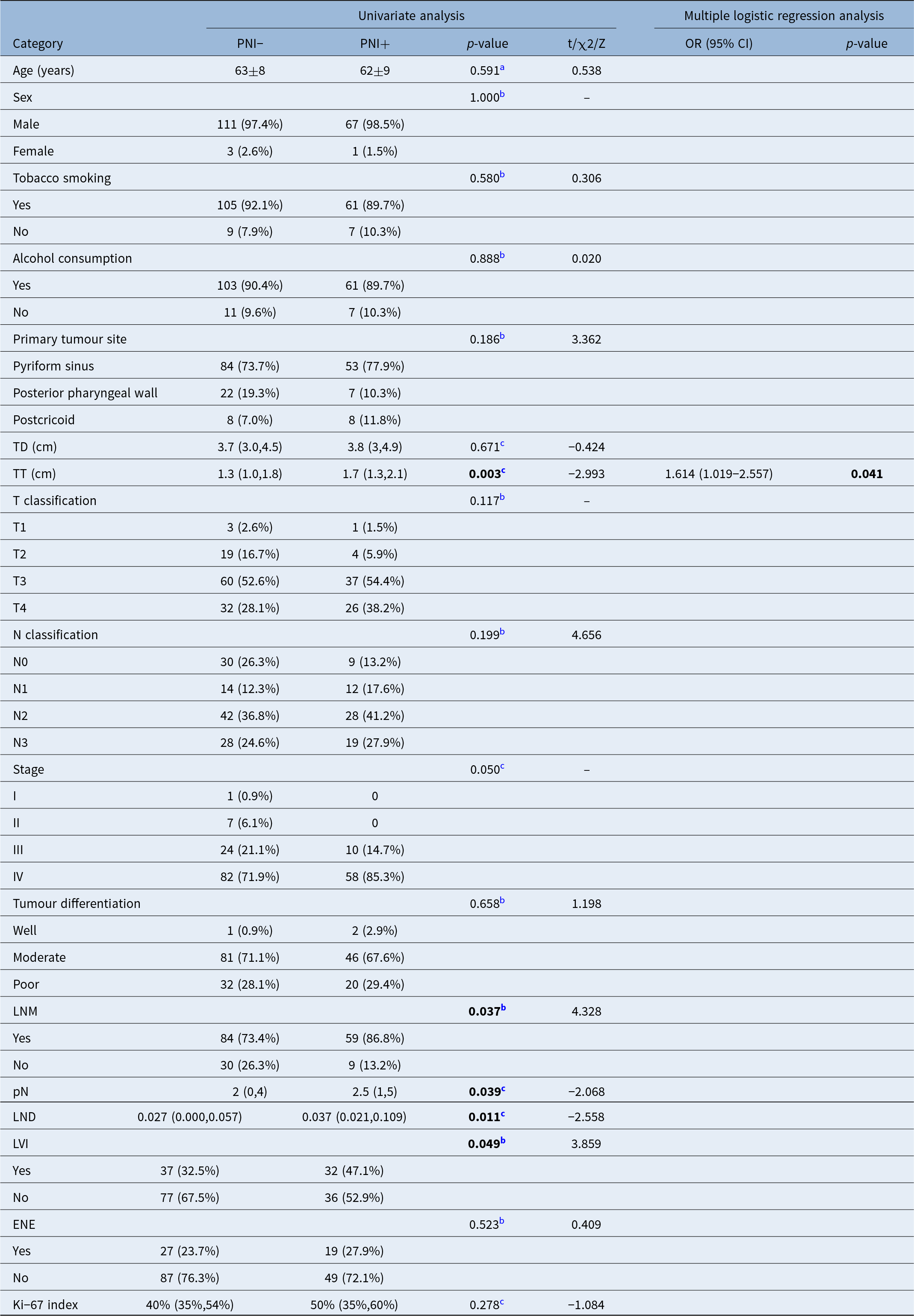
Table 1 provides an overview of the clinicopathological features of patients with HPSCC. The mean age was 62 years (range: 42–81 years), with 178 males and 4 females. The location of the primary tumour was as follows: 137 cases involving the pyriform fossa, 29 cases involving the posterior pharyngeal wall and 16 cases involving the post-cricoid area.
Univariate analysis and multiple logistic regression analysis of predictive factors for PNI; Quantitative data were expressed as mean ± standard deviation or median (q1,q3), qualitative data were described by n (%); PNI− = peri-neural invasion negative; PNI+ = peri-neural invasion positive; OR = odds ratio; CI = confidence interval; TD = tumour diameter; TT = tumour thickness; LNM = lymph node metastasis; pN = number of positive lymph nodes; LND = lymph node density; LVI = lymphovascular invasion; ENE = extranodal extension; Bold values denote p-value < 0.05

a Independent sample t-test
b Chi-square test or Fisher’s exact test
c Mann–Whitney U test
–No statistical value
Peri-neural infusion was noted in 68 of the 182 patients (37.4 per cent). In the PNI-positive group, the majority of patients (37; 54.4 per cent) were at the T3 stage, while only 1 (1.5 per cent) patient was at the T1 stage. In the PNI-positive group, most patients were at the N2 stage (41.2 per cent), whereas N0 stage was the least common, accounting for only 13.2 per cent. In the PNI-positive group, 58 (85.3 per cent) patients had stage IV disease, 10 (14.7 per cent) had stage III disease, and none had stage I or II disease. In the PNI-positive group, 46 (67.6 per cent) patients had moderately differentiated tumours, while only 2 patients (2.9 per cent) had well-differentiated tumours.
Relationship between peri-neural invasion and clinicopathological features
We observed a definite correlation between PNI and TT (p = 0.003), LVI (p = 0.049), LNM (p = 0.037), LND (p = 0.011), and pN value (p = 0.039), as shown in Table 1. The median value of TT in the PNI-positive and PNI-negative group was 1.7 cm (1.3 cm, 2.075 cm) and 1.3 cm (1 cm, 1.8 cm), respectively. Fifty-nine (86.8 per cent) patients had LNM in the PNI-positive group, and only 9 (13.2 per cent) patients had negative lymph nodes. No significant relationship was observed between positive PNI and other clinicopathological characteristics.
Risk factors of peri-neural invasion in hypopharyngeal squamous cell carcinoma
TT remained a significant risk factor for PNI in HPSCC (odds ratio = 1.614; 95 per cent confidence interval: 1.019–2.557; p = 0.041) in the multivariate analysis, as shown in Table 1.
Discussion
Although HNSCCs are similar in microstructure, they are heterogeneous in terms of location, aetiology and clinical characteristics. The prevalence of PNI in HNSCC is highly variable, with reported incidence rates ranging from 5.2 per cent to 90 per cent.Reference Schmitd, Scanlon and D’Silva7 This is because PNI incidence is related to the location and staging of the primary tumour. Oral cavity cancer, especially tongue cancer, has a high prevalence of PNI.Reference Nair, Singh, Pawar, Datta, Nair and Kane8 The occurrence of PNI increases in a stepwise fashion according to pathological tumour status,Reference Lee, Yang, Lin, Hsieh, Chen and Chang9 and the incidence of PNI in advanced tumours is high, seemingly unrelated to location.Reference Amit, Na’ara and Gil1° A previous study reported that 35.6 per cent of laryngeal and hypopharyngeal cancers were PNI-positive, including patients with stage III-IV.Reference Chirilă, Bolboacă, Cosgarea, Tomescu and Mureşan11 In a study by Joo et al.,Reference Joo, Cho, Lee, Kim and Kim4 25.7 per cent (27/105) of patients with HPSCC were PNI-positive. In the present study, we also exclusively included patients with HPSCC and the PNI rate was 37.4 per cent (68/182), higher than that reported by Joo et al. This may have been because we included more patients with advanced disease (174/182 versus 95/105).
There is a global consensus that LNM is an important prognostic factor and has a significant impact on survival rate. Consistently, LNM significantly negatively affects the prognosis and survival of patients with HPSCC: patients with HPSCC with LNM have worse disease-specific survival than those without LNM.Reference Layland, Sessions and Lenox12, Reference Roberts, Colevas, Hara, Holsinger, Oakley-Girvan and Divi13 The impact of LND and pN value on prognosis and survival of HNSCC has also been investigated in some studies. An LND greater than or equal to 0.07 was related to poorer overall survival and disease-specific survival.Reference Ye, Rao, Fan, Ji, Hu and Ying14 pN value demonstrated superior prognostic value compared to LND and the American Joint Committee on Cancer node staging in surgical patients.Reference Roberts, Colevas, Hara, Holsinger, Oakley-Girvan and Divi13 ENE is a well-known indicator of poor prognosis and a clear indication for the escalation of adjuvant therapy in patients with HNSCC.Reference Kann, Hicks, Payabvash, Mahajan, Du and Gupta15 The association between PNI and LNM in HPSCC was unclear in previous reports, and hence remains controversial. One study suggested that PNI was significantly related to node classification in HPSCC.Reference Joo, Cho, Lee, Kim and Kim4 However, other studies have indicated that no association exists between PNI and LNM.Reference Fagan, Collins, Barnes, D’Amico, Myers and Johnson5, Reference Soo, Carter, O’Brien, Barr, Bliss and Shaw6 Our results showed that the PNI was related to LNM, but had no association with node classification. We also verified the relationship between pN value, LND, ENE and PNI. In the present study, PNI was significantly correlated with LND and pN value in HPSCC. However, in the multivariate analysis, we found that neither LND nor pN value were independent predictors of PNI in HPSCC. Consistent with the results of Joo et al.,Reference Joo, Cho, Lee, Kim and Kim4 our results suggest that PNI is not related to ENE.
LVI refers to ‘the presence of tumour cells within definite endothelial-lined spaces, either lymphatic or blood vessels, as detected by hematoxylin and eosin staining or immunohistochemistry.’Reference Larson, Kemmer, Formeister, El-Sayed, Ha and George16 Similar to PNI, LVI is also an adverse pathological feature of HPSCC, associated with poor prognosis. LVI-positive patients with HPSCC experience an increased local and regional recurrence, and a decreased five-year overall survival, disease-free survival, and disease-specific survival.Reference Gang, Xinwei, LiXiao, Yu, Cheng and Pin17 However, due to a lack of literature, the relationship between LVI and PNI remains unclear. Joo et al.Reference Joo, Cho, Lee, Kim and Kim4 reported that PNI was not related to LVI. Our results indicate that LVI is associated with PNI, which is consistent with Samanta et al.Reference Samanta, Surendra, Suresh, Ajitesh, Maitree and Tapas18 In the present study, LVI positivity in the PNI-positive group was 47.1 per cent, higher than in the PNI-negative group (32.5 per cent).
Researchers have focused on the relationship between tumour size, tumour differentiation and PNI in HNSCC. One study suggested PNI was significantly associated with tumour differentiation and invasion depth in oral and oropharyngeal carcinoma.Reference Rahima, Shingaki, Nagata and Saito19 Other studies have suggested no relationship between the PNI and TD (the longest diameter in the resected specimen) or tumour differentiation in HNSCC.Reference Joo, Cho, Lee, Kim and Kim4, Reference Soo, Carter, O’Brien, Barr, Bliss and Shaw6 Samanta et al.Reference Samanta, Surendra, Suresh, Ajitesh, Maitree and Tapas18 reported that PNI was related to maximum tumour size and depth of invasion, but was not correlated with tumour differentiation in oral cavity carcinoma. Additionally, they did not clarify the method used for measuring depth of invasion. Our results showed that PNI in HPSCC was significantly associated with TT, but not with TD or tumour differentiation. TT remained the only independent risk factor for PNI of patients with HPSCC in the multivariate analysis. To our knowledge, this is the first study to investigate the correlation between PNI status and TT in HPSCC. TT reflects the ability of tumours to infiltrate deeper structures. As TT increases, the propensity of the tumour to extend to adjacent blood vessels, lymphatic vessels and nerves increases, and accordingly, the risk of PNI increases.
The Ki-67 index also reflects the invasiveness of tumours. In the current study, the correlation between PNI and the Ki-67 index was explored for the first time. We found a median Ki-67 index in the PNI-positive group of 50 per cent (35 per cent, 60 per cent), which appeared to be slightly higher than that in the PNI-negative group (40 per cent (35 per cent, 53.75 per cent)); however, the difference was not significant (p = 0.278).
The present study had certain limitations, such as its retrospective design, which was subject to selection bias, and its small sample size. Therefore, a larger prospective study should be conducted in the future to verify our findings. Lastly, because our study employed only one pathologist, inter-observer variability was not taken into consideration, and this could have affected the results.
Conclusion
The main modes of tumour cell dissemination in HPSCC are lymphatic and haematogenous metastasis, and PNI can potentially represent a third pathway. In the current study, 37.4 per cent of patients with HPSCC were PNI-positive, which was identified to be significantly associated with TT, LVI, LNM, pN value and LND. Multivariate analysis showed that TT was an independent risk factor for PNI, with the incidence of PNI increasing with TT. Therefore, it is crucial to include PNI analysis in post-operative histopathology reports for HPSCC to guide clinicians in choosing adjuvant therapy. Future investigations with larger sample sizes are needed to confirm the results of this study.
Acknowledgements
None.
Funding statement
This research received no specific grant from any funding agency, commercial or not-for-profit sectors.
Competing interests
The authors declare none.
Ethical standards
The study was conducted in accordance with the Declaration of Helsinki and was approved by the Institutional Review Board of Shandong Provincial ENT Hospital. The requirement for informed consent was waived because of the retrospective design.