

Milk is one of the most readily available and affordable protein sources and is nature's most complete food. Milk and milk products are essential dietary components for approximately six billion people, accounting for around 80% of the world's population today (Visioli and Strata, Reference Visioli and Strata2014). The bulk (83%) of the milk produced globally comes from bovines, and milk production employs around 750 million people, providing farmers with a daily income (FAO, 2022). Demands for milk are expected to increase due to growing populations, rising incomes, increasing urbanization, and westernization of diets, particularly in nations around the equator and the Global South (Salter, Reference Salter2017). Consequently, milk production and consumption are crucial to the food security and socioeconomic stability of several countries.
Milk production at both the individual animal and herd or farm level depends on several factors, including animal genetics, nutrition, and farming practices. The quality and quantity of milk, along with the related economics, are affected by mastitis (Bar et al., Reference Bar, Grohn, Bennett, Gonzalez, Hertl, Schulte, Tauer, Welcome and Schukken2008a, Reference Bar, Tauer, Bennett, Gonzalez, Hertl, Schukken, Schulte, Welcome and Grohn2008b; Bardhan and Sharma, Reference Bardhan and Sharma2013; Bonestroo et al., Reference Bonestroo, Fall, Hogeveen, Emanuelson, Klaas and van der Voort M2023; Das et al., Reference Das, Panda, Jena and Sahoo2018; Hadrich et al., Reference Hadrich, Wolf, Lombard and Dolak2018; Halasa et al., Reference Halasa, Huijps, Osteras and Hogeveen2007; Hogeveen et al., Reference Hogeveen, Huijps and Lam2011; Seegers et al., Reference Seegers, Fourichon and Beaudeau2003). Microbial infections primarily cause mastitis, and numerous bacteria have been identified as causative agents. These bacteria are broadly categorized into contagious or environmental pathogens based on their primary origin and transmission mode. However, most severe mastitis cases are associated with only about a dozen bacterial species, primarily Escherichia coli, Staphylococcus aureus, Streptococcus agalactiae, Streptococcus dysgalactiae, Streptococcus uberis, and more recently, Klebsiella pneumoniae and enterococci (Ashraf and Imran, Reference Ashraf and Imran2020). Among these, E. coli is the predominant environmental pathogen and the leading cause of acute clinical mastitis (Hogan and Larry Smith, Reference Hogan and Larry Smith2003; Shpigel et al., Reference Shpigel, Elazar and Rosenshine2008; Zaatout, Reference Zaatout2022).
The treatment of bovine mastitis involves antibiotic therapy. However, traditional antibiotic sensitivity testing (e.g., disc diffusion) takes 48–72 h, a critical time window that could be utilized for specific treatment if the test could be rapid. As a result, the treatment is often empirical, involving higher-order, broad-spectrum antibiotics. Over the last few years, the injudicious use of antibiotics and several other factors contributing to the inappropriate use and disposal of antimicrobials and other chemicals have led to a selective increase in the proportion of bacteria resistant to antimicrobials. Consequently, antimicrobial resistance (AMR) is now the most significant risk to global health, food security, and socioeconomic development. Without course correction, it is estimated that by 2050, AMR will be responsible for 10 million human fatalities and economic losses totalling USD 100 trillion (O'Neill, Reference O'Neill2014). Combating AMR, therefore, is an urgent need that requires a multipronged approach. One strategy is the discovery of newer antibiotics; however, antimicrobial discovery has declined precipitously over the last three to four decades.
Bacteriophages (phages) are viruses that specifically infect and replicate within bacteria. They have coevolved with bacteria for billions of years and are regarded as the most abundant and oldest biological entities (Hendrix, Reference Hendrix2002; Weitz et al., Reference Weitz, Poisot, Meyer, Flores, Valverde, Sullivan and Hochberg2013). The rise in AMR and the need for alternatives to antibiotics have renewed interest in phages and their enzymes, owing to their specificity and lack of side effects. There have been several instances of clearance of bacteria causing human intractable infections with the use of phages (albeit in combination with other antimicrobial therapy) under compassionate grounds (reviewed in Aranaga et al., Reference Aranaga, Pantoja, Martinez and Falco2022; Cui et al., Reference Cui, Watanabe, Miyanaga, Kiga, Sasahara, Aiba, Tan, Veeranarayanan, Thitiananpakorn, Nguyen and Wannigama2024; Hatfull et al., Reference Hatfull, Dedrick and Schooley2022; Pirnay, Reference Pirnay2020; Strathdee et al., Reference Strathdee, Hatfull, Mutalik and Schooley2023). Rapid advances are being made in developing phages as therapeutic agents and their use in clinical trials in humans (Chen et al., Reference Chen, Woolston, Grant-Beurmann, Robinson, Bansal, Nkeze, Permala-Booth, Fraser, Tennant, Shriver, Pasetti, Liang, Kotloff, Sulakvelidze and Schwartz2024; Kim et al., Reference Kim, Suh, Cullen, Perez Rodriguez, Dharmaraj, Chang, Li, Chen, Green, Lavigne, Pirnay, Bollyky and Sacher2025; Nir-Paz et al., Reference Nir-Paz, Onallah, Dekel, Gellman, Haze, Ben-Ami, Braunstein, Hazan, Dror, Oster, Cherniak, Attal, Barbosa, Dordio, Wagner, Jones-Dias, Neves, Barreto, Leandro, Corte-Real and Garcia2025; Tamma et al., Reference Tamma, Souli, Billard, Campbell, Conrad, Ellison, Evans, Evans, Greenwood-Quaintance, Filippov, Geres, Hamasaki, Komarow, Nikolich, Lodise, Nayak, Norice-Tra, Patel, Pride, Russell, Van Tyne, Chambers, FowlerJr, Schooley and Antibacterial Resistance Leadership2022; Uchechukwu and Shonekan, Reference Uchechukwu and Shonekan2024). On the other hand, phages have been commercialized to preserve and transport rapidly perishable commodities in the food industry (reviewed in Chaudhary et al., Reference Chaudhary, Kajla, Lather, Chaudhary, Dangi, Singh and Pandiselvam2024; Garvey, Reference Garvey2022; Gildea et al., Reference Gildea, Ayariga and Robertson2022; Narayanan et al., Reference Narayanan, Bhaskar and Han2024). Studies on the application of phages for bacterial infections in animals are comparatively fewer (reviewed in Choi et al., Reference Choi, Lee, Kwon, Kang, Kwak, Eor and Kim2024; Guo et al., Reference Guo, Luo, Hou, Zhou, Liu, Lei, Niu, Ran and Tan2024; Jiang et al., Reference Jiang, Liu, Lv, Zhou, Ran and Tan2024). One of the more explored areas has been bovine mastitis caused by S. aureus. On the other hand, very few studies have focused on mastitis caused by E. coli.
To develop phage-derived therapeutic options for mastitis, we initiated the isolation and characterization of phages against some of the major bacterial pathogens of bovine mastitis. This report describes the isolation, host strain tropism, growth characteristics, ultrastructure, temperature and pH stability, RFLP, genome analysis, and activity of bacteriophages in milk against bovine mastitis-associated E. coli.
Materials and methods
Sample collection
Seventy-five samples were collected from various environments in and around Hyderabad, India, between January and July 2021. Collection sites included cattle farms (cow dung, water runoff, soil, and milk from both healthy and mastitis-affected cows and buffaloes), drains from community living areas (such as hostels), sewage treatment plants, slaughterhouses, rivers, ponds, and canals. The details of the samples are provided in Table S1. The samples were stored at 4°C and processed within 24 h of collection.
Bacterial isolates
In this study, 31 E. coli isolates were included for the isolation and characterization of the phages: four of poultry origin, available in our laboratory (isolated from the state of Telangana, India), 26 of mastitis origin, of which 19 (isolated from the state of Karnataka, India) were obtained from Dr Shrikrishna Isloor, Karnataka Veterinary Animal and Fisheries Sciences University, Bangalore, seven (isolated from the state of Andhra Pradesh, India) were obtained from Dr Pankaj Suman, National Institute of Animal Biotechnology, Hyderabad, and one (DH5α) was a standard laboratory strain.
For reconfirmation, all E. coli isolates were cultured on eosin methylene blue (EMB) agar (HiMedia, Maharashtra, India) and their DNA was extracted using the boiling lysis method, as described previously (Queipo-Ortuno et al., Reference Queipo-Ortuno, De Dios Colmenero, Macias, Bravo and Morata2008). Briefly, 1 mL of overnight bacterial culture was centrifuged at 6000 × g for 5 min, and the resulting pellet was resuspended in molecular biology-grade water (HiMedia, Maharashtra, India). The suspension was then boiled at 100 °C for 10 min, followed by centrifugation at 6000 × g for 5 min. The supernatant containing the genomic DNA was collected and used as a template for polymerase chain reaction (PCR) for a fragment of the universal stress protein A (uspA) gene, as described earlier (Chen and Griffiths, Reference Chen and Griffiths1998), using the following primers: 5′-CCGATACGCTGCCAATCAGT-3′ (forward) and 5′-ACGCAGACCGTAGGCCAGAT-3′ (reverse). PCR was performed with an initial denaturation at 94°C for 5 min, 30 cycles of denaturation at 94°C for 10 s, annealing at 61°C for 10 s and extension at 72°C for 10 s, followed by a final extension at 72°C for 10 min. The additional 23 poultry isolates, which were multidrug-resistant and extended-spectrum β-lactamase (ESBL)-producing (see below for the phenotyping procedure), were obtained and confirmed similarly (data not shown) as part of a separate study conducted in our laboratory.
Antimicrobial susceptibility test
Antimicrobial susceptibility testing (AST) was performed using the Kirby–Bauer disc diffusion method on Mueller–Hinton agar (MHA) (HiMedia, Maharashtra, India), in accordance with the guidelines of the Clinical and Laboratory Standards Institute (CLSI) and the European Committee on Antimicrobial Susceptibility Testing (EUCAST). Bacterial suspensions were adjusted to a 0.5 McFarland standard and uniformly spread on MHA plates. Antibiotic discs were placed on the surface of the inoculated plates, which were then incubated at 37°C for 16–18 h. Escherichia coli ATCC 25922 was used as the quality control strain.
Two antibiotic panels were used for AST. Panel 1 included commonly used antibiotics: trimethoprim (1.25 µg/mL) + sulfamethoxazole (23.75 µg/mL), gentamicin (10 µg/mL), amikacin (30 µg/mL), doxycycline (30 µg/mL), cefpodoxime (30 µg/mL), enrofloxacin (5 µg/mL) and ampicillin (10 µg/mL). Panel 2 included third-generation cephalosporins used for extended-spectrum β-lactamase (ESBL) detection: cefotaxime (5 µg/mL), cefotaxime + clavulanic acid (5/10 µg/mL), ceftazidime (10 µg/mL) and ceftazidime + clavulanic acid (10/10 µg/mL).
The isolates were first subjected to AST with Panel 1 antibiotics. If an isolate showed resistance to cefpodoxime, it was suspected of being an ESBL producer and was further tested using Panel 2 for confirmation. On the other hand, isolates growing on EMB plates supplemented with cefotaxime (CX) were directly tested with both Panel 1 and Panel 2. Breakpoint criteria were employed according to EUCAST guidelines for trimethoprim-sulfamethoxazole and cefpodoxime of Panel 1 and all of Panel 2, as well as CSLI guidelines for the other antibiotics or combinations.
Isolation, amplification and purification of bacteriophages
Bacteriophages were isolated either by direct or enriched isolation using a previously described protocol (Pope et al., Reference Pope, Mavrich, Garlena, Guerrero-Bustamante, Jacobs-Sera, Montgomery, Russell, Warner and Hatfull2017) with some modifications. Only two liquid samples were subjected to direct isolation (Table S2). Both liquid and solid samples were used for the enriched isolation method. The liquid samples were filtered using a 0.45 μM syringe filter (TPP, Switzerland) and then centrifuged at 8000 × g for 10 min at 4°C to remove impurities. The solid samples were mixed with an equal volume of SM buffer (50 mM Tris–HCl, pH 7.5; 0.1 M NaCl, 8 mM MgSO4•7H2O and 0.01% [w/v] gelatin) and incubated in an orbital shaker incubator (250 rpm) for 2 h at 37°C to release the phage particles from solid matrix of the sample, and then centrifuged at 2000 × g for 10 min. The resuspended and clarified samples were filtered through a 0.45 μM syringe filter. For enrichment, two different combinations were used: DH5α with poultry-origin isolates (F1CX2, F2CX1, DWCX2 and DW3), and DH5α combined with poultry- and mastitis-origin isolates (1, 2, 5, 8, 9, 9a and 11) (Table S1). to increase the likelihood of isolating phages. The filtrate from each sample was mixed with a mixture of bacteria as mentioned above in equal volume with 2× Luria Bertani (LB) broth (HiMedia, Maharashtra, India) and incubated in a shaker incubator at 250 rpm and 37°C overnight. The cultures were centrifuged at 8000 × g for 10 min at 4°C, and the supernatant was filtered through a 0.22 μM syringe filter (TPP, Switzerland). To detect the presence of phages, a spot assay was performed with the same mixture of bacteria as above, on which the phages had been isolated. For this, 150 μL of bacteria growing in the log phase were spread evenly on LB agar plates (HiMedia, Maharashtra, India), and 10 μL of each filtered sample was dropped gently. After drying, the plates were incubated at 37°C overnight, and a clear zone was taken to indicate the possible presence of phage(s).
The positive samples were subjected to plaque purification using the double-layer agar (DLA) method (Kropinski et al., Reference Kropinski, Mazzocco, Waddell, Lingohr and Johnson2009). For this, the positive spots on LB agar plates were scraped with a sterile pipette tip, resuspended in 900 μL of SM buffer, and serially diluted tenfold in 1 mL volumes. The diluted mixture was then added to 150 μL of log-phase bacterial mixture and incubated for 15 min at room temperature. The mixture was then mixed with 4 mL of LB broth containing 0.7% (w/v) bacteriological-grade agar powder (HiMedia, Maharashtra, India), and maintained at 45°C. The mixtures were poured onto LB agar plates containing 1.5% agar as a double-layer agar, allowed to dry, and then incubated at 37°C overnight. A clear zone (plaque) was considered as an indication of the presence of phage. Isolated plaques were picked with a microtip, mixed with 100 μL of SM buffer, serially diluted, and then mixed with 150 μL of log-phase bacteria. The mixture was plated once again. The cycle was repeated three times.
A single plaque was picked from the last plaque-purified plate with a microtip to amplify the phages. A lower dilution (10−2 and 10−3) of the phage was used to create a webbed plate, as described previously (Poxleitner et al., Reference Poxleitner, Pope, Jacobs-Sera, Sivanathan and Hatfull2018). Seven microliters of SM buffer was added to the webbed plate and incubated for 2 h to allow the phage to diffuse into the buffer. The buffer was aspirated from the plate using a syringe and then filtered through a 0.22 μM filter. The filtrate containing the phage was titrated using the DLA method to calculate the plaque-forming units (PFU/mL) and stored at 4°C until use.
To prepare a large volume phage stock, bacteria were grown to an OD600 of 0.4–0.6, and 10 mL of the culture was added to 100 mL of LB broth. Following incubation at 37°C for 1 h, 100 μL of phage suspension was added and incubated at 37°C in a shaker incubator at 250 rpm for 24 h. The phage culture was centrifuged at 8000 × g for 10 min at 4°C, and the supernatant was filtered through a 0.22 uM filter. The stock was titrated as above to determine the PFU/mL.
Determination of host range
A modification of the spot assay protocol described previously (Hyman and Abedon, Reference Hyman and Abedon2009) was employed to determine host specificity against 31 isolates, comprising 26 of mastitis origin, four of poultry origin, and one laboratory strain (DH5α). Briefly, 150 μL of overnight bacterial culture was evenly spread on LB agar and allowed to dry. Then, 10 μL of phage stock was gently dropped on the plate, which was incubated at 37°C overnight. Multiple phages were screened simultaneously by dividing the plate into quadrants. A clear lysis zone was considered positive, and no lysis zone was considered negative.
One-step growth curve
A modified procedure amalgamating earlier described protocols (Hyman and Abedon, Reference Hyman and Abedon2009; Rezaei et al., Reference Rezaei, Elikaei, Barzi and Shafiei2022) was used for the one-step growth kinetics. The laboratory strain, DH5α, was used for this comparative assay, since all the phages could lyse this strain. Briefly, log-phase bacteria (OD₆₀₀ = 0.4–0.6) and phage were mixed so the multiplicity of infection (MOI; PFU/cell) was 0.1. The mixture was incubated at 37°C with shaking at 250 rpm for 10 min and then centrifuged twice at 12,000 × g for 2 min each. The resulting pellet was washed with SM buffer and resuspended in 20 mL of LB broth. The mixture was incubated at 37°C in a shaker incubator (250 rpm). A 100 μL sample was collected every 10 min starting from 20 min till 120 min, mixed with 100 μL of bacteria, and incubated for 2 h. The mixture was subjected to a DLA assay, and the PFU/mL was determined. The latent period (time from adsorption to host cell lysis) was identified based on the one-step growth curve, and the burst size was calculated as follows:
 \begin{align*}Burst\;size &= \frac{{highest\;titre\;in\;the\;plateau\;phase}}{{highest\;titre\;in\;the\;latent\;phase}},\\ &\qquad{\text{where the titre represents PFU/mL}}{\text{.}}\end{align*}
Transmission electron microscopy
Transmission electron microscopy was performed following a previously published protocol (Ackermann, Reference Ackermann2009) with some modifications. Bacterial lysate suspension containing a high titer of the phage (∼109 PFU/mL) was centrifuged at 21,130 x g for 30 min at 4°C, and the supernatant was discarded. The pellet was resuspended in 10 μL of SM buffer. Then, 10 μL of the phage lysate was applied to a carbon grid (Sigma-Aldrich, USA) and incubated at room temperature for 3 min. The grid was washed three times by touching the grid surface to deionized water, and excess water was removed by touching the grid to filter paper. Next, 5 μL of 2% uranyl acetate solution (Sigma-Aldrich, USA) was added onto the grid and incubated for 1 min at room temperature. The excess uranyl acetate was removed by washing three times. The grid was air-dried and observed using a JEM-1400 Flash (120kV) transmission electron microscope (JEOL, Japan).
DNA isolation and restriction fragment length polymorphism (RFLP) analysis
Phage DNA was isolated using a combination of previously described methods (Jakociune and Moodley, Reference Jakociune and Moodley2018; Sambrook and Russell, Reference Sambrook and Russell2001). Briefly, 500 μL of high-titered phage lysate were incubated for 90 min at 37°C with 50 μL of 10X DNase I buffer, 1 μL of DNase I (1 U/µL) (NEB, USA), and 1 µL of RNase A (10 mg/mL) (Merck Millipore, Germany) to degrade bacterial DNA and RNA. The activity of the nucleases was stopped by adding 20 μL of 0.5 M ethylenediamine tetraacetic acid (EDTA) (Sigma-Aldrich, USA) and incubating for 15 min at 75°C. To digest the phage capsid, 2 µL of Proteinase K (20 mg/mL) (Sigma-Aldrich, USA) and 50 µL of 10% sodium dodecyl sulfate (SDS) (Affymetrix, USA) were added and incubated for 2 h at 56°C. Phage DNA was extracted by adding an equal volume of phenol/chloroform/isoamyl alcohol (25:24:1) (Sigma-Aldrich, USA), followed by centrifugation at 10,000 × g for 10 min at room temperature. The aqueous phase was carefully collected and further purified by adding an equal volume of chloroform/isoamyl alcohol (24:1) (Sigma-Aldrich, USA), followed by another centrifugation at the same speed. The aqueous phase was collected again. To precipitate the phage DNA, 1/10 volume of 3 M sodium acetate (Sigma-Aldrich, USA) and 2.5 volumes of 100% ethanol were added, and the mixture was incubated overnight at − 20°C. The DNA was then pelleted by centrifugation at 4,668 × g for 10 min at room temperature, and the supernatant was discarded. The pellet was washed with 70% ethanol to remove residual salts and impurities, dried by heating at 50°C, dissolved in molecular-grade water and stored at −20°C for further use.
For RFLP analysis, the phage DNAs were digested with SmiI (SwaI) (Thermo Scientific, USA) according to the manufacturer's protocols. The reaction was incubated at 37°C for 16 h. The digested products were separated and visualized using agarose gel electrophoresis and iBright CL1500 Imaging System (Invitrogen, USA).
Stability assay
Assays to assess the stability of the phages at various temperatures and pH levels were conducted according to a previously described protocol (Liu et al., Reference Liu, Gao, Dong, Lu and Liu2020), with slight modifications. For temperature stability, 100 μL of the phage lysate was incubated at various temperatures (4, 25, 37, 40, 50, 60, 70 and 80°C) for 1 h. To determine pH stability, 100 μL of the phage lysate was incubated in 900 μL of SM buffer adjusted to various pH values (1, 3, 5, 7, 9 and 11, using either HCl or NaOH) at 37°C for 1 h. After incubation, the mixtures were immediately transferred to ice. The survival of the phages at various temperatures and pH levels was assessed by the DLA method, and the number of plaques was enumerated.
Sequencing and in silico analysis
The concentration and purity of genomic DNA were assessed using a Nanodrop spectrophotometer (Thermo Scientific, USA), and DNA integrity was confirmed through agarose gel electrophoresis. Library preparation was carried out using the QIASeq FX DNA Library Preparation protocol (Qiagen, Germany). Sequencing was performed on an Illumina NovaSeq 6000 (Illumina, San Diego, USA) platform using 150 bp paired-end chemistry. Quality assessment of the sequencing data was performed using FastQC3 v0.11.8 software, with adapter sequences trimmed and bases above Q30 retained. Low-quality bases were filtered out during read pre-processing for downstream analysis. De novo assembly was performed using the SPAdes v3.15.3 tool (Bankevich et al., Reference Bankevich, Nurk, Antipov, Gurevich, Dvorkin, Kulikov, Lesin, Nikolenko, Pham, Prjibelski, Pyshkin, Sirotkin, Vyahhi, Tesler, Alekseyev and Pevzner2012). The draft phage genome was further utilized for gene and protein prediction using Pharokka (Bouras et al., Reference Bouras, Nepal, Houtak, Psaltis, Wormald and Vreugde2023), an annotation tool for phage genomes and metagenomes. tRNAscan-SE v.2.0 (Lowe and Chan, Reference Lowe and Chan2016) was employed to identify genes encoding tRNAs. For the detection) of resistance genes and virulence factors, the ResFinder (Bortolaia et al., Reference Bortolaia, Kaas, Ruppe, Roberts, Schwarz, Cattoir, Philippon, Allesoe, Rebelo, Florensa, Fagelhauer, Chakraborty, Neumann, Werner, Bender, Stingl, Nguyen, Coppens, Xavier, Malhotra-Kumar, Westh, Pinholt, Anjum, Duggett, Kempf, Nykasenoja, Olkkola, Wieczorek, Amaro, Clemente, Mossong, Losch, Ragimbeau, Lund and Aarestrup2020; Camacho et al., Reference Camacho, Coulouris, Avagyan, Ma, Papadopoulos, Bealer and Madden2009) and VirulenceFinder (Joensen et al., Reference Joensen, Scheutz, Lund, Hasman, Kaas, Nielsen and Aarestrup2014; Malberg Tetzschner et al., Reference Malberg Tetzschner, Johnson, Johnston, Lund and Scheutz2020) platforms were used, respectively. PhageAI was used for life cycle prediction (Tynecki et al., Reference Tynecki, Guziński, Kazimierczak, Jadczuk, Dastych and Onisko2020). Blast analysis was performed to identify the closely related phages. The taxmyPHAGE tool was used to classify our phages into different genera and species based on genome sequence similarity (Millard et al., Reference Millard, Denise, Lestido, Thomas, Turner, Turner and Sicheritz-Ponten2024). A comparative analysis of the large terminase subunit and major capsid protein was performed using CLUSTAL W multiple sequence alignment, and phylogenetic trees were generated using MEGA X with the Maximum Likelihood method and a bootstrap value of 1000 (Kumar et al., Reference Kumar, Stecher, Li, Knyaz and Tamura2018). A graphical representation of the phage genome was generated using the Proksee server (Grant et al., Reference Grant, Enns, Marinier, Mandal, Herman, Chen, Graham, Van Domselaar and Stothard2023).
Phage activity in milk
A modification of a previously described protocol was used to assess the activity of the phages in milk (McLean et al., Reference McLean, Dunn and Palombo2013). Briefly, fresh cow milk was collected and pasteurized at 63°C for 30 min. Three mL of this pasteurized milk was mixed in separate tubes with (a) 100 µL of phage (107 PFU/mL) and 100 µL of bacteria (107 CFU/mL), (b) 100 µL of bacteria and 100 µL of LB, (c) 100 µL of phage and 100 µL of LB, or (d) 200 µL of LB alone. Following incubation at 37°C for 6 h, 100 µL samples were taken, serially diluted, and spread on LB agar plates. The plates were incubated overnight at 37°C, and the colonies were counted. The results were compared using a one-way ANOVA with Dunnett's correction for multiple comparisons.
Cellular toxicity assay
The MTT assay was used to assess the viability of Madin–-Darby bovine kidney (MDBK) epithelial cells exposed to phages, employing a previously described protocol (Torkashvand et al., Reference Torkashvand, Kamyab, Aarabi, Shahverdi, Torshizi, Khoshayand and Sepehrizadeh2024), with slight modifications. First, 25,000 cells were seeded in a 96-well plate (TPP, Switzerland) and grown in Dulbecco's Modified Eagle Medium (DMEM) (Gibco, USA) supplemented with 10% foetal bovine serum (FBS) (Gibco, USA) and 1% antibiotic-antimycotic solution (Gibco, USA) and incubated at 37°C for 24 h under 5% CO2. The medium was discarded, and the cells were washed with phosphate-buffered saline (PBS, pH 7.2). Phage dilutions ranging from 5 × 10⁴ to 5 × 10⁷ PFU/mL in DMEM were added, and the plates were incubated for 24 h. Afterwards, 10 µL of 5 mg/mL MTT [(3-(4,5-dimethylthiazol-2-yl)-2,5-diphenyltetrazolium bromide)] solution (HiMedia, Maharashtra, India) was added to each well, and the plates were incubated at 37°C for 4 h. The supernatant was carefully discarded, and formazan crystals were dissolved in dimethyl sulfoxide (DMSO) (SRL, Maharashtra, India). The plates were placed on an orbital shaker at room temperature for 30 min, and the absorbance was measured at 590 nm using a microplate reader (BioTek Synergy LX, Agilent BioTek, USA). Cell viability was calculated using the formula:
 \begin{align*}
\mathrm{viability}\,\,\%=\frac{\mathrm{OD}_{\mathrm{treatment}}-\mathrm{OD}_{\mathrm{blank}}}{\mathrm{OD}_{\mathrm{treatment}}-\mathrm{OD}_{\mathrm{blank}}}\times 100
\end{align*}
Results
All 31 bacterial isolates were confirmed as E. coli through PCR amplification of a 884 bp fragment of the uspA gene (Fig. S1). Twenty-three mastitis-origin and all four poultry-origin isolates were subjected to AST, using antibiotics from Panel 1. The results revealed varying susceptibility profiles among the isolates (Table S2). The highest level of susceptibility (89% of the isolates) was observed with gentamicin, followed by trimethoprim-sulfamethoxazole (85% of the isolates) and doxycycline (78% of the isolates). Notably, 22% of isolates resistant to doxycycline displayed intermediate resistance. Enrofloxacin was effective against 81% of the isolates, while 15% and 4% showed complete and intermediate resistance, respectively. Amikacin demonstrated moderate efficacy, with 59% of the isolates susceptible, 30% intermediate and 11% resistant. Resistance to ampicillin was more pronounced, with 26% of the isolates resistant and 74% susceptible. The highest resistance was recorded for ceftiofur, with 33% of isolates resistant and 67% susceptible.
For phage isolation, 75 samples were collected primarily in Hyderabad, India, from various environmental sources. Table S1 lists the details of the samples from which the phages were isolated, the isolation procedure, and the host strain used for each phage. Thirty-seven phages were isolated. DH5α served as the host for 13 phages, P16 – a mastitis-origin isolate – was used for one phage, and a combination of all four poultry-origin isolates was used to obtain the remaining 23 phages.
The lytic spectrum of the 37 phages was determined against 26 E. coli isolates of mastitis origin. Eleven phages were found to lyse 31–62% of the isolates, and 19 others exhibited lytic activity against 4–27% of the isolates, whereas seven phages did not lyse any of the isolates (Table S3). From these results, 11 phages that lysed a higher number of strains were selected for further study. During further culturing, one phage could not be further propagated for unknown reasons; hence, the remaining 10 were included in the subsequent analyses. Growth kinetics parameters, including latent period and burst size, were determined for all these phages. Seven phages obtained by propagating on DH5α (NHEP1, NHEP3, NHEP9, NHEP10, NHEP11, NHEP29 and NHEP36) exhibited a latent period of 50–60 min and a burst size between 40 and 69 PFU/mL, while three phages (NHEP2, NHEP4 and NHEP31) displayed a latent period and burst size of 60–90 min and 7–23 PFU/mL, respectively (Fig. 1, Table S4).
For each phage, DH5α cells were infected with 0.1 MOI (PFU/cell), incubated at 37°C for 10 min in a shaker incubator, and washed with SM buffer to remove free phage particles. Infected bacteria were incubated further at 37°C, and samples were collected at 10-minute intervals from 20 to 120 min, and subjected to plaque assay by the double-layer agar method. Each experiment was performed thrice. Data are presented as mean ± standard deviation (SD). Note that the y-axis scales differ for the various panels. No or small error bars indicate that the SD is low.

The seven phages, which exhibited faster growth and higher progeny output, were examined for their morphology using TEM. Six phages showed an icosahedral head and a short contractile tail typical of myoviruses, and one phage showed an icosahedral head and a long noncontractile tail suggestive of siphovirus morphology (Fig. 2). The respective sizes for head and tail, measured from five representative micrographs for each phage, were 84 ± 3 nm and 122 ± 4 nm for NHEP1, 48 ± 2 nm and 135 ± 6 nm for NHEP3, 108 ± 6 nm and 111 ± 7 nm for NHEP9, 67 ± 5 nm and 129 ± 4 nm for NHEP10, 75 ± 1 nm and 133 ± 3 nm for NHEP11, 89 ± 1 nm and 120 ± 3 nm for NHEP29 and 68 ± 2 nm and 131 ± 2 nm for NHEP36.
Ultrastructure of the phages. Phages were purified by high-speed centrifugation (21,130 × g for 30 min at 4°C), resuspended in SM buffer, applied to carbon grid, washed and dried. Then, the samples were stained with 2% uranyl acetate, washed, air-dried and visualized under a transmission electron microscope. Scale bars (nm) are indicated for each image.

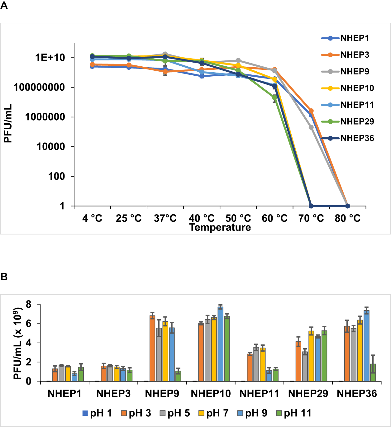
Stability assays were performed for all seven phages whose morphology was studied. All the phages exhibited complete stability as measured by viability without loss of titer at temperatures ranging from 4°C to 37°C. Stability decreased notably (by an order of magnitude) between 40°C and 60°C and reduced precipitously beyond 60°C. No viable phage could be recovered beyond 70°C in all cases (Fig. 3A). All the phages were completely stable at pH values ranging from 3 to 9. At pH 11, NHEP9, NHEP11 and NHEP36 titres were reduced by less than a log, while the other phages showed no reduction. No viable phages could be recovered at pH 1 (Fig. 3B).
Stability of phages at different temperatures and pH levels. Phage lysates were incubated at various temperatures (A) or in SM buffer adjusted to various pH levels (B) as indicated for 1 h. The mixtures were cooled immediately after incubation, and the titer (PFU/mL) was enumerated by plaque assay using the double-layer agar method. Each experiment was performed in triplicate. Data are presented as mean ± SD.

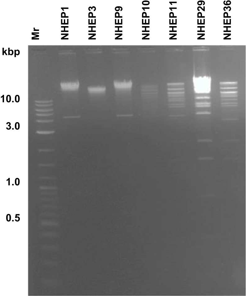
The same seven phages were subjected to RFLP with SmiI (SwaI), which revealed six distinct genotypic patterns. Phages NHEP1 and NHEP9 showed a similar pattern, whereas the genome of phage NHEP3 was not digested by the enzyme (Fig. 4).
Restriction fragment length polymorphism of phage DNA. Phage lysate was treated with DNase I and RNase A to eliminate host or released phage DNA and RNA, and the encapsidated phage DNA was extracted by phenol–chloroform method and acetate–alcohol precipitation. The phage DNA was dissolved in water, digested with SmiI (SwaI) enzyme for 16 h at 37°C and the products were visualized by agarose gel electrophoresis and UV transillumination.

The six phages showing unique RFLP patterns were subjected to whole-genome sequencing. A circular genome map of each phage is depicted in Fig. 5. All six phages possessed linear double-stranded DNA, with the genome size ranging from 44 to 169 kbp and GC content varying between 37.7% and 54.6%. The number of proteins encoded ranged from 67 to 280, while the tRNA count varied from 0 to 7 (Table S5). BLASTn analysis suggested that all six phages belonged to Class Caudoviricetes. The phage NHEP1 belonged to the Subfamily Vequintavirinae and Genus Vequintavirus, NHEP3 belonged to the Genus Dhillonvirus, and NHEP10, NHEP11, NHEP29 and NHEP36 belonged to the Family Straboviridae, Subfamily Tevenvirinae, and Genus Mosigvirus.
Genomic map of the phages. The genomes of the phages were subjected to paired-end sequencing and circular genomic organization was generated using the Proksee server. The outer circle (arrows depicted in blue), denotes predicted protein-coding sequences (CDS), while the inner circles (peaks/troughs) represent the GC content and skew.

The phylogenetic tree generated using conserved genes encoding the large terminase and the major capsid proteins supported the BLASTn analysis in that NHEP1 belonged to Vequintavirus, NHEP3 grouped under Dhillonavirus, and NHEP10, NHEP11, NHEP29 and NHEP36 belonged to Mosigvirus (Fig. 6A, B). Furthermore, classification by taxmyPHAGE analysis showed that all except NHEP1 could be new species in their respective genera (Fig. S2 A-F). It is worth noting that taxmyPHAGE can compare all phages classified by the International Committee on Taxonomy of Viruses (ICTV) and assigns genera and species using the ICTV cut-offs of 70% and 95% sequence identity, respectively. All the phages were predicted to follow a lytic life cycle, and genomic analysis did not reveal the presence of any virulence factors or AMR genes.
Phylogenetic relationship of the phages. The translated amino acid sequences of the large terminase subunit (A) and the major capsid protein (B) of our phages were aligned with sequences of the same proteins of related phages using Clustal W. Subsequently, maximum likelihood trees with 1,000 bootstraps were generated to depict the evolutionary relationships using MEGA X.

The six phages were then tested against 23 MDR and ESBL-producing E. coli isolates of poultry origin to evaluate whether the phages could target drug-resistant strains. These isolates had been obtained as part of another study and confirmed to be ESBL by phenotypic (AST with Panel 1 and 2) and genotypic (PCR) assays (data not shown). The results demonstrated that phage NHEP1 lysed 52% of the isolates, NHEP10 lysed 13%, NHEP11 lysed 9%, and NHEP29 lysed 17%. In contrast, NHEP3 and NHEP36 showed no lytic activity. These findings suggest that NHEP1 can potentially lyse MDR and ESBL E. coli strains (Table S6).
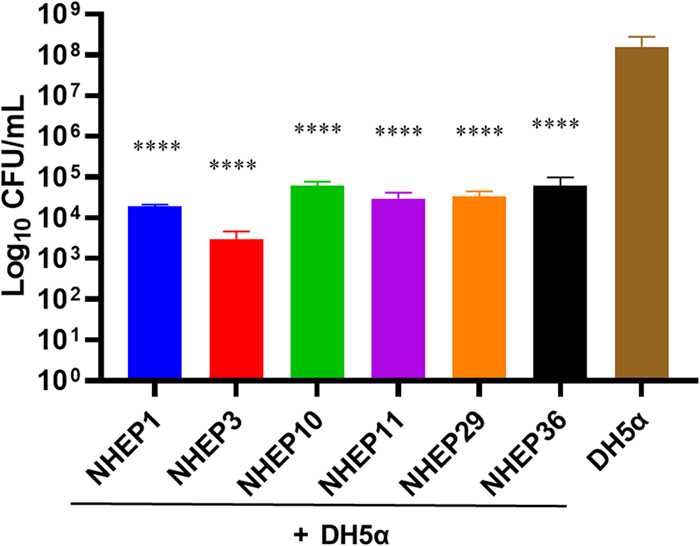
The antibacterial efficacy of the isolated phages was then evaluated in a milk matrix. The results using pasteurized milk demonstrated a significant reduction in bacterial load when treated with the bacteriophages. Specifically, a 3.5–4.5 log reduction in CFU/mL was observed with all the phages by 6 h of phage application. This reduction was consistent across multiple replicate experiments. Control samples (without phage treatment) showed no significant decrease in bacterial counts, confirming that the observed antibacterial activity was due to the bacteriophages (Fig. 7).
Assessment of phage activity in milk. Indicated phages (106 PFU) and bacteria (DH5α, 106 CFU) were incubated in 3 mL of pasteurized milk for 6 h at 37°C, followed by serial dilution and plating on LB agar. Colonies were counted after overnight incubation at 37°C. Error bars indicate SD compared to DH5α alone. *P-value < 0.05, **P < 0.01, ***P < 0.001, ****P < 0.0001. All the experiments were performed in triplicate technical samples and three independent biological replicates.

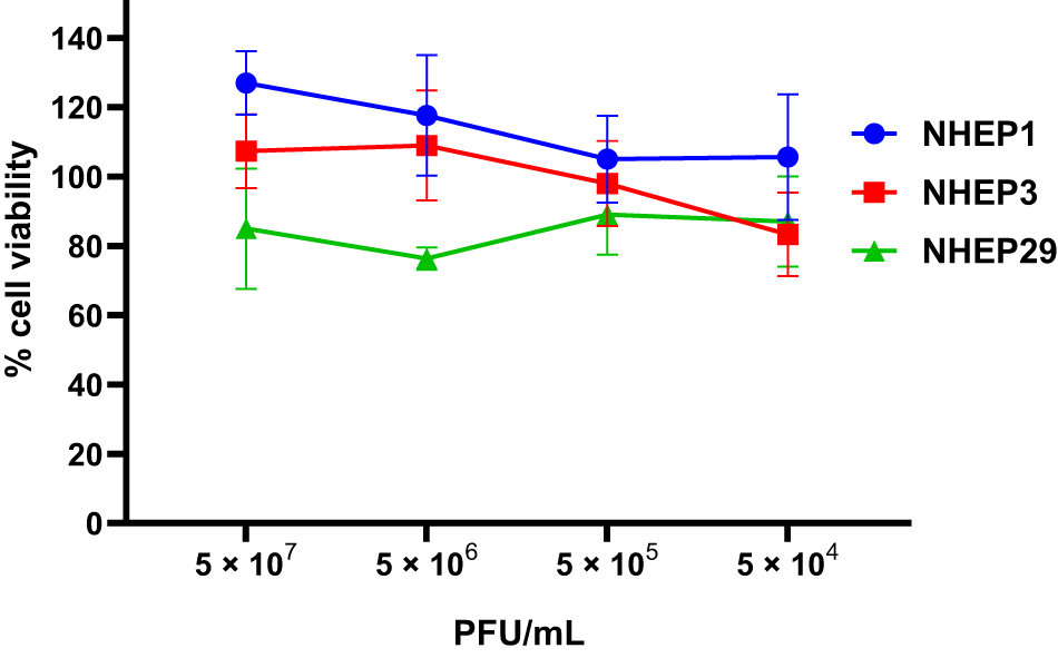
Three phages from each genus, NHEP1, NHEP3, and NHEP29, belonging to Venquintavirus, Dhillonvirus, and Mosigvirus genera, were then assessed for toxicity to mammalian cells. The results indicate that MDBK cells exposed to phages at concentrations of up to 5 × 10⁷ PFU/mL did not exhibit markedly different viability compared to untreated control cells. Whereas cells treated with NHEP1 and NHEP3 both displayed a slightly higher (18–27%) proliferation, cells incubated with NHEP29 showed reduced (15–24%) proliferation compared to untreated cells at the higher dose (5 × 10⁶ and 5 × 10⁷ PFU/mL) of phages. There was barely any difference in cell proliferation at the lower phage doses of 5 × 10⁵ and 5 × 10⁴ PFU/mL (Fig. 8).
Assessment of toxicity of phages to mammalian cells. MDBK cells were incubated with indicated concentrations (PFU/mL) of three different phages for 24 h. Cell viability was assessed by MTT assay, and absorbance values were recorded. All the values were normalized compared to untreated control cells, considered as 100% viability. TX-100 was used to achieve 100% cell death or lysis. Experiments were conducted in three independent trials, each with three technical replicates. Data are presented as mean + standard deviation.

Discussion
Bovine mastitis imposes a significant economic strain on the dairy sector, primarily due to bacterial pathogens. Dairying worldwide is dichotomous, with American, European, and some Asian nations adopting large-scale operations using high-yielding breeds of animals. In contrast, in many Asian and African countries, the operations are based on cooperative societies that pool milk from smallholder farmers who mostly rear animals with poor to moderate milk yield. Despite these dichotomies, the pathogens associated with intramammary infections are not vastly different. In addition, although the AMR profile of the organisms may differ from one region of the globe to another, it is evident that multidrug-resistant pathogens are circulating among humans and animals, with a high probability of horizontal genetic transfer of resistance determinants. Given this scenario, and considering that milk is the most widely consumed animal product worldwide, mitigating AMR in mastitis-associated pathogens is crucial for ensuring food security, public health, and socioeconomic development in the farming sector.
Broad-spectrum antibiotics are mainly used to treat bovine mastitis. However, the recent rise in resistance of pathogens to multiple antibiotics has prompted a reassessment of approaches to mitigating mastitis in the context of AMR. One such approach is the use of bacteriophages to infect and eliminate bacteria selectively. Bacteriophages operate independently of antibiotic resistance mechanisms. Many phages are also known to harbor depolymerase enzymes, aiding in breaking down biofilm, which is a mechanism for certain bacteria to resist the action of host anti-bacterial defenses and antimicrobials. However, phages are highly specific, to the extent that many phages are specific to certain strains of the same bacterial species. This would mean that a cocktail of multiple phages may be required to combat infections caused by a particular species of bacteria.
A major etiological agent of mastitis in cattle and buffaloes is E. coli. Several studies have focused on bacteriophages against bovine mastitis-associated S. aureus (reviewed in Anelopoulou et al., Reference Angelopoulou, Warda, Hill and Ross2019; El-Sayed and Kamel, Reference El-Sayed and Kamel2021; Li et al., Reference Li, Xu, Liang, Kastelic, Han, Tong and Gao2023; Nale and McEwan, Reference Nale and McEwan2023; Sharun et al., Reference Sharun, Dhama, Tiwari, Gugjoo, Iqbal Yatoo, Patel, Pathak, Karthik, Khurana, Singh, Puvvala, Amarpal, Singh and Chaicumpa2021), while very few studies have concentrated on coliphages. One study reported the utility of a single phage, showing several log reductions in bacterial count in pasteurized milk as early as two hours post-treatment (Imklin et al., Reference Imklin, Patikae, Poomirut, Arunvipas, Nasanit and Sajapitak2024). Another study showed that a cocktail of four phages was as effective as ceftiofur in vitro. The phage cocktail also decreased the bacterial load in raw milk and in buffer when emulsified to serve as a teat sealant and reduced the adhesion and survival of E. coli within eukaryotic cells (Porter et al., Reference Porter, Anderson, Carter, Donjacour and Paros2016). A third study reported the characterization of a single phage belonging to Tevenvirinae. This phage was stable between pH values of 4 and 12 and temperatures up to 62oC, and almost entirely labile at pH 2. The phage reduced bacterial load in a mouse model of mastitis caused by a mammary pathogenic E. coli strain (da Silva Duarte et al., Reference da Silva Duarte, Dias, Kropinski, Campanaro, Treu, Siqueira, Vieira, da Silva Paes, Santana, Martins, Crispim, da Silva Xavier, Ferro, Vidigal, da Silva and de Paula2018). A fourth study reported the isolation of three coliphages with a broad host spectrum. The phages lost viability significantly at temperatures between 60oC and 70oC and pH below 5 or above 10. A cocktail of all three phages was equivalent to antibiotic treatment in experimentally induced mastitis in cows (Guo et al., Reference Guo, Gao, Xue, Liu, Zeng, Cheng, Ma, Wang, Sun, Wang and Yan2021). A recent study also demonstrated the efficacy of seven phages in combination against S. aureus, E. coli, and P. aeruginosa in disinfecting the bacteria in the dairy environment (Xue et al., Reference Xue, Gao, Guo, Zhang, Zhao, Xia, Ma, Cheng, Wang, Sun, Wang and Yan2024). Another recent study reported the isolation and characterization of three phages against 96 colistin-resistant E. coli strains isolated from livestock and poultry (anal swabs and faeces) and their environment (soil and sewage). One phage that was extensively characterized belonged to the Dhakavirus genus of the Straboviridae family and did not carry genes for any virulence determinant. A combination of the phages was effective in rapidly and completely eradicating planktonic bacteria and eliminating preformed biofilms (Zhu et al., Reference Zhu, Xiao, Jia, Ni, Zhang, Fang and Hao2024). Studies from India have also reported on the isolation and limited in vitro characterization of phages against mastitis-associated E. coli (Badiyal et al., Reference Badiyal, Dhial, Singh, Dhar, Sharma and Verma2024; Mariramkumar, 2019; Pal, Reference Pal2023). A metagenomic study of bovine milk from indigenous and crossbred cows from India reported the presence of genetic sequences representing phages targeting S. aureus, Enterobacteria, Escherichia, and Yersinia (Bhatt et al., Reference Bhatt, Ahir, Koringa, Jakhesara, Rank, Nauriyal, Kunjadia and Joshi2012); however, no phages were isolated. Overall, these reports and our studies support the need for further investigations on expanding the repertoire of phages against E. coli and their characterization to develop therapeutics for bovine mastitis.
The bacteriophages we characterized could lyse E. coli isolates of mastitis origin to varying degrees, ranging from 0% to 62%. This heterogeneity is not unexpected, and it is also possible that some of the bacterial strains may develop resistance to specific phages. Considering this, we characterized in detail more than half a dozen phages in this report. Whereas the phages were originally obtained using host strains that were susceptible to several antibiotics, one phage (NHEP1) was later found to lyse more than half of the 23 MDR-ESBL isolates from our collection, suggesting that this phage could be evaluated against a broader spectrum of isolates. The availability of broad-spectrum phages could facilitate a shift towards applying either a phage-alone approach or an integrated therapy, where the treatment with phages can be combined with antibiotics or other antimicrobials, simultaneously or sequentially.
Understanding the genotypic and phenotypic properties of phages is the foundation for translating laboratory research into therapeutic applications. First, therapeutic phages are preferred to be lytic and not carry any detrimental virulence determinants. Genomic analysis revealed that our six sequenced phages were lytic and lacked toxin or AMR genes, suggesting a safe profile. However, understanding the dynamics of phage replication is crucial for choosing an appropriate phage. The variation we observed in the latent periods and the burst sizes among different phages suggests heterogeneity in their life cycles. Second, any phage projected for therapeutic use should be stable under varying environmental conditions, which can benefit applications under various physiological conditions during therapy and in situations that suit its dispatch and transport. The phages we studied were stable at a wide range of temperatures and pH conditions under controlled laboratory conditions, but need to be tested under various environmental conditions. Third, owing to the narrow spectrum of phages, it is essential to be prepared by establishing a collection of phages that could be used against many bacterial strains isolated from various hosts. Apart from mastitis, most of our phages lysed poultry-origin E. coli, demonstrating the versatility of our collection's small number of phages. Given the widespread prevalence of pathogenic E. coli strains in various diseases across humans and animals, applying these phages could extend beyond mastitis treatment (Oliveira et al., Reference Oliveira, Dias, Oliveira, Almeida, Fucinos, Sillankorva and Oliveira2024). However, since the emergence of resistance to specific phages is a common phenomenon, a more extensive collection of characterized phages is warranted. Fourth, the phages must be non-toxic and non-immunogenic. The six phages we tested did not affect the viability of MDBK cells. Finally, the phages must be tested in animal models and real-world scenarios. In this context, our results of phage activity in milk represent a first step and demonstrate that the phages could be formulated for infusion into the udder to treat bovine mastitis.
In conclusion, the isolation and characterization of bacteriophages targeting mastitis pathogens present a promising approach for addressing the challenges associated with AMR. The diverse origins, host range, growth kinetics, morphology, and genotypes of the isolated phages provide valuable information for a comprehensive understanding of their potential therapeutic applications. Further research, including in vivo studies in model organisms and animal clinical trials, will be crucial to validate their efficacy and safety. The findings from this study contribute to the growing body of knowledge supporting the exploration of bacteriophage therapy as a viable alternative for managing bovine mastitis. This underscores the potential of bacteriophages in a practical setting to reduce bacterial loads in the mammary gland, milk, dairy, and processing environments – a crucial factor in ensuring food safety and safeguarding public health by mitigating AMR.
Supplementary material
The supplementary material for this article can be found at https://doi.org/10.1017/S0022029925101349.
Acknowledgements
This work was primarily supported by a Core Research Grant from the National Institute of Animal Biotechnology and partially by an extramural grant (BT/PR54578/BSA/33/304/2024) from the Department of Biotechnology, Ministry of Science and Technology, Government of India, to NRH. SP is supported through a PhD fellowship from the University Grants Commission, Ministry of Education, Government of India. We thank Dr Shrikrishna Isloor, Professor and Head, Department of Veterinary Microbiology, Karnataka Veterinary Animal and Fisheries Sciences University, Bangalore, and Dr Pankaj Suman, National Institute of Animal Biotechnology, Hyderabad, for providing some of the bacterial isolates. The MDR-ELBL isolates were obtained as part of an extramural grant (BT/IN/Indo-UK/AMR/05/NH/2018-19) from the Department of Biotechnology, Ministry of Science and Technology, Government of India, to NRH.