Introduction
Mosquitoes are blood-sucking insects that can transmit many mosquito-borne diseases such as malaria, dengue fever, Zika, haemorrhagic fever, encephalitis and meningitis. They are responsible for infecting hundreds of millions of people and a large number of deaths each year (Liang et al., Reference Liang, Gao and Gould2015; Benelli, Reference Benelli2016; Pastula et al., Reference Pastula, Smith, Beckham and Tyler2016; Zhu et al., Reference Zhu, Zhang, Zhang, Zhao, Wang, Liang and Cheng2017). The World Health Organization (WHO) reported that in 2016, there were 216 million cases of malaria and 445000 deaths across 91 countries (WHO, 2017). The worldwide incidence of dengue fever has risen sharply in recent decades, and 390 million people get infected with dengue fever each year (Bhatt et al., Reference Bhatt, Gething, Brady, Messina, Farlow, Moyes, Drake, Brownstein, Hoen, Sankoh, Myers, George, Jaenisch, Wint, Simmons, Scott, Farrar and Hay2013). Culex pipiens pallens is the most abundant and widely distributed species of mosquito in China. This species is the main blood-sucking mosquito species in northern China and is primarily responsible for West Nile fever (Jiang et al., Reference Jiang, Wang, Guo, Zhang, Li, Dong, Xing and Zhao2014), bancroftian filariasis, and type B encephalitis (Cui et al., Reference Cui, Li, Zhao and Zou2013). Recently, a number of studies of C. pipiens pallens resistance to insecticides, especially pyrethroid insecticides, have been published (Liu et al., Reference Liu, Zhang, Qiao, Lu and Cui2011; Wang et al., Reference Wang, Li, Xing, Yu, Liu, Xue, Dong and Zhao2012).
There is an urgent need to devise effective methods of mosquito control to reduce the incidence and spread of these diseases. Chemical control is efficient and convenient for large-scale applications (Ma et al., Reference Ma, Li, Hu, Zhou, Sun, Ma, Zhu and Shen2017). In recent years, deltamethrin has been commonly used for indoor residual spraying and in insecticide-treated nets due to its low mammalian toxicity and high efficiency (White et al., Reference White, Lwetoijera, Marshall, Caron-Lormier, Bohan, Denholm and Devine2014). However, mosquitoes are gradually acquiring deltamethrin resistance, which has become a major obstacle in the control of mosquito-borne disease (Benelli, Reference Benelli2015; Siegwart et al., Reference Siegwart, Graillot, Blachere Lopez, Besse, Bardin, Nicot and Lopez-Ferber2015). There are three main causes for the development of resistance: metabolic resistance due to increased enzymatic activity, target-site resistance due to altered target protein bound to the pesticide, and cuticular resistance caused by decreased epidermal penetration (Hemingway et al., Reference Hemingway, Hawkes, McCarroll and Ranson2004), with studies showing the importance of metabolic resistance (Zhang et al., Reference Zhang, Meng, Yang, Li, Wang, Yang, Zhang, Li, Millar and Liu2016). Cytochromes P450 are often shown to be vital for the detoxification of pesticides in the metabolic resistance mechanism. A number of P450 genes have been reported to be overexpressed in resistant mosquitoes (Guo et al., Reference Guo, Huang, Zou, Liu, Tian, Ye, Guo, Sun, Zhou, Sun, Ma, Shen and Zhu2017). Recent studies on cytochromes P450 have focused on transcriptional regulation, but few have studied the post-transcriptional regulation.
MicroRNAs (miRNAs) are a group of non-coding single-stranded RNA molecules that are widely found in animals and plants. MiRNAs are encoded by endogenous genes and are approximately 22 nucleotides (nt) in length (Carthew, Reference Carthew2006). They negatively regulate gene transcription levels via base pairing with target sites in messenger RNA (mRNA) (Lucas and Raikhel, Reference Lucas and Raikhel2013; Ding et al., Reference Ding, Wang, Yang, Li, Cheng, Wang, Wang, Deng, Yang, Hua, Zhao and Fang2015; Sebastiani et al., Reference Sebastiani, Po, Miele, Ventriglia, Ceccarelli, Bugliani, Marselli, Marchetti, Gulino, Ferretti and Dotta2015). Generally, the miRNA ‘seed sequence’ (nt 2–8 at the 5′-end) binds to a complementary seed-matching site within the 3′- or 5′-untranslated regions of the mRNA, resulting in translational inhibition or mRNA degradation (Lucas and Raikhel, Reference Lucas and Raikhel2013; Kanokudom et al., Reference Kanokudom, Vilaivan, Wikan, Thepparit, Smith and Assavalapsakul2017; Lee et al., Reference Lee, Yeh, Ruan, Zhang, Hsu, Huang, Hsieh, Lin, Yeh, Liu and Gan2017). Previous studies on miRNAs and their role in deltamethrin resistance have found that miR-932 and miR-285 regulate deltamethrin resistance by targeting the cytochromes P450 family (Liu et al., Reference Liu, Tian, Guo, Ma, Zhou, Shen, Sun and Zhu2016; Tian et al., Reference Tian, Liu, Hu, Li, Guo, Zou, Liu, Hu, Guo, Ma, Zhou, Sun, Shen and Zhu2016). The miR-2~13~71 cluster (miR-2, miR-13 and miR-71) has been reported to be involved in deltamethrin resistance in mosquitoes (Guo et al., Reference Guo, Huang, Zou, Liu, Tian, Ye, Guo, Sun, Zhou, Sun, Ma, Shen and Zhu2017). In the previous research in our laboratory (Hong et al., Reference Hong, Guo, Wang, Hu, Fang, Lv, Yu, Zou, Lei, Ma, Ma, Zhou, Sun, Zhang, Shen and Zhu2014), which profiled miRNAs in a deltamethrin-resistant (DR) strain of C. pipiens, we identified miR-13664 to be highly expressed using Solexa high-throughput sequencing and miRDeep predictive analysis. However, the role of miR-13664 in insecticide resistance in mosquitoes remains unclear.
In the present study, we validated the role of miR-13664 in the DR strain by demonstrating that oversupply of miR-13664 decreased the transcript levels of a putative target gene cytochromes P450 314A1 (CpCYP314A1) in the DR strain. These results indicate that miR-13664 may be involved in the mechanism of pyrethroid resistance by regulating CpCYP314A1 expression.
Materials and methods
Mosquito strains
Two strains of C. pipiens pallens were used: the deltamethrin-sensitive (DS) strain (50% lethal concentration of deltamethrin LC50 = 0.04 mg L−1) and the DR strain (LC50 = 3.42 mg L−1). The DS strain was collected in the field from Tangkou in Shandong Province, and maintained in our laboratory without exposure to any insecticides. The DR strain was screened by culturing the field strains obtained from Tangkou for 80 generations. Prior to screening, the LC50 concentration was measured by larval impregnation, and the LC50 concentration was selected as the screening concentration for each subsequent generation. Deltamethrin solution was used immediately after preparation. In brief, the prepared deltamethrin solution was poured into a 1000 mL porcelain bowl, and approximately 1000 fourth instar larvae were placed in. After 24 h, the surviving larvae were transferred and reared in fresh dechlorinated water. Judgement of dead larvae: gently touching the larvae, those that did not move were considered as dead. To obtain DR strain, DS strain larvae were selected and exposed constantly to deltamethrin (LC50 concentration) and screened over 80 generations. All laboratory strains were fed at 28–30 °C at a relative humidity of 70–80% and with a 16 h light/8 h dark cycle. All adult mosquitoes were fed 10% glucose.
In addition, We collected mosquito larvae in the fields of Pingyin, Shanghe and Jining and raised them to adult mosquitoes in the laboratory. The feeding conditions were the same as above. After 3 days post-adult emergence, the female mosquitoes were selected and measured with WHO discriminating dose (0.05% deltamethrin film) according to standard WHO cones test. The mosquitoes were exposed to the film for 60 min. After 24 h recovery in the recovery cylinder, knocked down group were served as field-sensitive strains [field deltamethrin-sensitive (FDS)] and the survival group were served as field-resistant strain [field deltamethrin-resistant (FDR)].
RNA extraction and cDNA synthesis
According to the manufacturer's protocols, total RNA extraction was performed from pools of 10 individuals from adult female DR and DS mosquitoes at 3 days post-eclosion using TRIzol reagent (TaKaRa, Japan). RNA integrity was assessed by 1% agarose gel electrophoresis. The quality and quantity of the extracted RNA were determined using a Thermo Scientific™ NanoDrop 2000 spectrophotometer (NanoDrop, Wilmington, DE, USA). cDNAs were synthesized from mRNAs using a PrimeScript™ RT Master Kit (TaKaRa) and stem–loop primers were added using stem–loop method to synthesize cDNA from miRNA using the PrimeScript RT Kit (TaKaRa) according to the manufacturer's protocols, respectively (Tang et al., Reference Tang, Hajkova, Barton, Lao and Surani2006).
Quantitative reverse-transcriptase polymerase chain reaction analysis
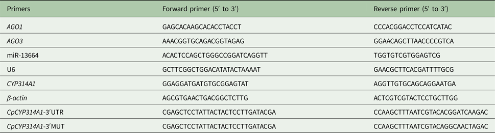
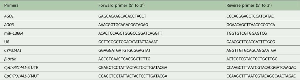
Quantitative reverse-transcriptase polymerase chain reaction (qRT-PCR) was carried out on a LightCycler® 96 instrument (Roche, Switzerland) based on the SYBR Green method, according to EvaGreen 2 × qPCR Master Mix-No Dye (ABM, Canada) protocols. The total reaction system consisted of a 20 µL mixture containing 10 µL of EvaGreen 2 × qPCR Master Mix-No Dye, 1 µL each of upstream and downstream primers, and 8 µL of cDNA. The relative expression levels of CpCYP314A1 and miR-13664 were calculated and compared with the expression levels of β-actin or U6 small nuclear RNA, respectively, which were used as the internal reference standards. Gene-specific primers were designed by Primer Premier 5.0 software and are listed in Table 1. The miRNA expression levels were calculated using the stem–loop RT-PCR method (Salone and Rederstorff, Reference Salone and Rederstorff2015). Assays were conducted in triplicate and repeated three times. Expression levels were calculated using 2−Δ Δct (Schmittgen and Livak, Reference Schmittgen and Livak2008) with the transcript level in DS strain set as 1.
List of primers for qRT-PCR and vector constructs
Plasmid construction and luciferase assay
The 3′-UTR region of CpCYP314A1 contained a potential miR-13664-binding site (CGGATCA). Two pairs of primers (Table 1) were designed to amplify this region with one pair introducing a mutation into this binding site. Primers were designed according to the transcript sequences of Culex quinquefasciatus (CPIJ001380) to amplify a 100 bp region upstream of CpCYP314A1. The 3′-UTR primer pair amplifies the native sequence, while in the 3′-UTR-MUT primer pair, four bases were altered (GGCAACT) in the seed region. The region of the CpCYP314A1 3′-UTR (CpCYP314A1 3′-UTR-WT) and mutant 3′-UTR (CpCYP314A1 3′-UTR–MUT) were PCR amplified from C. pipiens pallens cDNA. PCR was executed using the following cycle protocol: 94 °C for 5 min; 35 cycles of 94 °C for 30 s, 52 °C for 30 s, and 72 °C for 30 s; with a final step of 72°C for 7 min. The amplified PCR products were electrophoresed on 2% agarose gel, purified using a MiniBEST Agarose Gel DNA Extraction Kit Ver.4.0 (TaKaRa), and sequenced by Invitrogen.
The 3′-UTR-WT and 3′-UTR-MUT of CpCYP314A1 were cloned into pMIR-Report miRNA report vector using SacI and HindIII. MiR-13664 mimics were manufactured by GenePharma (Gene-Pharma, Shanghai, China). The negative control (NC) was an unrelated sequence designed according to other species, which would not interfere with the target genes in our study and were synthesized by GenePharma. HEK-293T cells were seeded in 24-well plates at a concentration of 1 × 10⁵ cells per well and incubated for 24 h. The recombinant plasmids (pMIR-UTR and pMIR-MUT) and miRNA mimics or NC were transfected into cells using FuGENE® HD Transfection Reagent (Promega, Madison, WI, USA). Briefly, the recombinant plasmids, miRNA mimics/NC and cell culture medium were gently mixed and incubated for 15 min at room temperature. The mixture was gently spread onto cells. After transfection for 48 h, the cells were lysed and subjected to luciferase assay that was implemented using the Dual-Luciferase Reporter Assay System (Promega) (Guo et al., Reference Guo, Huang, Zou, Liu, Tian, Ye, Guo, Sun, Zhou, Sun, Ma, Shen and Zhu2017).
Microinjection
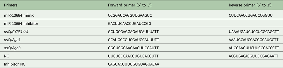
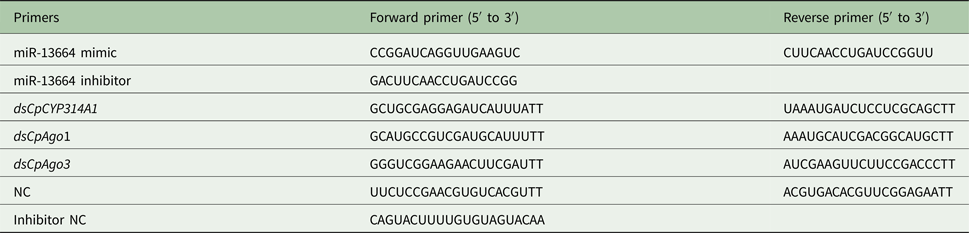
The miR-13664 mimic/inhibitor and dsRNAs of CpArgonaute 1 (CpAgo1), CpArgonaute 3 (CpAgo3) and CpCYP314A1 genes were procured from GenePharma (Table 2). The NC was designed to contain the same amount of scrambled small RNA (GenePharma; Table 2). One-day-old non-blood-fed female mosquitoes of both the DS and DR strains were anaesthetized on ice and then microinjected in the thorax. According to the standard methodology (Blandin et al., Reference Blandin, Moita, Kocher, Wilm, Kafatos and Levashina2002), the DR strain received injection of dsRNA against CpCYP314A1 or miR-13664 mimic (0.07 uL, 333 µm), while the DS strain received injection of miR-13664 inhibitor, CpAgo1 or CpAgo3. Mosquitoes that received an equivalent volume of diethylpyrocarbonate-treated water or NC were used for the two controls. Each group had three biological replicates. Three days after injection, total RNA was extracted from the injected female mosquitoes, and the efficiency of microinjection was verified by qRT-PCR. The Centers for Disease Control and Prevention (CDC) bottle bioassays were performed to test the mosquito's resistance to deltamethrin.
List of the miR-13664 mimic/inhibitor and dsCpCYP314A1, dsCpAgo1 and dsCpAgo3 primers
CDC bottle bioassay
CDC bottle bioassay is used as a monitoring tool to detect pesticide resistance (Aizoun et al., Reference Aizoun, Aikpon, Padonou, Oussou, Oke-Agbo, Gnanguenon, Ossè and Akogbeto2013). According to the laboratory protocol (Guo et al., Reference Guo, Huang, Zou, Liu, Tian, Ye, Guo, Sun, Zhou, Sun, Ma, Shen and Zhu2017), deltamethrin was dissolved in acetone to obtain a concentration of 10 mg mL−1 and diluted to a final concentration of 7.5 mg mL−1 for the DR strain and 2 µg mL−1 for the DS strain. Then, 1 mL of liquid was evenly applied to the surface of each experimental bottle. Another bottle coated with the same volume of acetone served as the control. For each bioassay, approximately 20 microinjected mosquitoes per bottle were exposed to deltamethrin. Then, mosquito mortality was recorded every 15 min for 2 h or until all the mosquitoes were dead. Each experiment was performed three times, with three biological replicates each.
Statistical analysis
In this study, statistical calculations were performed using SPSS (Version 20.0). Differences between populations were statistically analysed by the Student's t test (all values were represented as means ± s.d.). The χ 2 test was used to assess differences among the three independent experiments. A P value of <0.05 was considered statistically significant.
Results
Identification of the novel miRNA miR-13664
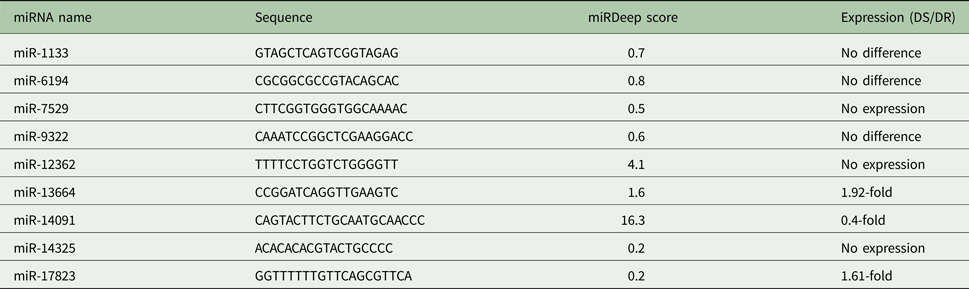
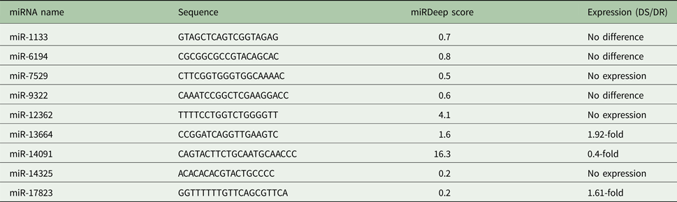
Based on the results of the Solexa high-throughput sequencing data obtained in a previous study in our laboratory (Hong et al., Reference Hong, Guo, Wang, Hu, Fang, Lv, Yu, Zou, Lei, Ma, Ma, Zhou, Sun, Zhang, Shen and Zhu2014), we used miRDeep prediction methods to identify novel miRNAs (Friedländer et al., Reference Friedländer, Chen, Adamidi, Maaskola, Einspanier, Knespel and Rajewsky2008). Briefly, nine miRNAs with relatively high scores from the miRNAs obtained from the miRDeep prediction analysis were selected, and their relative expression levels in DS and DR mosquitoes were analysed using quantitative PCR (Table 3). The results of the analysis revealed a significant difference in miR-13664 expression between the DS and DR strains; therefore, we chose to study the relationship between miR-13664 and deltamethrin resistance.
Relative expression levels of nine miRNAs with relatively high scores from the miRNAs obtained from the miRDeep prediction analysis in the DS and DR strains mosquitoes
Argonaute 1 is essential for miR-13664 biosynthesis
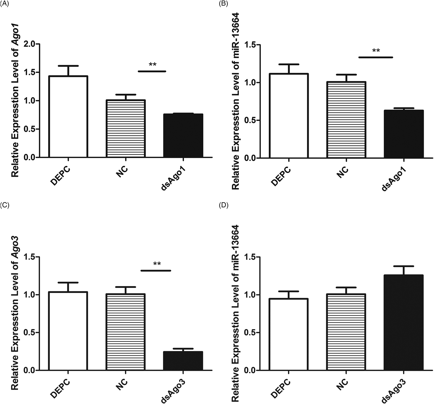
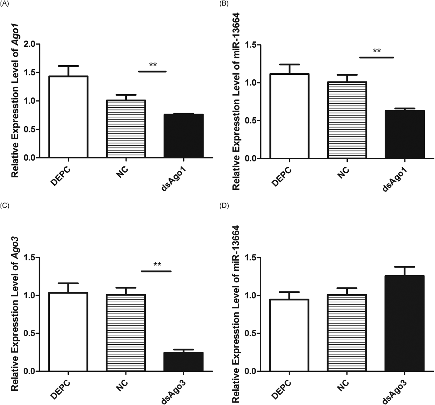
Argonaute 1 (Ago1) has been found to be involved in the miRNA pathways in insects (Hussain et al., Reference Hussain, O'Neill and Asgari2013; Lewis et al., Reference Lewis, Salmela and Obbard2016; Fu et al., Reference Fu, Dimopoulos and Zhu2017). Argonaute 3 (Ago3) is known to participate in the piRNA pathway (Campbell et al., Reference Campbell, Black, Hess and Foy2008; Luo and Lu, Reference Luo and Lu2017; Rogers et al., Reference Rogers, Situ, Perkins and Toth2017). We found that the miR-13664 transcript levels were 38% less in dsCpAgo1-injected mosquitoes compared with the NC control (Fig. 1A and B), whereas knockdown of dsCpAgo3 did not alter the expression of miR-13664 (Fig. 1C and D), indicating that Ago1 is necessary for the biosynthesis of the novel miRNA identified in this study, miR-13664.
MiR-13664 abundance is influenced by Ago1. (A) Ago1 transcript levels after dsCpAgo1 injection. (B) MiR-13664 abundance level after dsCpAgo1 injection. (C) Ago3 transcript level after dsCpAgo3 injection. (D) MiR-13664 abundance levels after dsCpAgo3 injection. Data are presented as means ± standard error (s.e.) of three independent experiments. **P < 0.01 compared with the negative control (NC).
Increased miR-13664 abundance in the laboratory and field-collected DS strains
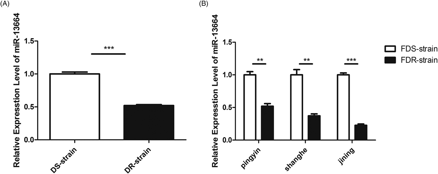
To verify the importance of miR-13664 in deltamethrin resistance in C. pipiens pallens, we used stem–loop qRT-PCR to verify the miR-13664 abundance in female DS and DR mosquitoes. As shown in Fig. 2A, miR-13664 expression was 1.92× higher in the laboratory DS strain than in the DR strain. To further analyse miR-13664 abundance in the field-collected C. pipiens pallens population, Pingyin, Shanghe and Jining strains were used in our experiment. As shown in Fig. 2B, the miR-13664 expression levels in FDS strains from Pingyin, Shanghe and Jining were 1.93-, 2.70- and 4.42-fold higher than the corresponding FDR strains from the same regions, suggesting that miR-13664 is related to deltamethrin resistance.
MiR-13664 abundance levels in the DS and DR strains of Culex pipiens pallens. (A) Laboratory mosquito populations. (B) Field-collected mosquito populations. Data are presented as means ± s.e. of three independent experiments. ***P < 0.001 compared with the DS strain.

MiR-13664 regulates deltamethrin resistance in mosquitoes
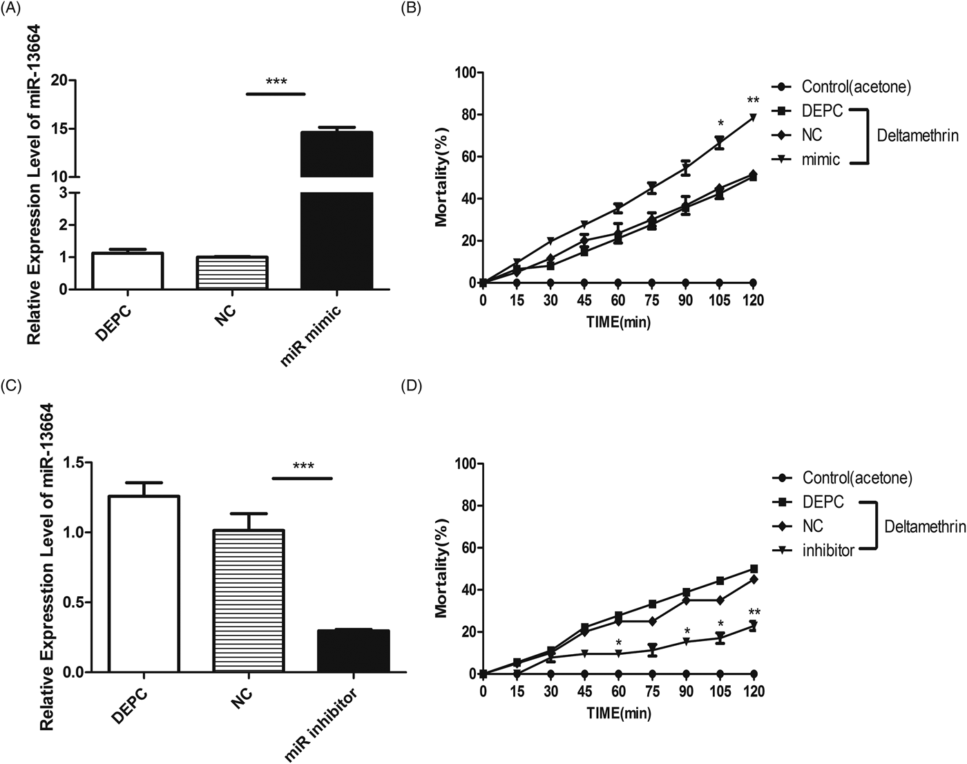
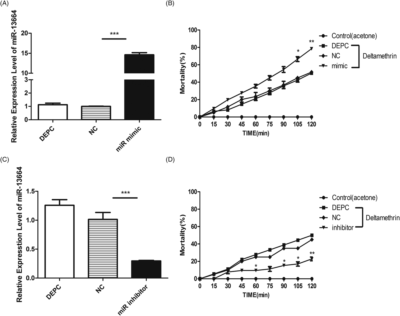
To investigate the role of miR-13664 in insecticide resistance, female DR and DS mosquitoes received thoracic microinjections of miR-13664 mimics and inhibitors 1 day after eclosion. After 3 days of recovery, the CDC bottle bioassay was conducted after microinjection and the miR-13664 expression level in the mosquitoes was determined by qRT-PCR to verify the injection efficiency (Fig. 3A and C). MiR-13664 oversupply in the DR strain was found to reduce the resistance of mosquitoes to deltamethrin (Fig. 3B), while knockdown of miR-13664 in the DS strain increased resistance to deltamethrin (Fig. 3D). Together, these results indicate that miR-13664 may be a potential regulator of insecticide resistance in C. pipiens pallens.
MiR-13664 is involved in mediating mosquito resistance to deltamethrin. (A) MiR-13664 abundance levels after miR-13664 mimic injection. (B) Mortality of mosquitoes exposed to deltamethrin in CDC bottles after 72 h by injection of miR-13664 mimic. (C) MiR-13664 abundance levels after miR-13664 inhibitor injection. (D) Mortality of mosquitoes exposed to deltamethrin in CDC bottles after 72 h by injection of miR-13664 inhibitor. Data are presented as mean ± s.e. of three independent experiments. *P < 0.05, **P < 0.01, ***P < 0.001.
MiR-13664 target gene prediction and identification
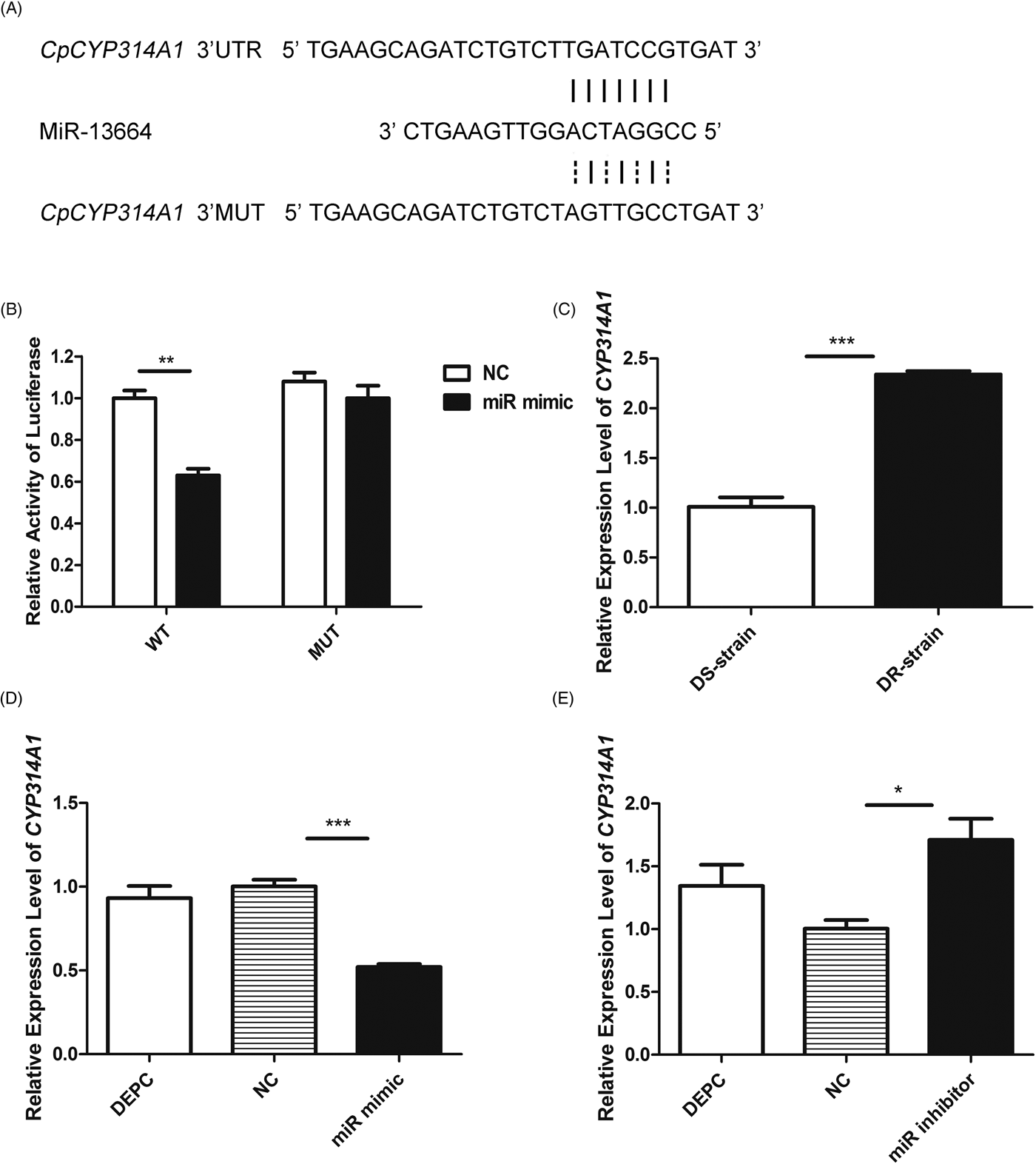
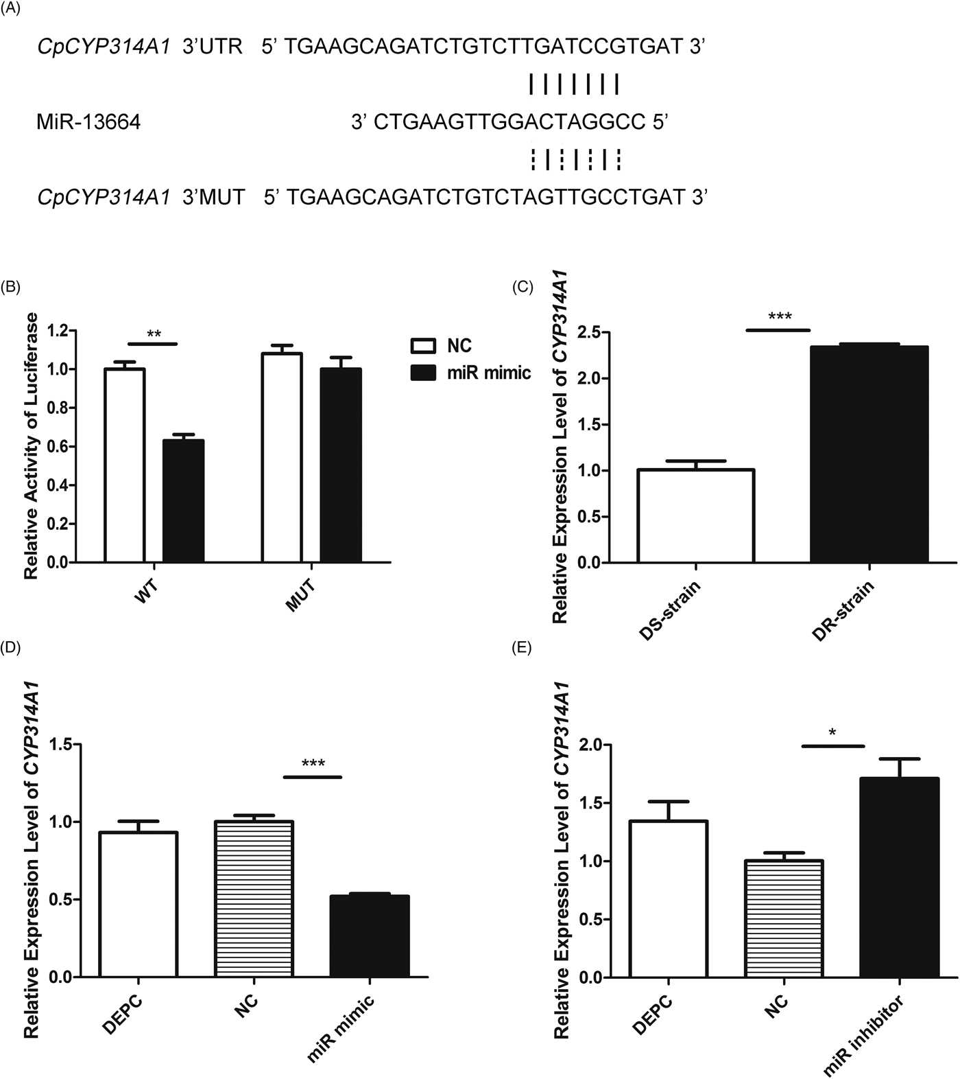
Mature miRNAs interact with the 3′-UTR or 5′-UTR to modulate target mRNA (Carthew, Reference Carthew2006). To identify the miRNA target gene in C. pipiens pallens, we aligned the 2–8 complementarily paired nt at the 5′-end of miR-13664 with the known mRNAs of C. quinquefasciatus (Yan et al., Reference Yan, Yang, Jiang, Cui, Ma, Qiao and Cui2012). A potential binding site for miR-13664 was found in the CpCYP314A1 3′-UTR region (Fig. 4A). Then, we measured the CpCYP314A1 transcript level in the DS and DR strains. Unlike the miR-13664 expression level, CpCYP314A1 expression was 2.32-fold higher in the DR strains than in the DS strains (Fig. 4C). MiR-13664 mimics induced a decrease in the CpCYP314A1 mRNA levels, while miR-13664 inhibitors increased their abundance (Fig. 4D and E). These results suggest that CpCYP314A1 may be the target gene of miR-13664. To confirm this, plasmids were inserted with the CpCYP314A1 3′-UTR and MUT of designated CpCYP314A1–3′-UTR (WT) and CpCYP314A1-3′-MUT (MUT), respectively, then cotransfected with the pGL4.7 control plasmid into HEK293T cells together with miR-13664 mimics or NC. Results of the luciferase assay revealed that miR-13664 reduced CpCYP314A1-3′-UTR expression by 37%, but did not affect the expression levels of CpCYP314A1-3′-MUT relative to the levels in the NC (Fig. 4B). The above results provide further evidence that CpCYP314A1 is the direct target of miR-13664.
CpCYP314A1 is a direct target of miR-13664. (A) The miR-13664 binding in the 3′-UTR of CpCYP314A1 mRNA and the constructed mutated vector sequence. (B) Luciferase activity in HEK293T cells cotransfected with CpCYP314A1-3′-UTR (WT) or mutant CpCYP314A1-3′-MUT (MUT) construct together with miR-13664. (C) CpCYP314A1 transcript levels in the DS and DR strains of Culex pipiens pallens. (D) CpCYP314A1 transcript levels after injection with miR-13664 mimic. (E) CpCYP314A1 transcript levels after miR-13664 inhibitor injection. Luciferase activity was assayed in HEK293T cells cotransfected with CpCYP314A1-3′-UTR (WT) or the mutant CpCYP314A1-3′-MUT (MUT) construct with miR-13664. Data are presented as means ± s.e. of three independent experiments. *P < 0.05, **P < 0.01, ***P < 0.001.
CpCYP314A1 plays a role in deltamethrin resistance of mosquitoes
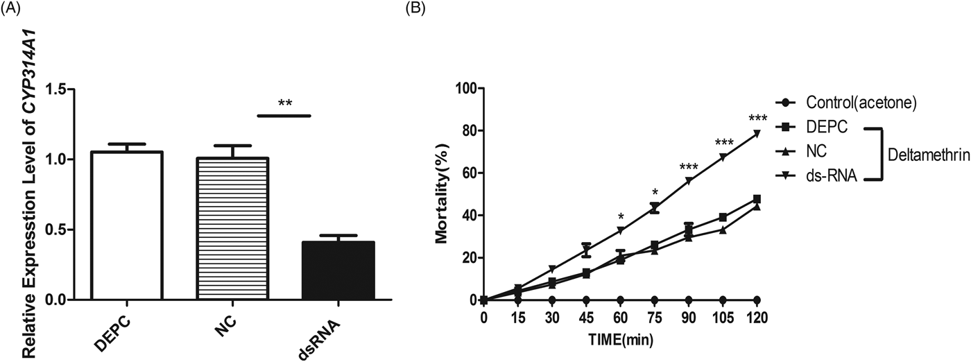
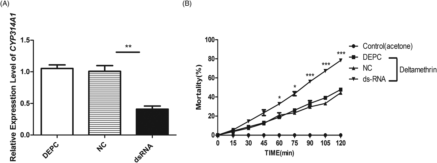
We microinjected DR strain mosquitoes with CpCYP314A1 dsRNA, performed a CDC bottle bioassay and determined the CpCYP314A1 expression levels by qRT-PCR to verify injection efficiency. The qRT-PCR results showed that knockdown efficiency of CpCYP314A1 was 59% compared with the NC (Fig. 5A). CDC bottle bioassays showed that knockdown of CpCYP314A1 can reduce the resistance of mosquitoes to deltamethrin (Fig. 5B). As shown in Fig. 5B, the mortality in the knockdown group was increased by 11.83, 20.03, 26.5, 33.97 and 33.97% at 60, 75, 90, 115 and 120 min, respectively, compared with the control group.
CpCYP314A1 affects deltamethrin resistance in DR mosquitoes. (A) Expression levels of CpCYP314A1 after microinjection with CpCYP314A1 dsRNA. (B) Microinjected mosquito mortality observed after exposure to deltamethrin in CDC bottles for 2 h. Data are presented as the means ± s.e. of three independent experiments. *P < 0.05, **P < 0.01, ***P < 0.001.
Discussion
The vast majority of miRNAs originate in the endogenous locus and are transcribed into primary miRNA (pri-miRNA) transcripts by RNA polymerase II. The classical miRNA pathway converts pri-miRNA hairpins into approximately 22 nt miRNAs by sequential cleavage with the help of two dsRNA-binding proteins by two RNase III enzymes, Drosha and Dicer. One of the strands of a mature duplex miRNA is loaded onto the Argonaute protein, which is responsible for the activity of the small RNA, to form the ribonucleoprotein complex miRNA-mediated silencing complex (miRISC). The bound strand in miRISC is called the guide strand, while the unbound strand that is subsequently degraded is called the passenger strand. The guide strand directs miRISC to the mRNA target via incomplete base pairing, resulting in decreased gene expression through mRNA destabilization and/or translational inhibition (Chorostecki et al., Reference Chorostecki, Moro, Rojas, Debernardi, Schapire, Notredame and Palatnik2017; Fu et al., Reference Fu, Dimopoulos and Zhu2017). Ago1 is reported to be involved in miRNA pathways (Hussain et al., Reference Hussain, O'Neill and Asgari2013; Lewis et al., Reference Lewis, Salmela and Obbard2016; Fu et al., Reference Fu, Dimopoulos and Zhu2017). In our study, we found that miR-13664 is involved in deltamethrin resistance and that the miR-13664 expression level is decreased following microinjection of mosquitoes with dsCpAGO1, but microinjection of mosquitoes with dsCpAGO3 did not alter miR-13664 expression. Therefore, we predicted miR-13664 to be an miRNA.
In recent years, miRNA has become the focus of life science research. In humans, chemoresistance mechanisms are closely related to miRNA regulation (Han and Chen, Reference Han and Chen2015; Chen et al., Reference Chen, Cao and Feng2017; Li et al., Reference Li, Zeng, Hu, Yan, Liu and You2017). As important post-transcriptional regulators, miRNAs themselves are regulated by other genes. Studies have found numerous binding sites for transcription factors in the promoter regions of some miRNAs, which themselves are the main targets of other miRNAs. Interestingly, these transcription factor promoter regions contain active sites that interact with hormones (such as ecdysone and auxin), and a network relationship is established between the physiological needs of biology and the regulation of genes, constituting a delicate feedback path, which coordinates the constant application of biological changes to the external environment (Liu et al., Reference Liu, Yang and Liu2013). In insects, miRNAs, miR-7a and miR-8519 are involved in diamide insecticide resistance in Plutella xylostella and they function by upregulating ryanodine receptor expression (Li et al., Reference Li, Guo, Zhou, Gao and Liang2015). In C. pipiens pallens, our laboratory found that miR-92a is involved in deltamethrin resistance by targeting the epidermal protein CpCPR4 (Ma et al., Reference Ma, Li, Hu, Zhou, Sun, Ma, Zhu and Shen2017), and for the first time reported the relationship between miR-2~13~71 clusters and deltamethrin resistance (Guo et al., Reference Guo, Huang, Zou, Liu, Tian, Ye, Guo, Sun, Zhou, Sun, Ma, Shen and Zhu2017). Pesticide resistance is a complex multi-gene genetic phenomenon (Zou et al., Reference Zou, Chen, Zhong, Shen, Zhang, Guo, Wang, Yu, Lv, Lei, Ma, Ma, Zhu and Yan2015), and here we explored the relationship between miR-13664 and deltamethrin resistance in C. pipiens pallens to explore in detail the mechanism underlying resistance to insecticides. We found that microinjecting DR mosquitoes with miR-13664 mimics resulted in an increased susceptibility of these mosquitoes to deltamethrin, whereas microinjecting DS mosquitoes with the miR-13664 inhibitor resulted in increased resistance to deltamethrin. These findings indicate that miR-13664 is involved in the regulation of deltamethrin resistance in C. pipiens pallens.
The ‘seed sequence’ (5′-nt 2–8) of an miRNA is known to modulate the inhibition or degradation of targeted mRNA translation by interacting with 3′- or 5′-UTR (Breving and Esquela-Kerscher, Reference Breving and Esquela-Kerscher2010; Shi et al., Reference Shi, Wang, Liu, Qi and Yu2017). In this study, we compared the seed sequence of miR-13664 with the C. quinquefasciatus database and found that CpCYP314A1 was completely complementary to the seed sequence. Cytochromes P450 are involved in the metabolism of compounds in conjunction with endogenous or exogenous compounds, including insecticides (Elzaki et al., Reference Elzaki, Miah, Wu, Zhang, Pu, Jiang and Han2017). Cytochromes P450 protect insects from insecticides by metabolizing insecticides (Gong et al., Reference Gong, Li, Feng and Liu2017). CYP6Z1 overexpression has been detected in pyrethroid and DDT-resistant strains that metabolize DDT (Chiu et al., Reference Chiu, Wen, Rupasinghe and Schuler2008). CYP6P3 and CYP6M2 have appeared to be most widely overexpressed in resistant field populations (Djouaka et al., Reference Djouaka, Bakare, Coulibaly, Akogbeto, Ranson, Hemingway and Strode2008; Muller et al., Reference Muller, Warr, Stevenson, Pignatelli, Morgan, Steven, Yawson, Mitchell, Ranson, Hemingway, Paine and Donnelly2008a; Reference Muller, Chouaibou, Pignatelli, Etang, Walker, Donnelly, Simard and Ranson2008b). Importantly, they have been shown to metabolize permethrin and deltamethrin (Muller et al., Reference Muller, Warr, Stevenson, Pignatelli, Morgan, Steven, Yawson, Mitchell, Ranson, Hemingway, Paine and Donnelly2008a; Reference Muller, Chouaibou, Pignatelli, Etang, Walker, Donnelly, Simard and Ranson2008b; Stevenson et al., Reference Stevenson, Bibby, Pignatelli, Muangnoicharoen, O'Neill, Lian, Müller, Nikou, Steven, Hemingway, Sutcliffe and Paine2011). Previous studies have shown that three of the four CYP6F subfamily genes are associated with deltamethrin or cisplatin detoxification in Locusta migratoria (Guo et al., Reference Guo, Wu, Zhang, Ma, Guo, Zhu and Zhang2016). In the present study, CpCYP314A1 was overexpressed in the DR strains and CpCYP314A1 knockdown C. pipiens pallens mosquitoes were more susceptible to deltamethrin compared with those with this gene. These results indicate the involvement of CpCYP314A1 in deltamethrin resistance in C. pipiens pallens.
MiRNAs are involved in the regulation of target gene expression levels by post-transcriptional mechanisms. Here, miR-13664 mimics were found to reduce luciferase activity in HEK293T cells. Meanwhile, miR-13664 knockdown resulted in increased CpCYP314A1 expression, indicating that miR-13664 could regulate CpCYP314A1 in vitro or in vivo. In summary, miR-13664 expression is decreased in C. pipiens pallens DR strains and potentially plays a role in deltamethrin resistance by regulating CpCYP314A1. This study provides new information on the mechanism by which C. pipiens pallens individuals exhibit resistance to deltamethrin, providing a new scientific basis for the treatment of mosquito resistance.
Acknowledgments
We would like to thank the native English-speaking scientists of Elixigen Company (Huntington Beach, California, USA) for editing our manuscript.
Financial support
This work was supported by the National Institutes of Health of USA (NIH) (grant number 2R01AI075746); the National Natural Science Foundation of China (grant numbers 81772227, 81471984, 81672056, 81672058) and the Priority Academic Program Development of Jiangsu Higher Education Institutions.
Conflicts of interest
None.
Ethical standards
Not applicable.