Introduction
The Fontan circulation is a definitive palliation for single-ventricle CHD in which systemic venous blood is routed passively to the pulmonary arteries, unloading the single functional ventricle and improving systemic oxygenation. Despite these physiological benefits, Fontan physiology carries recognised long-term risks and therefore demands careful candidate selection, precise anatomic planning, and lifelong multidisciplinary follow-up.Reference Rychik, Atz and Celermajer1,Reference Gewillig and Brown2
Conceived by Francis Fontan and Eugène Baudet in the late 1960s (with parallel work by Guillermo Kreutzer), the operation was first applied to tricuspid atresia and subsequently adapted to a broad spectrum of single-ventricle anatomies.Reference Fontan and Baudet3,Reference Kreutzer, Galíndez, Bono, De Palma and Laura4 Surgical techniques evolved from the classic atriopulmonary connection—predisposing to atrial dilation, arrhythmia, and suboptimal haemodynamics—to the total cavopulmonary connection: initially the lateral tunnel and, most commonly today, an extracardiac conduit, which minimises atrial exposure and promotes more streamlined cavopulmonary flow.Reference de Leval, Kilner, Gewillig and Bull5–Reference Ali, Bouhout, Khairy, Bouchard and Poirier8
Nevertheless, surgical Fontan completion usually requires a repeat sternotomy and often cardiopulmonary bypass. In patients with prior operations or higher-risk physiology, the perioperative burden can be substantial, which has driven the adoption of staged and hybrid strategies to mitigate risk.Reference Hausdorf, Schneider and Konertz9 Transcatheter Fontan completion has therefore emerged as a minimally invasive alternative for selected anatomies. After the initial human report by Hausdorf and colleagues in 1996 describing surgical preconditioning with later interventional completion, subsequent groups refined catheter-based strategies that replicate the haemodynamic intent of an extracardiac Fontan; more recently, the Brisbane team described a simplified interventional extracardiac Fontan that closely mirrors standard surgical anatomy.Reference Hausdorf, Schneider and Konertz9–Reference Sallehuddin, Mesned, Barakati, Fayyadh, Fadley and Al-Halees17
At our institution, we implemented a standardised, anatomy-guided transcatheter completion protocol along these lines. This study details patient selection, pre-procedural planning, device configuration, and periprocedural management and reports early outcomes.Reference Jalal, Gewillig and Boudjemline21
Materials and methods
This study included five consecutive patients who underwent transcatheter Fontan completion between 2022 and 2025. Institutional Research Ethics Board approval was obtained prior to data collection, and written informed consent was obtained from parents or legal guardians.
Patient selection
Patients were selected on the basis of clinical and anatomic suitability for catheter-based completion. Within a single selection framework, three clinical scenarios were prospectively prioritised: late presenters who had reached the usual age for Fontan without prior completion (newly diagnosed or previously lost to follow-up) in whom a direct Glenn-plus-Fontan operation was not desired; patients who had undergone multiple staged palliation up to a bidirectional Glenn and had achieved appropriate age and somatic growth for conduit placement; and patients in whom significant atrioventricular valve regurgitation was identified around the time of Glenn assessment but a combined valve repair and Fontan in the same surgical sitting was not preferred. All candidates underwent comprehensive pre-procedural evaluations, including echocardiography, cross-sectional imaging, and diagnostic cardiac catheterisation. Haemodynamic eligibility required acceptable systemic oxygenation and ventricular function, an unobstructed superior cavopulmonary pathway, a feasible inferior vena cava-to-pulmonary artery alignment for conduit landing, and no prohibitive pulmonary vascular disease or distal pulmonary artery stenosis. Patients who did not meet these criteria, had uncontrolled infections, or had inadequate vascular access for large-bore delivery were referred for surgical completion.
Pre-procedural preparation
All patients had previously undergone a bidirectional Glenn procedure using a standardised surgical technique that connected the superior vena cava to the pulmonary arteries as part of the staged approach for managing single-ventricle physiology. During the Glenn operation, surgical preparations for future transcatheter Fontan completion were made to optimise anatomical alignment and flow dynamics for the second-stage intervention (Figure 1, Supplementary Video S1).
Surgical preparation at bidirectional Glenn for subsequent transcatheter extracardiac Fontan completion. The SVC is anastomosed to the pulmonary artery confluence (Glenn). A short, straight extracardiac conduit (“Fontan tube”) is positioned alongside the right pulmonary artery, with its superior end closed by a thin, radiographically visible membrane that separates the conduit from the PA bifurcation. At the staged catheter procedure, this membrane is intentionally traversed, and a covered stent is deployed to complete the total cavopulmonary connection. Blue arrows indicate SVC-to-PA flow; red arrows indicate IVC inflow within the conduit and atrium. Schematic; not to scale. SVC = superior vena cava; IVC = inferior vena cava; RPA = right pulmonary artery; LPA = left pulmonary artery.

Following Glenn surgery, patients were followed for at least 6 months to allow for physiological stabilisation and tissue healing. Pre-intervention imaging included transthoracic echocardiography, cardiovascular MRI, and CT, enabling detailed evaluation of anatomy, ventricular function, and flow patterns. A multidisciplinary team of paediatric cardiologists, cardiac surgeons, and radiologists reviewed each case to determine eligibility for catheter-based Fontan completion, after which patients were scheduled for intervention in the catheterisation laboratory.
Data collection and analysis
Clinical and procedural variables were prospectively captured, including demographics, cardiac anatomy, prior surgical details, catheterisation findings, device configuration, technical success, complications, and early post-procedural outcomes with subsequent follow-up results. To assess the physiological impact of transcatheter completion, key haemodynamic parameters—Fontan pressures, transpulmonary gradients, and systemic oxygen saturation—were compared descriptively before and after the procedure. Descriptive statistics summarised baseline characteristics and outcomes; continuous variables are reported as mean ± SD or median (IQR) as appropriate, and categorical variables as counts and percentages. Analyses were performed in SPSS v25 (IBM, Armonk, NY, USA).
Follow-up
All patients were followed in our dedicated Fontan Clinic, which provides comprehensive multidisciplinary surveillance tailored to single-ventricle physiology. The observation period ranged from 6 to 36 months. Routine assessments included clinical evaluation, transthorasic echocardiography, and laboratory testing (complete blood count, electrolytes, renal and liver function, serum proteins), with abdominal ultrasonography to surveil liver status. Additional investigations—N-terminal pro–B-type natriuretic peptide (NT-proBNP), faecal α-1 antitrypsin, fat-soluble vitamins, and immunoglobulin profiles—were obtained when clinically indicated. Cardiopulmonary exercise testing was performed during convalescence, and patients were routinely seen by a dietitian and a psychologist as part of an integrated care pathway.
How I did it
Surgical preparation
All patients underwent a primary or redo median sternotomy using standard techniques, with meticulous dissection to maintain haemodynamic stability and avoid dysrhythmias. After exposing the right and left pulmonary arteries, aorta, inferior vena cava, superior vena cava, and the free wall of the common atrium, the intended Fontan conduit size was determined. A polytetrafluoroethylene tube was prepared, and the planned atrial crossing point was identified. A pericardial (or CorMatrix) patch sized to the tube diameter was sewn into the conduit to create two compartments and to provide a controlled plane for later transcatheter traversal. At the pulmonary arterial end, a small rim of the isolating membrane was deliberately preserved to facilitate a secure anastomosis to the pulmonary artery without compromising luminal calibre.
Systemic heparinisation was administered, and cardiopulmonary bypass was instituted via aortic, bicaval, and common-atrial cannulation when required by the index plan; in later cases, when anatomy permitted, limited pump-free preparation (membrane suturing and site marking) was favoured to streamline the subsequent catheter procedure without prolonging ICU or ward stay. The Glenn anastomosis was fashioned on the beating heart by sewing the transected superior vena cava end-to-side to the superior border of the right or left pulmonary artery. The inferior vena cava was disconnected from the atrium, and the distal polytetrafluoroethylene-conduit anastomosis was completed on the beating heart.
After aortic cross-clamping and Del Nido cardioplegia, the common atrium was opened for any necessary intracardiac procedures (e.g., valve repair or atrial septectomy). A fenestration matching the inferior vena cava diameter was created between the conduit and the atrial free wall, while the patch segment near the superior vena cava anastomosis was left closed. In a subset, the anticipated conduit–pulmonary artery entry site was marked with a temporary pacing wire to aid fluoroscopic localisation during the catheter procedure. Following meticulous de-airing, the cross-clamp was removed and the proximal conduit anastomosis completed at the inferior border of the right pulmonary artery or left pulmonary artery, thereby preserving Glenn physiology with inferior vena cava flow to the atrium and superior vena cava flow to the pulmonary arteries (Figure 1, Supplementary Video S1).
Catheterisation procedure
Under general anaesthesia with endotracheal intubation, patients were prepared as for surgical Fontan, including invasive monitoring (arterial line and urinary catheter) and transoesophageal echocardiography when indicated. Percutaneous vascular access was obtained via the femoral vein and, as required, the femoral artery and internal jugular vein. Baseline angiography used simultaneous superior vena cava and inferior vena cava injections through 5-Fr pigtail catheters to delineate the pulmonary artery bed, hepatic venous inflow orientation, and any stenoses; mean pulmonary artery pressure and pulmonary vascular resistance were measured to reconfirm suitability.
The procedure began with traversal of the surgically placed membrane separating the proximal and distal conduit segments. A 5.2-Fr JR4 catheter (Cordis®, Miami Lakes, FL) was advanced from the jugular sheath. A chronic total occlusion-type guidewire was used to perforate the membrane; when this was unsuccessful, electrosurgical assistance (radiofrequency/electrocautery) was employed, with energy settings recorded in the procedural log. The JR4 catheter was advanced over the wire, followed by a 0.035″ hydrophilic wire into the distal conduit. Through femoral venous access, a snare captured the wire within the inferior vena cava to establish a jugular–femoral loop, after which the hydrophilic wire was exchanged for an Amplatzer Extra-Stiff guidewire positioned through a long femoral sheath into the proximal conduit. Contrast injection confirmed the intended stent landing zone. A balloon-expandable covered stent mounted on a size-matched balloon was deployed to seal the fenestration and create an unobstructed inferior vena cava-to-pulmonary artery channel. Final angiography and pressure measurements verified complete membrane exclusion, preserved branch pulmonary artery flow, and satisfactory cavopulmonary gradients (Figure 2, Supplementary Video S2).
Catheter-based extracardiac Fontan completion. ( a,b) Fluoroscopic sequence depicts baseline cavopulmonary angiography; ( c) membrane traversal with a CTO-type wire (electrocautery when required); ( d,e) snare loop formation and wire exchange, and covered stent deployment to establish an unobstructed inferior vena cava-to-pulmonary artery pathway; and ( f) completion angiography confirms a satisfactory result. CTO.

Results
Five patients underwent transcatheter Fontan completion-related interventions between 2022 and 2025. Four were male and one female; the median age at catheterisation was 7 years (range, 4–17). Two children had prior palliation before the staged Fontan preparation surgery: Patient 1 had two Blalock–Taussig shunts (one via sternotomy and one via thoracotomy), and Patient 5 had two pulmonary artery bands with an atrial septectomy; the remaining patients had no pre-Glenn procedures. Baseline diagnoses and index surgical details (including cardiopulmonary bypass/aortic cross-clamp times) are summarised in Table 1. Total hospital stay was a median of 5 days (range, 4–6; Table 2). Resting oxygen saturation at follow-up was a median of 96% (range, 94–97; Table 3).
Baseline demographics, diagnoses, and index surgical procedures

Data are shown as n (%) or median (range), as appropriate.
AVSD = atrioventricular septal defect; BCPC = bidirectional cavopulmonary connection; BT = Blalock–Taussig (shunt); CPB = cardiopulmonary bypass; DILV = double-inlet left ventricle; DOLV = double-outlet left ventricle; DORV = double-outlet right ventricle; LV = left ventricle; PA = pulmonary artery; PS = pulmonary stenosis; RPA = right pulmonary artery; VSD = ventricular septal defect.
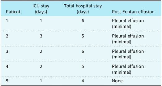
Post-catheter course: ICU and total length of stay, and effusion surveillance

Data are shown as n (%) or median (range), as appropriate.
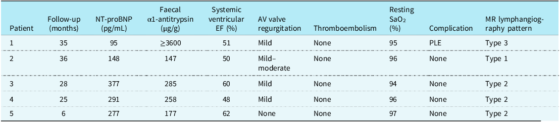
Follow-up clinical, laboratory, and imaging findings

Data are shown as n (%) or median (range), as appropriate.
AV = atrioventricular; EF = ejection fraction; NT-proBN = N-terminal pro–B-type natriuretic peptide; PLE, protein-losing enteropathy; SaO2 = arterial oxygen saturation.
During the Fontan preparation operation, in addition to establishing the bidirectional cavopulmonary connection and preparing the conduit tract, two patients underwent pulmonary artery banding and two underwent pulmonary artery reconstruction (right-sided in Patient 1 and as part of the index procedure in Patient 5). Cardiopulmonary bypass/aortic cross-clamp times were 279/94, 144/24, 110/42, 97/16, and 135/97 minutes for Patients 1–5, respectively. The intratubular membrane materials were CorMatrix (n = 1), native pericardium (n = 2), porcine pericardium (n = 1), and bovine pericardium (n = 1). In two patients, an 18-mm polytetrafluoroethylene extracardiac conduit was used, whereas three patients received a 20-mm polytetrafluoroethylene conduit (Table 1).
The interstage interval from surgery to catheterisation was a median of 7 months (range, 6–9). Procedural compositions and times are listed in Table 4. Median procedure time was 140 minutes (range, 120–180). Traversal of the surgically placed membrane required adjunct electrocautery in one patient and balloon predilatation in one (both in Patient 2). Antegrade pulmonary flow was closed simultaneously in two patients (Patients 1 and 4), and right pulmonary artery stenting was performed in one (Patient 1).
Catheter-based interventions performed for Fontan completion

Data are shown as n (%) or median (range), as appropriate.
PA = pulmonary artery; RPA = right pulmonary artery.
Post-catheter course is presented in Table 2. The median ICU stay was 2 days (range, 1–3), and total hospital stay was 5 days (range, 4–6). Minimal pleural effusions occurred in four patients (Patients 1–4), none requiring drainage; Patient 5 had no effusion. A transient desaturation following balloon dilatation occurred in one case and resolved prior to discharge.
At follow-up (median 28 months, range 6–36), all patients were clinically stable (Table 3). Resting oxygen saturation had a median of 96% (range, 94–97). NT-proBNP ranged from 95 to 377 pg/mL, and faecal α-1 antitrypsin ranged from 147 to ≥ 3600 µg/g. One late-presenting child (Patient 1, 35-month follow-up) had markedly elevated α-1 antitrypsin consistent with protein-losing enteropathy, remained asymptomatic, and required no targeted therapy. Across the cohort, systemic ventricular ejection fraction ranged from 48 to 62% (median 51%), and atrioventricular valve regurgitation was none-to-mild in four and mild-to-moderate in one. No thromboembolic events or other Fontan-related complications (e.g., plastic bronchitis) were observed.
Discussion
Transcatheter Fontan completion has become a practical option for certain single-ventricle patients.Reference Hausdorf, Schneider and Konertz9–Reference Galantowicz and Cheatham13 This is done using a hybrid approach, starting with surgical preparation during the Glenn stage, followed by a fully percutaneous second stage. The idea builds on early concepts and experiences with nonsurgical completion. In our method, an extracardiac polytetrafluoroethylene conduit is placed during Glenn surgery. At the same time, a membrane accessible by catheter is created. Later, when the child is physiologically ready, we cross this membrane in a controlled way and create a tunnel supported by covered stents. This follows established techniques that avoid the atrium and use standardised extracardiac pathways.Reference Marcelletti, Corno, Giannico and Marino7
A common decision in single-ventricle care is whether late-presenting patients should undergo a single combined Glenn and Fontan operation or follow a staged pathway. In our centre, located in a region where late referrals are common, we have encountered many patients who miss the ideal timing for Fontan. Based on these experiences, we believe it is safer not to perform Glenn and Fontan in the same setting for such patients. They often carry higher perioperative risk, and delaying Fontan until after circulatory and somatic stabilisation provides a safer window for intervention. As we began separating these procedures, we started asking: why not complete Fontan in these patients using a hybrid transcatheter approach? This led us to adopt a strategy where Glenn surgery includes surgical preparation for future catheter-based Fontan completion. It offers a less invasive alternative and remains faithful to the original concept—first preparing the body and then completing the pathway without open-heart surgery. For example, one patient with no prior palliation—whose family had deferred surgery because of procedural anxiety—was not late for Fontan per se but was late for the Glenn stage; accordingly, this case was included in our cohort, and Fontan completion was performed as a single open-heart operation without any complication. Since adopting this approach, we have observed a noticeable reduction in both mortality and morbidity in this high-risk group of Fontan patients.
Another scenario we frequently encounter involves atrioventricular valve regurgitation that becomes significant after the Glenn procedure but before Fontan completion. From our experience with single-ventricle patients, we have learned that it is safer and more effective to repair the atrioventricular valve and prepare for transcatheter Fontan completion during the same surgical session. By doing so, we allow time for the patient’s haemodynamics to stabilise and for pulmonary vascular resistance to decrease. This approach eliminates the need for reoperation and makes it possible to complete the Fontan later using a less invasive, catheter-based technique.
There are two main strategies for transcatheter Fontan completion: performing the entire procedure percutaneously without prior surgical preparation or planning a transcatheter intervention in a surgically prepared patient.Reference Galantowicz and Cheatham13–Reference Gerelli, Van Steenberghe, Patel, Van Aerschot and Boudjemline20 We follow the second approach, as our experience has shown that it offers safer and more predictable outcomes.Reference Prabhu, Anderson, Ward, Karl and Alphonso19,Reference Gerelli, Van Steenberghe, Patel, Van Aerschot and Boudjemline20 When a fully percutaneous Fontan is attempted in a child who has undergone a Glenn procedure without prior preparation, the procedure often becomes longer and more complex and carries a higher risk of complications while also requiring greater operator expertise.Reference Gerelli, Van Steenberghe, Patel, Van Aerschot and Boudjemline20 In contrast, placing a short, straight extracardiac conduit with a controlled membrane during the initial surgery not only simplifies the later intervention but also significantly reduces these risks. In line with this, we found membrane crossing to be consistently feasible using high-penetration chronic total occlusion wires, without any associated complications. These advantages were anticipated in early concept papers and later supported by technical studies. Reference Prabhu, Anderson, Ward, Karl and Alphonso19,Reference Gerelli, Van Steenberghe, Patel, Van Aerschot and Boudjemline20
In our study, early and mid-term outcomes were favourable, with all procedures technically successful, no major complications or thromboembolic events, and only minimal, self-limited effusions, similar previous studies.Reference Hausdorf, Schneider and Konertz9,Reference Sidiropoulos, Ritter, Schneider and Konertz11,Reference Sallehuddin, Mesned, Barakati, Fayyadh, Fadley and Al-Halees17 Recovery was rapid, with a median ICU stay of 2 days and hospital stay of 5 days, consistent with prior hybrid experiences. At a median follow-up of 28 months, oxygenation and ventricular function remained stable. One child showed lab signs of protein-losing enteropathy but stayed asymptomatic without treatment, highlighting the importance of long-term follow-up.
One common question in this patient group is what to do if fenestration becomes necessary. In surgical Fontan, fenestration is often created preemptively in patients with higher-risk physiology. Similarly, in our transcatheter Fontan completion approach, fenestration can be created percutaneously when clinically required. Although none of our patients require it during the study period, having this option remains a core advantage of the platform and aligns with existing technical guidance.Reference Jalal, Gewillig and Boudjemline21
Although theoretical application at the time of the Glenn in early infancy (∼6 months) can be envisaged, we do not advocate its use at that age. Rapid somatic growth would soon outsize a fixed-diameter extracardiac conduit, predisposing to flow limitation, thrombosis, and early re-intervention. Moreover, in infants with a small inferior vena cava, implanting a relatively large conduit introduces substantial calibre mismatch that perturbs cavopulmonary flow. Accordingly, we reserve this approach for late-presenting Glenn-stage candidates and older children, in whom growth rates are slower and conduit durability is more predictable.
Limitations
These include a small, heterogeneous cohort and follow-up that is modest relative to the lifetime course of Fontan physiology. Implementation also presupposes co-located congenital surgical and interventional expertise, advanced imaging, and hybrid capability—factors that may restrict generalisability and have already been highlighted in foundational reports.Reference Galantowicz and Cheatham13,Reference Konstantinov, Benson and Caldarone14 Prospective, preferably multicentre, studies are needed to refine patient selection, standardise membrane-crossing and covered-stent techniques, and determine the long-term comparative effectiveness versus surgical completion.
Conclusion
A staged surgical preparation followed by transcatheter extracardiac Fontan completion was feasible and safe in carefully selected single-ventricle patients, with reliable membrane traversal, low periprocedural morbidity, and stable clinical status at follow-up. These results support a resternotomy-avoiding approach and highlight the need for larger multicentre comparisons to standardise technique and clarify long-term haemodynamic, lymphatic, and end-organ outcomes relative to surgical completion.
Supplementary material
The supplementary material for this article can be found at https://doi.org/10.1017/S1047951125110998.
Acknowledgements
None.
Financial support
This research received no specific grant from any funding agency, commercial or not-for-profit sectors.
Competing interests
The authors declare none.
Ethical standard
The authors assert that all procedures contributing to this work comply with the ethical standards of the relevant national guidelines on human experimentation and with the Helsinki Declaration of 1975, as revised in 2008, and were approved by the institutional committee.