


Anatomic variations in patients with double inlet ventricle
Double inlet ventricle exists when the greater part of both atrioventricular junctions is supported by the same ventricular chamber.3 Since this definition (Fig. 1) is independent of atrial, atrioventricular valvar, and ventricular morphology, there is great potential for variability amongst patients unified because they exhibit this particular atrioventricular connection. Patients can be anticipated, therefore, to have any of the four arrangements of the atrial appendages (Fig. 2), although obviously the usual arrangement will be encountered most frequently.4 The atrioventricular junctions can be guarded by two separate valves, best described as the right and left valves,5 or by a common valve (Fig. 3). One of the two atrioventricular valves, or the common valve, can override the crest of the muscular ventricular septum, when there is one big and one small ventricle, as long as the greater part of both junctions remain committed to the dominant ventricle. Very rarely, both atrioventricular valves can be found in overriding position. When an atrioventricular valve does override in these circumstances, then almost always there is straddling of the valvar tension apparatus, but sometimes the atrioventricular junctions can override in the absence of straddling.6 The key morphologic feature, therefore, is that both junctions are supported in their greater part by the dominant ventricle. The most important differentiating feature is the morphology of that dominant ventricle (Fig. 2). This is most frequently of left morphology, as shown by the fine criss-crossing nature of the apical trabeculations, and then there is an incomplete right ventricle found antero-superiorly within the ventricular mass,3 sometimes to the right (Fig. 4), most frequently to the left (Fig. 5), but always antero-superiorly relative to the dominant ventricle (Figs 4 and 5). The second commonest pattern is for the dominant ventricle to have coarse apical trabeculations, identifying it as being a morphologically right ventricle. In this variant, there is again a second incomplete ventricle to be found, usually to the left, and rarely to the right, but always postero-inferiorly, and always with the fine criss-crossing apical trabeculations of the morphologically left ventricle (Fig. 6). In the rarest variant of double inlet ventricle, only a solitary chamber is found within the ventricular mass.7 This chamber always has coarse apical trabeculations (Fig. 7). In the clinical setting, the chamber may well be a right ventricle, the incomplete left ventricle being so small that it cannot be identified. Autopsy studies, nonetheless, have shown that double inlet can also exist when the solitary ventricle is of indeterminate morphology due to failure of development of the ventricular septum. The solitary ventricle of indeterminate pattern is found most frequently in the setting of isomerism of the right atrial appendages, but can rarely be encountered when there is usual atrial arrangement (Fig. 7). In the hearts with one big and one small ventricle, the communication between the chambers is another feature of clinical significance, since depending on the ventriculo-arterial connections, stenosis at the level of the interventricular communication can produce obstruction to flow into either the pulmonary or the systemic pathways, or rarely both.
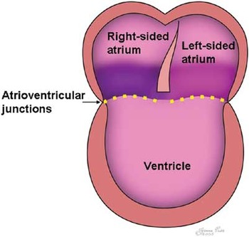
Figure 1. The figure shows how double inlet ventricle will exist whenever both segments of atrial myocardium (purple and pink shadings) are connected to the same ventricle across the atrioventricular junctions (yellow dotted line). The arrangement is independent of the morphology of the atriums and the ventricle, and also independent of the valvar morphology (see Figure 3).
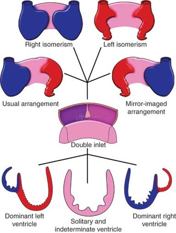
Figure 2. The figure shows how double inlet ventricle can exist with any of the four possible arrangements of the atrial appendages (morphologically right appendage shown in blue, and the morphologically left appendage in red), and with any of three ventricular morphologies, the morphologically right ventricle shown in blue, the morphologically left ventricle in red, and a solitary and indeterminate ventricle in pink.

Figure 3. The figure shows how double inlet ventricle is independent of the presence of two separate atrioventricular valves or a common atrioventricular valve. There will always be double inlet as long as the right atrial vestibule (purple) and the left atrial vestibule (pink) are joined to the same ventricle across the atrioventricular junctions (yellow dotted lines).

Figure 4. The incomplete right ventricle in this heart with double inlet left ventricle and discordant ventriculo-arterial connections is positioned antero-superiorly and on the right shoulder of the dominant left ventricle.
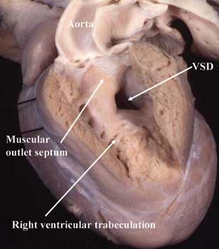
Figure 5. In this heart with double inlet left ventricle and discordant ventriculo-arterial connections, the incomplete right ventricle is positioned antero-superiorly and on the left shoulder of the dominant left ventricle. VSD: ventricular septal defect.

Figure 6. The essence of double inlet right ventricle is that the incomplete left ventricle is positioned postero-inferiorly, being left-sided in this specimen. Note that the left atrioventricular junction is connected to the dominant right ventricle despite the presence of straddling on one papillary muscle across the ventricular septum.

Figure 7. In this heart with usual atrial arrangement and double inlet, the trabeculations are uniformaly coarse throughout the apical component. It was not possible to find a second ventricle. PT: pulmonary trunk; AV: atrioventricular.
Another obvious feature of clinical importance, therefore, is the ventriculo-arterial connection. When there is a dominant left ventricle and an incomplete left ventricle, the ventriculo-arterial connections are usually discordant, although concordant ventriculo-arterial connections are by no means infrequent, this latter arrangement producing the so-called “Holmes Heart”8 (Fig. 8). On rare occasions, both outflow tracts can originate for either the dominant left or the incomplete right ventricle, while a common arterial trunk can also rarely be encountered. When the right ventricle is dominant, then double outlet from the dominant ventricle is the commonest pattern encountered. Concordant connections can sometimes be found, but discordant connections are exceedingly rare, as is the presence of a common arterial trunk. Indeed, to the best of our knowledge, these latter ventriculo-arterial connections have never been described in the setting of double inlet right ventricle, but they must be anticipated to exist. When there is a solitary ventricle, then the only possible connections across the ventriculo-arterial junctions are double outlet or a common arterial trunk. Double outlet is the expected arrangement, although if there is isomerism of the right atrial appendages, there will likely be co-existing subpulmonary atresia or severe stenosis.

Figure 8. The incomplete right ventricle (RV) in this heart with double inlet left ventricle has a long infundibulum supporting the pulmonary trunk. The morphology is remarkably similar to that seen in “classical” tricuspid atresia. This is the so-called “Holmes Heart”, with concordant ventriculo-arterial connections. VSD: ventricular septal defect.
It is atresia or stenosis of one or other outflow tract which is the clinically most significant associated malformation. When the stenosis is subaortic, typically at the level of the ventricular septal defect in the setting of a dominant left ventricle, then coarctation or interruption of the aortic arch should be anticipated to exist. In the setting of pulmonary atresia, then the source of pulmonary arterial blood is another important associated lesion, and typically this will be through the arterial duct. Other associated malformations may exist, such as juxtaposition of the atrial appendages, but are infrequent.
The most significant differential diagnosis is a huge ventricular septal defect. If present, this is important to recognise, since it may well potentiate to biventricular surgical repair. The key is to find the rudimentary apical septum separating the ventricular inlet components, with the coarse right ventricular trabeculation to one side, and the fine criss-crossing left ventricular trabeculations to the other side (Fig. 9). It may also be possible to septate a dominant left ventricle to produce biventricular surgical repair,9 but this is much more difficult than creating separate pulmonary and systemic circulations in the presence of a huge ventricular septal defect.

Figure 9. This specimen shows a huge ventricular septal defect, with a rudiment of the ventricular septum interposed between the right and left atrioventricular valves (AV). The different apical trabeculations to the right and left of the septal remnant show that this specimen exhibits biventricular atrioventricular connections. RV: right ventricle.
Conclusions
As described elsewhere in our supplement, for very many years hearts with double inlet left ventricle were considered the paradigm of “single ventricle”,10, 11 despite the fact that such hearts unequivocally possess a second chamber within the ventricular mass. For us, the solution to this paradox in terms of nomenclature was resolved when we came to appreciate that it was the atrioventricular connection, rather than the ventricular mass, which was truly univentricular.12 There are still some, nonetheless, who continue to argue that the ventricular mass itself is univentricular, suggesting that the second chamber is no more than the infundibulum of the right ventricle.13 Despite these assertions, there is a wealth of evidence that shows that the chamber, in reality, is the incomplete right ventricle. Thus, the septum separating the small chamber from the dominant ventricle usually possesses two discrete parts, one being the outlet septum but the other representing the apical ventricular septum. This interpretation is supported by the fact that this apical part of the septum is nourished by septal perforating arteries,14 and carries the atrioventricular conduction axis on its crest.15 Furthermore, it is possible to trace the cord-like right bundle branch into the apical part of the small chamber. And it is only when the small chamber is recognised as having right ventricular morphology that it becomes logical to describe discordant and concordant ventriculo-arterial connections in this setting. Thus, all the morphological evidence of which we are aware points to the small chamber being the incomplete right ventricle, and not simply the infundibulum. Irrespective of such niceties, it remains necessary to distinguish patients having double inlet to a dominant left ventricle from those with double inlet right ventricle, and those with a solitary and indeterminate ventricle. For the morphologist, this distinction is readily made by examining the nature of the apical ventricular trabeculations, but this is not easy to achieve in the clinical situation. In our experience, the position of the apical septum serves to distinguish between the variants. The septum is positioned antero-superiorly within the ventricular mass when the left ventricle is dominant, but postero-inferiorly when the right ventricle is the dominant chamber. The septum will be lacking in the presence of a solitary and indeterminate ventricle, while the presence of a hypoplastic apical septum interposing between the right and left atrioventricular valves will serve to distinguish those patients with a huge ventricular septal defect who may be amenable to biventricular repair. Locating the apical ventricular septum also provides the necessary information required to predict the course of the atrioventricular conduction axis.
Acknowledgements
The research on which this review is based was supported by grants from the British Heart Foundation together with the Joseph Levy Foundation.
Research at the Institute of Child Health and Great Ormond Street Hospital for Children NHS Trust benefits from R&D funding received from the NHS Executive.



