Small RNAs are a family of regulatory noncoding RNAs up to 40 nucleotides in length that can induce gene silencing through specific base-pairing with target mRNA molecules. Apart from their major function of gene regulation (Ref. Reference Bartel and Chen1), small RNAs in plants defend genomes against random integration of transposable elements and attack from invasive nucleic acids such as viruses (Ref. Reference Zamore2); this mechanism of defence against viral infection may also occur in mammals (Ref. Reference Kumar3). MicroRNAs (miRNAs) represent a major class of these small regulatory RNAs.
Following transcription of miRNA genes, one or two miRNAs can be generated from a single hairpin-loop precursor RNA (Ref. Reference Lau4), although some precursor molecules are known to contain more than six hairpin loops, referred to as miRNA clusters (Ref. Reference He5). miRNAs bind to complementary sequences within the 3′ untranslated region (3′ UTR) of their target mRNA transcript and, by virtue of proteins associated with the miRNA, usually direct target cleavage (if there is perfect complementarity with the target), or translational repression without cleavage of target (if partial complementarity with target) (Ref. Reference Bartel6). The ‘seed’ region (nucleotides 2–7) at the 5′ end of the miRNA is often sufficient for specificity and functionality of the miRNA (Ref. Reference Brennecke7).
Hundreds of miRNA genes are predicted to be present in mammals, with each miRNA apparently regulating multiple mRNAs, and multiple miRNAs regulating each mRNA (Refs Reference Leung and Sharp8, Reference Lim9, Reference Lim10). miRNAs are proposed to be involved in regulating at least a third of all genes within the human genome (Ref. Reference Zamore and Haley11) although, of the hundreds of miRNAs identified to date, the biological function(s) of only very few has been elucidated (Ref. Reference Rajewsky12).
miRNA discovery
The first miRNA, lin-4, was identified in 1993 during a genetic screen for mutants that disrupt developmental timing in Caenorhabditis elegans (Ref. Reference Lee, Feinbaum and Ambros13). The lin-4 gene was shown to produce a pair of small RNAs of approximately 61 and 22 nucleotides in length, with the larger being the precursor of the smaller. Both RNAs contained sequences complementary to sites in the 3′ UTR of lin-14 mRNA, suggesting that lin-4 regulates lin-14 translation by an antisense RNA–RNA interaction (Refs Reference Ha, Wightman and Ruvkun14, Reference Olsen and Ambros15). A second C. elegans miRNA, let-7, was discovered in 2000 (Ref. Reference Reinhart16); let-7 is also involved in developmental timing and represses expression of the lin-41 and hbl-1 mRNAs (Refs Reference Slack17, Reference Lin18, Reference Vella19). let-7 and lin-41 are phylogenetically conserved among a wide variety of multicellular organisms, indicating that these small RNAs could represent a general mechanism for post-transcriptional regulation (Ref. Reference Lau4).
Since these initial discoveries, many miRNAs have been identified in single-celled and multicellular organisms, including plant and mammalian cells (a database of known and predicted endogenous miRNAs is available at http://www.sanger.ac.uk/Software/Rfam/mirna). Although the exact number of miRNA genes in the human genome has yet to be determined, current estimates range to approximately 800 (http://microrna.sanger.ac.uk/sequences/). It is thought that many new miRNA genes may have evolved through duplication and mutation, with the number of gene duplications possibly correlating with the level of complexity of the organism (Refs Reference Zamore and Haley11, Reference Takuno and Innan20). Furthermore, RNA editing (i.e. site-specific modification of an RNA sequence to yield a product differing from that encoded by the DNA template) has been reported in at least 6% of human miRNAs, which may further increase the diversity of miRNAs and their targets (Ref. Reference Blow21).
miRNA biogenesis
Mammalian miRNA genes are generally transcribed by RNA polymerase (pol) II (Ref. Reference Lee22). However, recent reports show that human miRNAs mir-515-1, mir-517a, mir-517c and mir-519a-1 of the C19MC loci are transcribed by RNA pol III (Ref. Reference Borchert, Lanier and Davidson23), and bioinformatic analysis suggests that miRNA sequences containing upstream Alu, tRNA and mammalian-wide interspersed repeat (MWIR) sequences may also be transcribed by RNA pol III (Ref. Reference Borchert, Lanier and Davidson23). These transcripts are subsequently capped, polyadenylated and spliced, generating primary miRNA transcripts (pri-miRNAs) (Ref. Reference Cai, Hagedorn and Cullen24). The pri-miRNAs contain hairpin-loop domains from which mature miRNAs, contained within one arm of the hairpin-loop, are produced. In a limited number of cases a mature miRNA can be produced from either arm of the hairpin-loop; in these events the miRNAs can be named in different ways – for example, mir-458-3p and mir-458-5p, or mir-202 and mir-202* (with the less predominantly expressed miRNA designated by the asterisk) (Ref. Reference Lau4).
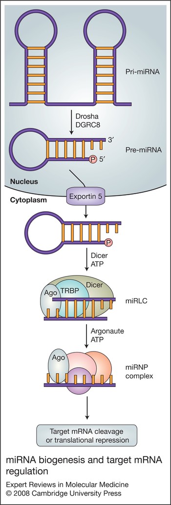
Pri-miRNAs are cleaved by the ‘microprocessor complex’, which comprises the double-stranded-RNA-specific RNase-III–type endonuclease Drosha (RNASEN) and its cofactor DGCR8 (Refs Reference Denli25, Reference Gregory26, Reference Han27) (Fig. 1). DGCR8 apparently functions to recognise the hairpin-loop of pri-miRNAs and to orientate the catalytic RNase III domain of Drosha to ensure correct cleavage, which releases hairpin-shaped precursor miRNAs (pre-miRNAs) of approximately 70 nucleotides in length (Ref. Reference Lee28) (Fig. 1). Cleavage by Drosha introduces staggered cuts on each side of the RNA helix stem, resulting in a 5′ phosphate and a two-nucleotide overhang at the 3′ end (Ref. Reference Lee28). In flies and nematodes, several functional miRNAs have been discovered that bypass the general biogenesis pathway. These miRNAs, known as ‘mirtrons’, are generated from spliced intronic sequences and have similar structural characteristics to pre-miRNAs; they enter the traditional miRNA biogenesis pathway at this stage, bypassing Drosha-mediated cleavage (Ref. Reference Ruby, Jan and Bartel29).
miRNA biogenesis and target mRNA regulation. Primary microRNA (pri-miRNA) generated from transcription in the nucleus is cleaved by Drosha (in conjunction with DGCR8) to generate precursor miRNA (pre-miRNA), which is translocated across the nuclear membrane by the action of exportin 5. In the cytoplasm, pre-miRNA is cleaved by Dicer with cofactor TRBP (transactivation-responsive RNA-binding protein) and argonaute protein (Ago), which together make up the complex miRLC [miRNA RISC (RNA-induced silencing complex) loading complex] to produce a double-stranded miRNA duplex. This is then unwound by the helicase armitage (not shown), releasing single-stranded mature miRNA. Mature miRNA becomes assembled into miRNPs (miRNA-containing ribonucleoprotein particles), which always include an argonaute protein. A number of other proteins may be – but are not always – involved in miRNP function; these include gemin3, gemin4, vasa intronic gene product (VIG), fragile-X-related protein (dFXR), tudor-SN, fragile X mental retardation protein (FMRP) and survival of motor neuron protein (SMN). miRNA guides miRNP to its mRNA target and, depending on the level of complementarity, can initiate cleavage or translational repression of mRNA target (see Fig. 2).
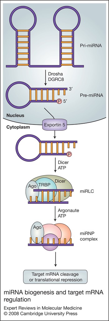
Translocation of pre-miRNAs across the nuclear envelope to the cytoplasm is facilitated by the nuclear transport protein exportin-5 (Fig. 1), which recognises the two-nucleotide 3′ overhangs on the pre-miRNA hairpin. Upon arrival in the cytoplasm, pre-miRNAs are cleaved by a second double-stranded-RNA-specific RNAse-III-type endonuclease, Dicer (DICER1) (Fig. 1), which acts in conjunction with a double-stranded-RNA-binding protein partner, transactivation-responsive RNA-binding protein (TRBP/TARBP2P) (Refs Reference Jiang30, Reference Chendrimada31, Reference Saito32). In human cells, TRBP recruits argonaute protein (Ago2/EIF2C2); together Dicer, TRBP and Ago2 form the miRNA RISC loading complex (miRLC; RISC stands for ‘RNA-induced silencing complex’) (Refs Reference Gregory33, Reference Maniataki and Mourelatos34). Cleavage of the pre-miRNA by Dicer produces an approximately 22 nucleotide double-stranded miRNA duplex – one strand of which will become the mature miRNA. The duplex is then unwound by the DEAD-box helicase armitage, releasing the single-stranded mature miRNA (Refs Reference Nykanen, Haley and Zamore35, Reference Cook36).
The Ago2-bound mature miRNA then becomes assembled into effector complexes termed miRNA-containing ribonucleoprotein particles (miRNPs) (Ref. Reference Mourelatos37) (Fig. 1). Several forms of miRNPs exist that differ in size and composition, but each form of miRNP contains a member of the argonaute protein family. The major function of miRNAs is to guide the miRNP complex to its target mRNA, where its associated argonaute protein mediates the effect (Ref. Reference Pillai, Artus and Filipowicz38). Several other miRNP components have been identified, including gemin3 (DDX20), gemin4, vasa intronic gene product (VIG), fragile-X-related protein (dFXR), and the tudor staphylococcal-nuclease-domain-containing protein (tudor-SN) (Refs Reference Caudy39, Reference Caudy40). Gemin3 is a putative DEAD-box RNA helicase, which may function in the unwinding of the mRNA target (Ref. Reference Nykanen, Haley and Zamore35), but the precise role of the other proteins in RNA-silencing events remains unclear. Although miRNAs function primarily in the cytoplasm, one miRNA, mir-29b has been found to localise in the nucleus; this is likely due to a hexanucleotide terminal motif in the 3′ region that directs the mature miRNA to be imported back into the nucleus after it is processed in the cytoplasm (Ref. Reference Hwang, Wentzel and Mendell41).
Mechanism(s) of miRNA action
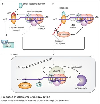
In mammals, miRNAs usually exhibit partial complementarity with their mRNA targets; perfect or near-perfect base pairing is quite rare in these organisms, but is predominantly found in plant miRNAs. Partial complementarity of miRNA to mRNA usually leads to translational inhibition (Ref. Reference Zeng, Yi and Cullen42), although animal miRNAs can also induce target degradation despite the lack of perfect complementarity (Refs Reference Bagga43, Reference Jing44, Reference Giraldez45). Several proposed models exist for the mechanism of translational repression, including miRNAs repressing translation at both pre-initiation and post-initiation stages (Fig. 2), and effects on mRNA stability (decapping and deadenylation) and compartmentalisation into translationally repressive sites (Fig. 2); it still remains to be deciphered which of these model mechanisms are cause and consequence of translational repression.
Proposed mechanisms of miRNA action. MicroRNAs (miRNAs) can inhibit translation at pre- and post-initiation stages. (a) At pre-initiation stages, the miRNP complex may affect m7G-cap-dependent translation through competition of the argonaute protein with the eIF4G initiation complex for binding to the cap structure; argonaute proteins also recruit eIF6, which prevents large ribosomal subunit binding to the small subunit. (b) At postinitiation stages, miRNPs may cause ribosomes to terminate translation prematurely, generating truncated polypeptides, or recruit proteolytic enzymes that degrade the polypeptide chain as it emerges from the ribosome. Repressed mRNAs arising from these models can then be transported to P-bodies for storage or degradation: the miRNP complex recruits GW182 protein; the latter subsequently recruits deadenylase enzyme CCR4–NOT1; the mRNA is then decapped by DCP1–DCP2, and degraded by exonuclease activity of XRN1.

miRNAs affecting initiation steps only affect cap-dependent translation, possibly through m7G cap recognition (Refs Reference Humphreys46, Reference Wang47, Reference Mathonnet48, Reference Wakiyama49, Reference Pillai50). Argonaute proteins contain structural similarities to the cap-binding protein eIF4E, and thus it has been suggested that translational repression may occur due to competition between argonaute and eIF4E for binding to the cap structure (Ref. Reference Kiriakidou51) (Fig. 2a). Argonaute proteins are also thought to recruit eIF6, which binds to the large ribosomal subunit, preventing binding of the small subunit and thus inhibiting mRNA translation (Ref. Reference Chendrimada52) (Fig. 2a).
Much evidence also exists for post-initiation mechanisms of repression, which affect both cap-dependent and cap-independent translation (Ref. Reference Petersen53). Polysome profile experiments indicate that, under conditions of translational repression, target mRNAs are fully loaded with ribosomes (Refs Reference Olsen and Ambros15, Reference Seggerson, Tang and Moss54), a number of which are engaged in active translation (Ref. Reference Petersen53), suggesting that translation initiation and elongation phases are not compromised. Two possible theories were suggested to explain these findings. The ribosome ‘drop-off’ theory suggests that ribosomes engaged in translation of miRNA-associated mRNAs are prone to terminate translation prematurely (Fig. 2b). Alternatively, association of active ribosomes with repressed mRNAs could also be explained by the ability of miRNP complex to recruit proteolytic enzymes to degrade the nascent polypeptide as it emerges from the ribosome (Ref. Reference Olsen and Ambros15) (Fig. 2b). Conflicting evidence exists on the role of proteolytic enzymes in miRNA function, as targeting of reporter proteins and the use of proteinase inhibitors have shown no effect on translational repression (Refs Reference Pillai50, Reference Petersen53).
miRNAs are apparently also involved in regulating mRNA stability and induction of decay of repressed mRNA targets. Argonaute proteins, miRNAs and their repressed target mRNAs have recently been shown to be compartmentalised in cytoplasmic foci called P-bodies (Refs Reference Pillai50, Reference Behm-Ansmant55, Reference Sen and Blau56, Reference Liu57, Reference Jakymiw58, Reference Meister59). These are sites of translational repression and mRNA decay; they are rich in factors associated with these processes, and are lacking in ribosomes or any other factors associated with translation initiation (Ref. Reference Parker and Song60). It is proposed that P-body proteins may participate in the formation of a repressive complex on the target mRNA, which could eventually lead to mRNA aggregation into P-bodies (Ref. Reference Pillai, Bhattacharyya and Filipowicz61). Within P-bodies, miRNA/mRNA-bound argonaute protein recruits GW182 protein (TNRC6A), which subsequently recruits deadenylating enzyme CCR4–NOT1 (CNOT1), and this is followed by mRNA decapping by DCP1–DCP2 enzyme – thereby affecting stability of repressed mRNA. Repressed mRNAs are then degraded by 5′ to 3′ exonuclease activity of XRN1 (5′-exoribonuclease 1) (Refs Reference Bagga43, Reference Behm-Ansmant55, Reference Liu57, Reference Schmitter62, Reference Rehwinkel63) (Fig. 2c). In addition to facilitating mRNA degradation, P-bodies may function as temporary storage sites for repressed mRNAs; once protein synthesis has been stimulated, repressed mRNAs may re-enter translation (Ref. Reference Brengues, Teixeira and Parker64).
Although miRNAs generally negatively regulate their target mRNAs, miRNA-associated proteins can play a role in AU-rich element (ARE)-mediated translational activation of tumour necrosis factor α (Ref. Reference Vasudevan and Steitz65). The miRNAs mir-369-3 and let-7 function in the recruitment of these proteins to the ARE sites in a sequence-specific manner (Ref. Reference Vasudevan, Tong and Steitz66). It is thought that miRNAs function in translation activation under the quiescence phase of the cell cycle and translation inhibition during the proliferation phase of the cell cycle (Ref. Reference Vasudevan, Tong and Steitz67), although the mechanisms of miRNA-mediated translation activation remain unclear. mir-122 has also been shown to enhance replication of hepatitis C virus, but it is unclear whether this occurs by similar mechanisms of ARE activation (Ref. Reference Jopling68).
Technologies for miRNA identification and analysis
Both computational prediction and experimental analysis have been used successfully to identify and analyse miRNAs.
Computational analysis (e.g. applying MirScan software) involves candidate miRNA prediction, based on known structural features, followed by experimental analysis to validate the existence of the predicted sequence (Ref. Reference Yoon and De Micheli69). Computational approaches have greatly contributed to miRNA target analysis. Based on the realisation that the ‘seed’ nucleotides within the 5′ region of miRNAs are of significant functional relevance, bioinformatics approaches have been developed and applied to predict direct targets of specific miRNAs, by searching for seed complementarity in mRNA 3′ UTRs (Refs Reference Bentwich70, Reference Kiriakidou71, Reference Lewis72, Reference Farh73, Reference Bentwich74, Reference Stark75, Reference Sood76, Reference Maziere and Enright77). As a result of the short seed sequence (nucleotides 2–7), numerous potential mRNA targets are generally predicted for each miRNA. Binding studies and functional analysis are necessary to determine true mRNA targets.
Experimental analysis involves the identification of a small RNA sequence, followed by bioinformatic analysis to determine if this sequence fulfils the defined structural characteristics of a miRNA (Refs Reference Zilberstein78, Reference Berezikov, Cuppen and Plasterk79). De novo identification of miRNAs generally involves sequencing of size-fractioned cDNA libraries. To achieve this, small RNAs (approximately 20–28 nucleotides) are isolated from denaturing gels and, following attachment of 5′ and 3′ adapters to the RNAs, reverse-transciptase (RT)-PCR is performed. The resulting cDNAs are cloned to form a cDNA library. Individual clones are subsequently sequenced to establish the genomic origin of the small RNA.
In addition to identifying new miRNAs, large-scale cDNA cloning may be used to evaluate the relative expression levels of miRNAs in a range of specimens. However, global profiling of miRNAs most frequently utilises microarrays (Refs Reference Lim9, Reference Krichevsky80, Reference Babak81, Reference Barad82, Reference Liu83, Reference Miska84, Reference Sun85, Reference Thomson86, Reference Baskerville and Bartel87, Reference Monticelli88, Reference Shingara89, Reference Wienholds90, Reference Castoldi91) or the RNA-primed array-based Klenow enzyme (RAKE) assay (Ref. Reference Nelson92). TaqMan low-density microarrays (TLDAs) have proven popular for such studies (http://www.appliedbiosystems.com/index.cfm). Bead-based flow cytometry assays have also been developed for miRNA analysis, whereby beads are coupled to probes (<100 probes) representing individual miRNAs. Following incubation with the specimen of interest, the beads are analysed by flow cytometry for identification and quantification of expressed miRNAs (Ref. Reference Lu93). Methods used for validation of results from global analysis – or for analysis of small numbers of miRNAs – include qRT-PCR, northern blotting, dot blotting, RNase protection assay, and a modified invader assay (Refs Reference Allawi94, Reference Kim and Nam95).
The functional relevance of miRNAs may be investigated using pre-miRNAs (Pre-miRTM miRNA precursors) or miRNA inhibitors (Anti-miRTM miRNA inhibitors) (see http://www.ambion.com). Antisense technologies have also been used successfully to regulate miRNA levels in vitro and in vivo (Refs Reference Krutzfeldt96, Reference Davis97, Reference Esau98). Simultaneous expression of multiple miRNAs by RNA pol III is being investigated, as RNA pol III can achieve higher expression levels compared with expression driven by RNA pol II; as miRNA-mediated mRNA silencing is dose-dependent, this mechanism would possibly increase the chances of producing hypomorphic phenotypes (Ref. Reference Zhou, Huang and Xia99).
miRNAs in normal and pathological conditions
miRNAs have been implicated in regulation of cellular processes such as differentiation (Ref. Reference Esau100), proliferation, apoptosis (Ref. Reference Brennecke101), metabolism (Ref. Reference Krutzfeldt and Stoffel102), haematopoiesis (Ref. Reference Chen103), cardiogenesis (Ref. Reference Zhao, Samal and Srivastava104), morphogenesis and insulin secretion (Ref. Reference Poy105), in addition to acting in several feedback loops involved in signal transduction pathways (Ref. Reference Carthew106). miRNAs are vital for cell survival: elimination of miRNA maturation by Dicer knockout leads to embryonic lethality in mice (Ref. Reference Harfe107). miRNAs are involved in such a wide variety of cellular processes that it is likely their dysregulation or abnormal expression could lead to a range of disease states. miRNAs have already been implicated in the pathogenesis of several human diseases, such as neurological disorders, cancer, and viral and metabolic diseases (Ref. Reference Esau98).
Neurological disorders
Spinal muscular atrophy (SMA), a progressive neurodegenerative disease, is caused by deletion or loss of function mutations in the SMN (survival of motor neuron) protein (Ref. Reference Dostie108). SMN is a component of the miRNP complex that performs the effector functions of the miRNA pathway (Ref. Reference Mourelatos37). Fragile X syndrome is caused by inactivation of the gene FMR1, and hence silencing of the fragile X mental retardation protein (FMRP), which is also associated with miRNP complex formation (Ref. Reference Jin109). These studies indicate that disruptions in the miRNP machinery and hence miRNA activity can lead to disease states. Tourette syndrome is associated with a single-nucleotide polymorphism (SNP) in the 3′ UTR of the SLITRK1 gene, which is the binding site of mir-189; this SNP hence modifies the interaction of mir-189 (Ref. Reference Abelson110). In addition, mir-134 regulation of LIMK1 in hippocampal neurons controls spine development and possibly also contributes to synaptic development, maturation and plasticity (Ref. Reference Schratt111); thus, dysregulation of mir-134 could potentially lead to complications in these processes.
Cancer
Many miRNA genes are thought to reside at chromosomal breakpoints or fragile sites associated with cancer (Ref. Reference Calin112). The mir-15/16 cluster is located at one such site and is deleted in the majority of B cell chronic lymphocytic leukaemias (B-CLLs) (Ref. Reference Calin113), as well as mantle cell lymphomas and prostate cancers (Ref. Reference Stilgenbauer114), suggesting that mir-15/16 may function as tumour suppressors. Members of the let-7 family also located at fragile sites (Ref. Reference Calin112) are frequently deleted in cancer patients, leading to elevated levels of the oncogene product RAS (Ref. Reference Johnson115). Some miRNAs have also been shown to possess oncogenic potential; the mir-17-92 cluster, which contains six miRNAs, is located at a chromosome site that is amplified in a range of cancers and overexpression leads to accelerated tumour development in mouse B cell lymphoma models (Ref. Reference He5). Overexpression of the individual miRNAs from the cluster did not reveal the same oncogenic potential, indicating that interaction between a range of miRNAs could be necessary for the development of disease phenotypes. mir-155, which is elevated in Burkitt lymphoma, also acts as an oncogene, with overexpression in B cells leading to development of pre-B-cell lymphomas (Ref. Reference Costinean116). These putative miRNA tumour suppressors and oncogenes represent a potential set of miRNA therapeutic targets. Microarray profiling of miRNAs in tumour tissues and cell lines has identified miRNA differentially expressed in different tumour types, indicating potential use of tumour miRNA profiling in cancers for prediction of developmental lineage, differentiation state, and prognosis (Ref. Reference Lu93).
Viral disease
Host mir-32 expression restricts infection of the primate foamy virus 1 (PFV-1), with inhibition of mir-32 leading to doubling of the PFV-1 proliferation rates in host cells (Ref. Reference Lecellier117). PFV-1 encodes the Tas protein, which is known to be a suppressor of RNA silencing (Ref. Reference Lecellier117), thereby removing the growth limitation inflicted by mir-32 by disrupting the silencing machinery. Many viruses encode similar suppressors of RNA silencing – for example, the Tat protein from human immunodeficiency virus 1 (HIV-1) (Ref. Reference Bennasser118) and the B2 protein from Nodamura virus (Ref. Reference Sullivan and Ganem119).
miRNAs represent an efficient mechanism for viruses to use to manipulate host machinery, as they require less space on the viral genome than alternative protein products. Viral miRNAs can target both viral and host mRNAs for repression. Twelve miRNAs from the Karposi sarcoma-associated herpesvirus (KSHV) genome expressed in cells led to the downregulation of a number of genes including thrombospondin 1 (THBS-1), which is a known tumour suppressor and antiangiogenic factor. It is thought that these KSHV miRNAs may contribute directly to pathogenesis of KSHV by downregulation of THBS-1 (Ref. Reference Samols120). The simian virus 40 (SV40) encodes a miRNA that is perfectly complementary to transcripts coding viral T antigens, leading to their degradation (Ref. Reference Sullivan121). This destruction of viral T antigens aids the virus in evading immune detection by the host. The hepatitis C virus (HCV) enchances replication via a novel interaction of abundantly expressed mir-122 with the 5′ UTR of the viral genome (Ref. Reference Jopling68). Interferons (IFNs) are key molecules involved in eliciting the antiviral response once an infection has been detected (Ref. Reference Cullen122). IFN-β has recently been implicated in the activation of several miRNAs in mammals that have antiviral properties against HCV (Ref. Reference Pedersen123), and treatment also leads to reduced mir-122 expression (Ref. Reference Pedersen123), which limits HCV replication (Ref. Reference Jopling68). These studies identify a number of different miRNAs that could be therapeutically targeted to hinder viral infection, aid host detection of infection, and prevent viral manipulation of host machinery.
miRNAs relevant to diabetes
Diabetes mellitus is a metabolic disorder in which insulin either is not secreted in sufficient amounts from β-cells or does not efficiently stimulate its target cells. Despite high glucose levels, cells starve, as a result of impaired glucose entry into cells. Current treatments for diabetes cannot efficiently control glycaemic levels, resulting in episodes of hyper- and hypoglycaemia (Ref. Reference Nathan124), which increases the possibility of developing secondary complications such as retinopathy, nephropathy and neuropathy (Ref. 125). In the search for more-targeted molecular therapies, miRNAs implicated in insulin secretion and diabetic complications have recently attracted attention.
miRNAs associated with β-cell insulin secretion
Recent experimental work has revealed a limited number of miRNAs – including mir-375, mir-124a and mir-9 – associated with various subcellular events involved in glucose-stimulated insulin secretion (GSIS) (Refs Reference O'Driscoll, Gammell and Clynes126, Reference O'Driscoll127, Reference Dowling128). In addition, bioinformatic analysis has indicated potential miRNA target sites in a range of other mRNAs encoding proteins involved in exocytosis – including VAMP2 (vesicle-associated membrane protein 2), SNAP25 (synaptosomal-associated protein 25kDa), syntaxin-1, Rab27a (member of the RAS oncogene family), granuphilin (SYTL4) and MyRIP (myosin VIIA and Rab interacting protein). Some miRNAs (mir-153, mir-1, mir-133, mir-200 and mir-34) have predicted target sites in several of these functionally related genes (e.g. mir-153 and mir-1 have putative target sites in VAMP2 and SNAP25) (Ref. Reference Abderrahmani129). Although the miRNA target sites identified by bioinformatics have yet to be experimentally validated, this gives an insight into the potential extent of complex networking of molecules involved in exocytosis regulation.
mir-375
Selective cloning of small RNAs 21–23 nucleotides in length from the β-cell line MIN-6 and the α-cell line TC1 led to the identification of mir-375, a miRNA specific to pancreatic islet cells. Gain- and loss-of-function experiments on mir-375 indicated it was involved in GSIS in β-cells, with overexpression resulting in reduced GSIS and, conversely, knockout of expression resulting in enhanced GSIS (Ref. Reference Poy105). mir-375 apparently acts on the later stages of exocytosis to reduce insulin secretion.
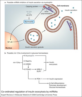
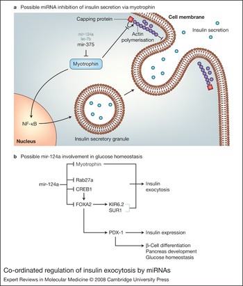
Based on sequence information, myotrophin has been confirmed as a target of mir-375 action (Table 1); mir-375 mediates repression via a single target site in the 3′ UTR of the myotrophin mRNA (Ref. Reference Poy105). Myotrophin is involved in vesicle transport in neurons and in neurotransmitter release but its function in pancreatic β-cells has not been clearly defined (Refs Reference Dunn130, Reference Yamakuni131, Reference Antonin, Riedel and von Mollard132). Myotrophin (via its three consecutive ankyrin repeats) interacts with the capping protein CP (also known as CapZ or β-actinin). This myotrophin–CP interaction inhibits CP-regulated actin polymerisation (Ref. Reference Taoka133), thereby allowing access of secretory granules to exocytotic site (Fig. 3a). Myotrophin also acts in the nucleus as a transcription factor to activate nuclear factor κB (NF-κB), a critical component in maintaining GSIS in β-cells (Refs Reference Hammar134, Reference Norlin, Ahlgren and Edlund135) (Fig. 3a). It is not yet clear whether mir-375-induced inhibition of myotrophin translation and the corresponding reduction of GSIS are mediated by the CP or NF-κB pathway, or a combination of both. Myotrophin is also the predicted target of repression for two other miRNAs: mir124 and let-7b (Ref. Reference Krek136). The function of let-7b in GSIS of β-cells still remains to be established.
Co-ordinated regulation of insulin exocytosis by miRNAs. (a) Possible microRNA (miRNA) inhibition of insulin secretion via myotrophin. Overexpression of the myotrophin-targeting miRNA mir-375 results in reduced glucose-stimulated insulin secretion, which can be explained through cytoplasmic and/or nuclear actions of myotrophin. Myotrophin interacts with capping protein to inhibit actin polymerisation. Inhibition of actin polymerisation allows access of insulin granules to the cell membrane for exocytosis. In addition, myotrophin interacts with transcription factor NF-κB, which controls expression of several genes critical for glucose-stimulated insulin secretion. Myotrophin also contains putative binding sites for the miRNAs let-7b and mir-124a. (b) Possible mir-124a involvement in glucose homeostasis. Mir-124a targets Rab27a and also FOXA2 (directly and indirectly via CREB1). Myotrophin has also been identified as a potential mir-124a target. FOXA2 may influence several targets relevant to diabetes via PDX-1, including insulin mRNA levels and possibly also KATP channel subunits KIR6.2 and SUR1 (involved in regulated insulin release). It also plays a role in β-cell differentiation, pancreas development and glucose homeostasis.
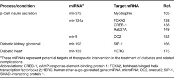
miRNAs implicated in β-cell insulin secretion and diabetic complications, and their mRNA targets

aThese miRNAs represent potential targets of therapeutic intervention in the treatment of diabetes and related complications.
Abbreviations: CREB-1, cAMP-response-element-binding protein 1; FOXA2, forkhead/winged helix transcription factor boxa 2; HERG, human ether-a-go-go related gene; miRNA, microRNA; OC2, onecut 2; SIP-1, SMAD-interacting protein 1.
More recently, knockdown of mir-375 in zebrafish embryos has revealed a role for this miRNA in pancreatic islet development (Ref. Reference Kloosterman137). When morpholino oligonucleotides were injected into one-cell-stage embryos, resulting in a knockdown of mir-375 activity during the first four days of development, insulin staining showed the formation of an islet at 24 h post fertilisation but by day 3 the islet had fallen apart and insulin-positive cells were scattered (Ref. Reference Kloosterman137). The original formation of an islet at 24 h suggests that mir-375 expression is not essential in early endocrine formation, but more so for maintenance of tissue identity at a later stage. It has not yet been deciphered whether this scattered islet phenotype occurs as a result of mir-375 action on myotrophin expression or whether other mir-375 targets are involved.
mir-124a
Mir-124a exists in three different isoforms – mir-124a1, 2 and 3 – encoded on chromosome 14, 3 and 2, respectively, in the mouse genome. The isoform mir-124a2 is differentially expressed during pancreas development, with a sixfold upregulation at embryonic stage e18.5 compared with e14.5 (Ref. Reference Baroukh138). e18.5 is the critical stage for β-cell differentiation, indicating that mir-124a2 might be significant in this process.
Using PicTar (http://pictar.bio.nyu.edu/cgi-bin/PicTar) (Ref. Reference Krek136) and miRanda (http://www.microrna.org/mammalian/index_new.html) (Ref. Reference Enright139) bioinformatics tools, the forkhead/winged helix transcription factor boxa2 (FOXA2) mRNA was identified as a potential target of mir-124a (Table 1). This relationship was subsequently confirmed by over- and underexpression of mir-124a2 in MIN6 murine pancreatic β-cells, using Pre-Mir and Anti-Mir technology (Pre-miRTM miRNA precursors and Anti-miRTM miRNA inhibitors; see http://www.ambion.com). CREB-1 (cAMP-response-element-binding protein), a stimulus-inducible transcription factor, was also predicted as a potential target of mir-124a regulation, and mir-124a2 over- and underexpression correspond with decreasing and increasing levels of CREB-1, respectively (Ref. Reference Baroukh138) (Table 1). As FOXA2 is a target of CREB-1 regulation (Ref. Reference Zhang140), this suggests that FOXA2 expression may be regulated by mir-124a2 directly as well as indirectly (via CREB-1) (Fig. 3b).
FOXA2 is an upstream regulator of the homeobox protein PDX-1 (Refs Reference Wu141, Reference Lee142). PDX-1 is essential for β-cell differentiation, glucose homeostasis and pancreas development (Refs Reference Jonsson143, Reference Offield144) (Fig. 3b), and the human orthologue (insulin promoter factor; IPF1) is mutated in a proportion of early-onset type 2 diabetic patients (Ref. Reference Stoffers145). Manipulation of FOXA2 expression, by overexpression or inhibition of mir-124a2, corresponds with a decrease and increase in PDX-1 mRNA levels, respectively (Ref. Reference Baroukh138). PDX-1 regulates expression of the insulin gene; consequently, overexpression and inhibition of mir-124a2 leads to a decrease and increase in insulin mRNA levels, respectively (Ref. Reference Baroukh138).
Further downstream targets of FOXA2 regulation are the KATP channel subunits SUR1 (sulphonylurea receptor 1) and KIR6.2 (inward rectifier K+ channel member 6.2) (Ref. Reference Lantz146), which are critical for regulated insulin release; mutations in either of these genes can lead to persistent hyperinsulinaemic hypoglycaemia of infancy (PHHI) in humans (Ref. Reference Meissner, Beinbrech and Mayatepek147). Overexpression of mir-124a2 leads to increased Ca2+ levels within the cell (Ref. Reference Baroukh138). Knockdown of the SUR1 and KIR6.2 subunits results in impaired KATP channels, causing a build-up of K+ ions within the cell, which stimulates opening of voltage-gated calcium channels, thereby allowing Ca2+ ions to enter the cell (increased Ca2+ ions usually stimulate exocytosis). Thus, reduced expression of KATP channel subunits could explain the increase in cytosolic free Ca2+ concentrations following transfection with mir-124a2. FOXA2 deficiency in mice leads to loss of GSIS and excessive insulin release in response to amino acid stimuli (Ref. Reference Lantz146). However, mir-124a2-induced reduction in FOXA2 levels has not shown as dramatic an effect on GSIS as seen in the FOXA2-null mouse (Ref. Reference Baroukh138).
Rab27A, which is also involved in GSIS (Ref. Reference Kasai148), has recently been shown to be the target of mir-124a action via a binding site in the 3′ UTR of Rab27A mRNA (Ref. Reference Lovis, Gattesco and Regazzi149). Mir-124a also indirectly regulates expression of several other components of the exocytotic machinery in MIN6-B1 cells, including SNAP25, Rab3A, synapsin 1A (SYN1) and NOC2 (nucleolar complex associated 2) (Ref. Reference Lovis, Gattesco and Regazzi149). Overexpression of mir-124a in these cells leads to reduced GSIS. In the same study, mir-96 was identified as a regulator of granuphillin and NOC2, and its expression in MIN6-B1 cells leads to a reduction in stimulated insulin secretion (Ref. Reference Lovis, Gattesco and Regazzi149).
mir-9
Mir-9 is expressed predominately in neurons in both human and mouse models (Refs Reference Lagos-Quintana150, Reference Landgraf151), and to a lesser extent in pancreatic β-cells in rat and mouse models (Ref. Reference Plaisance152). Onecut2 transcription factor (OC2), which negatively regulates granuphilin (also known as SLP4/SYTL4) expression, has been identified as a mir-9 target (Table 1). A basal level of mir-9 expression is needed to maintain optimum onecut2 expression levels for normal β-cell function (Ref. Reference Plaisance152), but mir-9 overexpression in rat INS-1E β-cells leads to a reduced GSIS in these cells (Ref. Reference Plaisance152).
Granuphilin associates with insulin secretory granules (Ref. Reference Wang153) and promotes targeting of these granules to the plasma membrane (Ref. Reference Torii154); however, it is a negative modulator of exocytosis as it imposes a constraint to inhibit fusion until the correct signals are received by the cell (Ref. Reference Torii154). Overexpression of mir-9 leads to increased levels of granuphilin expression due to the removal of the repressive effects of onecut2 on the granuphilin promoter (Ref. Reference Plaisance152), and hence reduced GSIS is observed as a result of its negative effects on exocytosis. Granuphilin-null mice also show impaired GSIS, with reduced quantity of insulin granules docked to the β-cell membrane, and conversely exhibit increased insulin exocytosis in response to stimulus (Ref. Reference Gomi155).
Binding partners of granuphilin include the GTP-binding proteins Rab3/Rab27, the SNARE-binding protein Munc-18 and the tSNARE protein syntaxin-1, which are involved in exocytosis of secretory granules in pancreatic β-cells (Refs Reference Plaisance152, Reference Coppola156, Reference Torii157). mir-9-induced reduction of exocytosis does not occur through manipulation of Rab3, Rab27 and SNARE proteins such as SNAP25, VAMP-2 and syntaxin-1, as the expression levels of these key exocytosis proteins are unchanged in mir-9-transfected cells relative to control cells (Ref. Reference Plaisance152). However, it is as yet unknown whether the mir-9-mediated reduction of secretagogue-stimulated exocytosis via granuphilin occurs through downstream manipulation of Munc-18 activity. The effect of granuphilin on Munc-18 is not alone sufficient to mediate such a profound knockdown of stimulus-induced exocytosis (Ref. Reference Coppola156), suggesting that granuphilin and possibly mir-9 have additional targets that participate in this process.
miRNAs associated with diabetic kidney glomeruli
Diabetic nephropathy – generally defined as urinary albumin excretion of >300 mg per 24 h or abnormal renal function characterised by abnormality in serum creatinine, creatinine clearance, or glomerular filtration rate – is the most common cause of kidney failure in patients with diabetes. The abnormal renal function is thought to arise largely from accumulation of extracellular matrix (ECM) proteins in the mesangial cells, hypertrophy of glomerular and tubular elements, and thickening of the glomerular and tubular basement membranes (Refs Reference Schleicher and Olgemoller158, Reference Ziyadeh159).
ECM proteins such as collagen 1α1 and 1α2 are positively regulated by transforming growth factor β (TGF-β), which is upregulated in mesangial cells under diabetic conditions (Refs Reference Reeves and Andreoli160, Reference Sharma and Ziyadeh161). TGF-β is known to upregulate ECM proteins via SMAD transcription factors and mitogen-activated protein kinases (MAPKs) (Refs Reference Poncelet and Schnaper162, Reference Tsuchida163, Reference Chin164, Reference Hayashida165); in addition, recent work has revealed TGF-β downregulates the E-box repressor proteins δEF1 and SMAD-interacting protein 1 (SIP1), which mediate repression of collagen expression at its E-box element (Ref. Reference Kato166). δEF1 can also repress SMAD proteins (Ref. Reference Postigo167).
Several miRNAs, including mir-192, -194, -204, -215 and -216, are preferentially expressed in the kidney, as compared with other tissues (Ref. Reference Sun85). Using computational miRNA target predictions from miRNA databases (http://cbio.mskcc.org; http://microrna.sanger.ac.uk/index.shtml) the E-box repressor SIP1 was shown to contain a potential target site for mir-192 and mir-215 regulation.
Using a luciferase reporter system, SIP1 was validated as a target of mir-192 regulation (Table 1), but not of mir-215. TGF-β treatment induces mir-192 expression. TGF-β-induced mir-192 expression or mir-192 transfection can decrease SIP1 levels, while mir-192 inhibitor increases SIP1 levels (Ref. Reference Kato166). The mechanism of TGF-β regulation of mir-192 expression is not completely understood. The mir-192 promoter contains a binding site for the proto-oncogene ETS-1 (Ref. Reference Sun85), which is also induced by TGF-β expression (Ref. Reference Liu168), representing a possible mechanism of TGF-β regulation of mir-192 expression.
mir-192 overexpression leads to repression of translation of its target SIP1, thereby increasing levels of collagen expression. Repression of δEF1 using short hairpin RNA (shRNA; for stable transfection of siRNA) shows similar effects, resulting in increased levels of collagen expression; however, double transfection of a mir-192 mimic and δEF1 shRNA shows a much larger increase in collagen expression than either achieved separately, suggesting that these two mechanisms act synergistically in the control of collagen expression (Ref. Reference Kato166).
In vivo analysis of type 1 and type 2 diabetic mice showed elevated levels of mir-192, TGF-β and collagen 1α2 in the renal glomeruli (Ref. Reference Kato166), suggesting the possible involvement of mir-192-mediated collagen expression in the pathogenesis of diabetic nephropathy, or other diabetic complications where TGF-β levels are raised.
miRNAs associated with diabetic heart
Cardiovascular disease is the principal cause of death in more than 60% of diabetic cases, with an annual mortality of approximately 5.4%, thereby decreasing life expectancy by up to 10 years (Refs Reference Donnelly169, Reference Duncan170). For diabetic patients, the most prominent cardiac electrical disturbance is an abnormal QT interval, which is associated with increased risk of sudden cardiac death (Refs Reference Casis and Echevarria171, Reference Veglio, Chinaglia and Cavallo-Perin172). QT interval is the total duration for ventricular depolarisation and repolarisation of cardiac myocytes, which is controlled by the flow of inward and outward ion currents. Increasing inward currents and/or decreasing outward currents lead to prolonged QT interval. The outward currents occur via a number of K+ channels.
Human ether-a-go-go related gene (HERG) encodes one of these channels – the rapid delayed rectifier K+ current channel (IKr). HERG is downregulated in diabetic hearts, thereby contributing to slowed repolarisation and prolonged QT interval (Refs Reference Zhang173, Reference Zhang174). HERG expression is downregulated at the post-transcriptional level: HERG mRNA levels remain constant, while HERG protein levels are reduced by 60% in diabetic heart as compared with nondiabetic/control heart (Refs Reference Zhang173, Reference Zhang174, Reference Xiao175).
mir-1 and mir-133 are specifically expressed in adult cardiac and skeletal muscle tissues, and upregulated in rabbit diabetic heart tissue and also in ventricular samples from human diabetic patients (Refs Reference Zhao, Samal and Srivastava104, Reference Chen176). Using a luciferase reporter plasmid and western blotting, HERG mRNA was shown to be a target of mir-133 action (Table 1), while mir-1 had no effect on HERG expression (Ref. Reference Xiao175). IKr, the channel for rapid delayed rectifier K+ current, was shown to be underexpressed in diabetic hearts and healthy hearts transfected with mir-133, while transfection of a mir-133 inhibitor AMO-133 partially rectified the depression of IKr in diabetic hearts, and completely rectified expression of IKr in mir-133-transfected healthy hearts (Ref. Reference Xiao175).
Serum response factor (SRF) is a cardiac transcription factor highly overexpressed in diabetic hearts (Ref. Reference Xiao175). SRF is essential for expression of mir-1 and mir-133 (Refs Reference Zhao, Samal and Srivastava104, Reference Chen176). SRF siRNA or the SRF inhibitor distamycin reduced expression of mir-1 and mir-133 in diabetic cardiac myocytes. Transfection of SRF siRNA into cardiac myocytes of diabetic hearts resulted in increased levels of IKr expression (Ref. Reference Xiao175). It still remains to be seen whether use of AMO-133 or SRF siRNA in vivo increases IKr expression sufficiently to correct or reduce prolonged QT interval in diabetic subjects.
mir-133 is also known to repress expression of KCNQ1, which is involved in the formation of the slow delayed rectifier K+ current channel (IKs) (Ref. Reference Luo177), although it is currently unknown whether this channel plays a role in the development of long QT syndrome in diabetic patients.
Clinical implications/applications
Until recently, miRNAs had not been considered as classical therapeutic targets, as they do not code for proteins. Initial studies aimed at exploiting miRNAs as a form of therapy have shown promising results. Following intravenous injection of modified antisense oligonucleotides (termed antagomirs) into mice, in vivo inhibition of four miRNAs – mir-16, mir-122, mir-192 and mir-194 – has been successfully demonstrated (Ref. Reference Krutzfeldt96). This approach resulted not only in blockage of target miRNAs, but also in their degradation in most organs analysed, including liver, kidney, heart, lung, intestine, bone marrow, muscle, skin, fat, ovaries and adrenals. Lack of effect observed in brain is possibly due to restricted diffusion of charged nucleic acids across the blood–brain barrier. Alternative approaches to targeting miRNAs therapeutically by inhibiting Drosha, Dicer or other miRNA pathway components are being investigated. Conversely, where reduced miRNA expressed is associated with a disease phenotype and increased expression of relevant miRNA could be of potential therapeutic relevance to rescue disease phenotype, introduction of miRNA mimics is being investigated. However, suitable expression vectors have yet to be identified for the safe delivery and maintenance of such effects long-term (Ref. Reference Esau and Monia178).
Research in progress and outstanding research questions
The importance of miRNAs in normal and pathological conditions is still being realised. Recent studies have clearly indicated an association between dysregulated expression of these short RNAs in regulated and defective insulin secretion from β-cells and in diabetic kidney and heart disease. Recently, overexpression of a specific miRNA (mir-29) – which is upregulated in diabetic rats – has been found to have a functional role in insulin resistance (Ref. Reference He179) and, furthermore, analysis of murine pancreas development has indicated a unique miRNA profile to be necessary during pancreas development for generation of normal β-cells (Ref. Reference Lynn180). So, while studies associating miRNAs with diabetes are so far limited in numbers, they suggest important roles for miRNAs as potential biomarkers and possibly therapeutic targets. More extensive studies investigating the expression and functional relevance of miRNAs in both type 1 and type 2 diabetes will undoubtedly increase our understanding of these complex conditions and will hopefully aid in the identification of novel therapeutic targets and interventions.
Acknowledgements and funding
The authors acknowledge support from Ireland's Higher Educational Authority Programme for Research in Third Level Institutes (PRTLI) Cycle 3, the Health Research Board, and Dublin City University's Research Fellowship Award.