Introduction
Seed dormancy is an innate seed property that defines the environmental conditions in which the seed acquires the capability to germinate (Finch-Savage and Leubner-Metzger, Reference Finch-Savage and Leubner-Metzger2006) and, therefore, has an ecological significance in tuning the timing of germination and in the build-up of a seed bank (Long et al., Reference Long, Gorecki, Renton, Scott, Colville, Goggin, Commander, Westcott, Cherry and Finch-Savage2015). Dormancy is present throughout the higher plants and in all major climatic regions (Finch-Savage and Leubner-Metzger, Reference Finch-Savage and Leubner-Metzger2006) as it is an important trait for seed survival and persistence, which allows plants to disperse germination of their seeds through time, thereby avoiding the risk of complete germination failure at a single time at which adverse germination conditions could materialize for a plant population (Long et al., Reference Long, Gorecki, Renton, Scott, Colville, Goggin, Commander, Westcott, Cherry and Finch-Savage2015). Thus, it is particularly relevant to the survival and persistence of weeds and, therefore, to weed management (Long et al., Reference Long, Gorecki, Renton, Scott, Colville, Goggin, Commander, Westcott, Cherry and Finch-Savage2015).
‘Red rice’ is the name used for the heterogeneous group of the weedy rices, which are congeneric to crop rice with phenotypic similarity (Vaughan et al., Reference Vaughan, Ottis, Prazak-Havey, Bormans, Sneller, Chandler and Park2001; Ziska et al., Reference Ziska, Gealy, Burgos, Caicedo, Gressel, Lawton-Rauh, Avila, Theisen, Norsworthy, Ferrero, Vidotto, Johnson, Ferreira, Marchesan, Menezes, Cohn, Linscombe, Carmona, Tang and Merotto2015). It has sometimes been referred to as Oryza sativa f. spontanea (Gianinetti et al., Reference Gianinetti, Laarhoven, Persijn, Harren and Petruzzelli2007). The weedy rices are commonly, though not exclusively, characterized by a red caryopsis (Delouche et al., Reference Delouche, Burgos, Gealy, de San Martin, Labrada, Larinde and Rosell2007; Ziska et al., Reference Ziska, Gealy, Burgos, Caicedo, Gressel, Lawton-Rauh, Avila, Theisen, Norsworthy, Ferrero, Vidotto, Johnson, Ferreira, Marchesan, Menezes, Cohn, Linscombe, Carmona, Tang and Merotto2015), and the red rice genotype studied in this work has indeed a red caryopsis at maturity (Gianinetti and Vernieri, Reference Gianinetti and Vernieri2007). The caryopsis is the fruit of grasses in which the seed coat firmly adheres to the pericarp. In rice, the caryopsis coat is composed of the pericarp, seed coat (tegmen) and nucellar layers (Bechtel and Pomeranz, Reference Bechtel and Pomeranz1977). The caryopsis is further enclosed by the hull to form a spikelet (Ellis et al., Reference Ellis, Hong and Roberts1985), which is the dispersal unit (or disseminule, i.e. the detachable plant part capable of being disseminated) that in weedy rices shatters at maturation. These rices show various degrees of seed dormancy and can have much stronger dormancy than the cultivated rice (Delouche et al., Reference Delouche, Burgos, Gealy, de San Martin, Labrada, Larinde and Rosell2007; Ziska et al., Reference Ziska, Gealy, Burgos, Caicedo, Gressel, Lawton-Rauh, Avila, Theisen, Norsworthy, Ferrero, Vidotto, Johnson, Ferreira, Marchesan, Menezes, Cohn, Linscombe, Carmona, Tang and Merotto2015).
From a physiological point of view, seed dormancy is considered as the temporary failure of an imbibed and metabolically active seed (from here on, the term ‘seed’ will be used in a wide, non-botanical sense, and generically referred to either the spikelet or the caryopsis, depending on which is used for the germination tests) to complete germination under favourable conditions (Bewley et al., Reference Bewley, Bradford, Hilhorst and Nonogaki2013). It is a useful trait for the weedy rices, as it ensures the survival of the weed population even when destructive events occur that kill all the vegetating plants (Delouche et al., Reference Delouche, Burgos, Gealy, de San Martin, Labrada, Larinde and Rosell2007; Ziska et al., Reference Ziska, Gealy, Burgos, Caicedo, Gressel, Lawton-Rauh, Avila, Theisen, Norsworthy, Ferrero, Vidotto, Johnson, Ferreira, Marchesan, Menezes, Cohn, Linscombe, Carmona, Tang and Merotto2015). Weeding of rice fields infested by red rice is one such event, and the seed bank, which can exist due to dormancy, allows the weedy rice to re-infest the paddy (Delouche et al., Reference Delouche, Burgos, Gealy, de San Martin, Labrada, Larinde and Rosell2007; Ziska et al., Reference Ziska, Gealy, Burgos, Caicedo, Gressel, Lawton-Rauh, Avila, Theisen, Norsworthy, Ferrero, Vidotto, Johnson, Ferreira, Marchesan, Menezes, Cohn, Linscombe, Carmona, Tang and Merotto2015). Many studies have therefore been devoted to investigating the dormancy of red rice (Chao et al., Reference Chao, Horvath, Anderson and Foley2005; Ziska et al., Reference Ziska, Gealy, Burgos, Caicedo, Gressel, Lawton-Rauh, Avila, Theisen, Norsworthy, Ferrero, Vidotto, Johnson, Ferreira, Marchesan, Menezes, Cohn, Linscombe, Carmona, Tang and Merotto2015). Even though the dispersal unit is the spikelet, physiological studies often utilize the dehulled kernel (the caryopsis) to avoid interference due to the hull, which can act as a physical barrier to the penetration of chemicals (Footitt and Cohn, Reference Footitt and Cohn1995; Cohn, Reference Cohn1996). Seed dormancy can be broken, or released, by a number of treatments, the simplest of which is dry-afterripening, that is, storing the dry seed at 20–50°C for some to several weeks (Cohn and Hughes et al., Reference Cohn and Hughes1981; Delouche et al., Reference Delouche, Burgos, Gealy, de San Martin, Labrada, Larinde and Rosell2007; Gianinetti and Cohn, Reference Gianinetti and Cohn2007, Reference Gianinetti and Cohn2008).
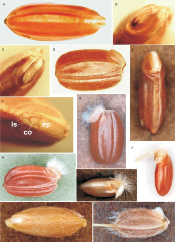
Normally, at the beginning of rice seed germination, the lateral scales (also called auricles; Saha, Reference Saha1957; Tateoka, Reference Tateoka1964), the epiblast and the coleorhiza swell, causing the pericarp to split open (S.X. Xu et al., Reference Xu, Xu and Han1983; X.B. Xu et al., Reference Xu, Han and Zee1989) along the ventral junction of the caryopsis coat (i.e. the fused seed coat/pericarp) covering the embryo (Suzuki et al., Reference Suzuki, Taniguchi and Maeda1991; Fig. 1A). Frequently, the ventral junction starts fissuring above the epiblast (Fig. 1B) and the splitting then extends along the junction to the coleorhizal end of the embryo, at the ventrobasal tip of the caryopsis (Fig. 1C). Indeed, for non-dormant dehulled caryopses the first visible signal of germination is pericarp splitting (Footitt and Cohn, Reference Footitt and Cohn1995; Gianinetti and Vernieri, Reference Gianinetti and Vernieri2007). Notably, the lateral scales, the epiblast and the coleorhiza are all extensions of the embryo collar, i.e. the grass hypocotyl that is expanded to form a bulging tissue that covers and envelops both the shoot and the radicle primordia (Tillich, Reference Tillich2007). The embryo collar can be recognized by its ability to develop particular epidermal hairs, the rhizoids (see below), which are shoot (hypocotylar) trichomes and not root hairs (Tillich, Reference Tillich2007). These epidermal hairs form on the hypocotyl or on the collet region (the hypocotyl–radicle transition zone) and facilitate seedling anchorage to the substratum and water uptake well before the development of root hairs (Parsons, Reference Parsons2009; Sliwinska et al., Reference Sliwinska, Mathur and Bewley2012).
Normal germination (at 30°C) of non-dormant (afterripened for 16 weeks at 30°C) red rice dehulled caryopses. (A) The splitting of the caryopsis coat along the ventral junction above the embryo (ventral view of the seed, the embryo is on the right, the whitish bulging collar is visible through the splitting of the brick-red-coloured coat; about 22 h of imbibition). Frequently, (B) the ventral junction starts loosening above the epiblast (i.e. the area at the centre of the figure). Then (C) the splitting extends to the coleorhizal end of the embryo, at the ventrobasal tip of the caryopsis (20× magnification), while (D) the embryo is slightly protruding (lateral view). Thereafter, (E) the embryo collar begins to swell such that the caryopsis coat covering the embryo is separated in two split-halves (ventral view; 26 h; a slight excess of water prevented the formation of collar rhizoids). At 20× magnification, (F) the lateral scales (ls), the epiblast (ep) and the tip of the coleoptile (co) are clearly distinguishable (ventral view; 28 h). Under aerated conditions, (G) in a few hours after the splitting of the ventral junction, many rhizoids grow on the embryo collar, which is sheltered on both sides by an embryo coat split-half (32 h). At this stage, (H) the embryo often appears entirely hidden by a dense tuft of thin rhizoids. Then, (I) the radicle elongates first (40 h), while (J) the coleoptile growth lags (48 h). Visible germination of a spikelet (2 d of imbibition; a slight excess of water prevented the formation of collar rhizoids): (K) ventral view of the dispersal unit (the lower glume, i.e. the one above the embryo, had been removed); (L) lateral view, the coleoptile is slightly protruding and is shielded on the right by the epiblast.

After breaking apart the caryopsis coat, the embryo collar swells and protrudes (Fig. 1D) and the caryopsis coat above the embryo is separated in two split-halves (Fig. 1E), while the coleoptile begins to emerge through the Y-shaped gap between the lateral scales and the epiblast (Xu et al., Reference Xu, Xu and Han1983; Fig. 1F). When the embryo collar swells further, the two split-halves of the caryopsis coat fully open and rhizoids very quickly develop on each part of the collar, namely the lateral scales, the epiblast and the coleorhiza (Figure 1G, H; see also the supplementary video). However, if the seeds are submerged in water, no epidermal hairs would develop (Xu et al., Reference Xu, Xu and Han1983; Suzuki et al., Reference Suzuki, Taniguchi and Maeda1991). Thereafter, the shoot, or acrospire (the coleoptile with the plumule it envelops), begins to grow out of the Y-shaped gap and of the ventral slit of the caryopsis coat. Under aerated conditions, this is accompanied by the prompt growth of the radicle (primary seminal root), which, unlike the coleoptile, has to break through the coleorhiza (as the radicle is a shoot-born root; Tillich, Reference Tillich2007). Once the radicle has penetrated through the coleorhiza, it elongates faster than the coleoptile, at least in aerobic conditions (Xu et al., Reference Xu, Xu and Han1983; Fig. 1I, J). The relative development of the coleoptile and the radicle is regulated by means of phytohormones, such ethylene (Gianinetti et al., Reference Gianinetti, Laarhoven, Persijn, Harren and Petruzzelli2007).
This work aims to characterize an anomalous form of germination observed when red rice dormant dehulled caryopses are incubated in water for a long time. This phenomenon, described here for the first time, shows peculiar features that reveal how the caryopsis coat contributes to maintain dormancy and how its failure affects the transition from dormancy to germination.
Materials and methods
Plant material
A population of straw-hulled red rice (Gianinetti and Vernieri, Reference Gianinetti and Vernieri2007; Gianinetti et al., Reference Gianinetti, Laarhoven, Persijn, Harren and Petruzzelli2007) was multiplied in a greenhouse. The seeds were harvested when showing shattering capability. After harvest, the spikelets were dried for 1 d at 35°C and stored at –18°C to preserve dormancy. Samples of the seeds were afterripened in closed containers at 30°C for 16 weeks to obtain non-dormant seeds before manual dehulling. In these seeds, 50% pericarp splitting is reached at about the 18th hour of imbibition in water (not shown). The initial viability of the seeds was estimated as the sum of the percentage of seeds germinating following dry-afterripening (98.8%; 250 seeds tested) plus those germinating after a viability test (Gianinetti et al., Reference Gianinetti, Laarhoven, Persijn, Harren and Petruzzelli2007) of the afterripened seeds that had not germinated, and it amounted to 99.6%.
Experimental set-up
Germination was performed at 30°C in disposable round Petri dishes (9-cm diameter; enclosed in a humidity box). For each treatment, replicated dishes were prepared by placing 20 caryopses on two filter-paper discs with 5 ml of water. During long incubation periods, water was added when necessary, without ever letting the dishes dry out (i.e. seeds were always kept fully imbibed). Unless otherwise specified, incubation was in the dark, although inspection (about twice a week) and documentation required exposure to the light. However, separate experiments were set up for documentation and for precise assessment of the germination time course (which was obtained as average of 14 Petri dishes; that is, 280 caryopses). During incubation, besides either pericarp splitting or tearing of the caryopsis coat, the first growth stage (S1, Counce et al., Reference Counce, Keisling and Mitchell2000) was recorded for each seed when the rootlet or coleoptile were actively growing to ≥1 mm (minimal visible seedling growth). Although growth stage S1 is a conventional stage at which germination is recorded, it is also the usual sign that, from a physiological point of view, this process has concluded, and it therefore signals the onset of seedling growth as well as the conventional end of germination (Gianinetti et al., Reference Gianinetti, Laarhoven, Persijn, Harren and Petruzzelli2007).
The imbibition medium maintained a pH of 5.5–6 (assayed throughout the experiment with universal indicator paper; Riedel-de Haën, Seeize, Germany). Examination of the seeds was performed with the aid of a magnifier, and plastic (PMP) tweezers were used for handling the seeds. After long incubations, in one experiment a microscopic growth of a blue-green mould was detected on some seeds in some dishes. To block such infection, the seeds of each infected dish were transferred to two new Petri dishes with 5 ml of water: one for the uninfected seeds and one for the infected ones, and the latter were soaked in a saturated solution of sodium bicarbonate (adapted and modified from Latifa et al., Reference Latifa, Idriss, Hassan, Amine, El Hassane and Abdellah2011) for 5 min, blotted, and then placed into the new dish (without rinsing). Evidently, the resulting pH of the imbibition medium was increased after this treatment, but with time (>5 months) it returned to slightly acidic values. The caryopses treated with the bicarbonate solution did not show any subsequent growth of the blue-green mould, but they turned darker in 1–2 d. However, no significant difference in germination was observed in these dishes with respect to the others (not shown).
Fluridone, an inhibitor of abscisic acid (ABA) synthesis, was added to the incubation medium at a final concentration of 10 μM, just before incubation (Gianinetti and Vernieri, Reference Gianinetti and Vernieri2007). For this treatment, some caryopses (three replications of 10 seeds each) were transferred to new Petri dishes containing the fluridone solution after 9 months of incubation in water.
Image collection and data analysis
Regular photographs were taken with an Olympus E-510 digital camera using a 50-mm 1:2 macro, an extension tube EX-25 and a combined HOYA HMC 52-mm Close-Up lens set (+1, +2, +4), and employing a ring flash RF-11. Details were photographed with a BX51TF microscope (Olympus Corporation, Tokyo, Japan) equipped with a Microscope Digital Camera System DF50, and captured with Viewfinder Lite 1.0 software (Pixera Co., Osaka, Japan).
Distribution fitting was performed with Systat 12 software (SPSS Inc., Chicago, Illinois, USA) by using the maximum likelihood procedure for the estimation of the distribution parameters from the probability density distribution (additional details are given in the supplementary statistical notes).
Results
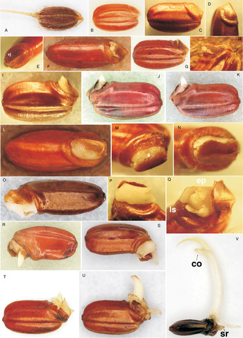
In contrast to the prompt germination (about 1–2 d for dehulled caryopses, and 2–5 d for spikelets, at 30°C) of non-dormant seeds (dry-afterripened for ≥16 weeks at 30°C), fully dormant seeds remain ungerminated and can persist in the Petri dishes for long times. Most spikelets of the studied red rice population can stay imbibed and dormant for a year or more when incubated in Petri dishes (Fig. 2A). However, dormant dehulled caryopses (Fig. 2B) progressively germinated over the same span of time (Fig. 3A). In particular, along several months of incubation (Fig. 3A) an increasing number of dehulled caryopses showed a germination that became anomalous in various respects, as documented here. Some interesting inferences can be obtained from this.
Imbibed (at 30°C) dormant red rice seed. (A) Spikelet (1 year of incubation). (B) Dehulled caryopsis (2 weeks of incubation). The scutellum of dehulled caryopses gradually swells and forms an annular bulge around the embryo base (pictures taken after about 1 year of incubation): (C) lateral view, (D) ventro-lateral view of the embryo, (E) viewed from above the embryo [the ventral junction (vj) is clearly visible]. Subsequently, (F) the caryopsis coat covering the embryo is torn apart from the side (more or less extensively) and, often, (G) the embryo tip sticks out and can remain at such a stage for many days (about 41 d, in the case of the seed in the figure). (H) The 20× magnification shows the detail of the coleoptile that juts out. Sometimes, (I) the tearing of the caryopsis coat extends along one side of the embryo toward the coleorhiza (20× magnification; ≤ 3 d in this stage), and the seed can remain at such a stage for several days: (J) a caryopsis at this stage for about 24 d (the edges of the tear have darkened); (K) the same caryopsis after about 52 d at this stage, the acrospire has minimally extended and the tear has barely widened (this caryopsis had germinated 3 d later). In every case, after some time (1 d to a couple of months), (L) the embryo collar swells and the caryopsis coat is suddenly lifted from the side; (M, N) details at 20× magnification of different caryopses. Thereby, embryo growth is resumed and germination is elicited: (O) the collar rapidly becomes prominent and the caryopsis coat above the embryo is rotated on one side (≤90°) like a hinged lid that is opened. Even under aerated conditions, (P) most often collar rhizoids do not develop, and therefore (Q) the epiblast (ep) and the lateral scales (ls) remain clearly visible (20× magnification). (R) The seminal radicle emerges through the coleorhiza, causing (S) the coat above the embryo to rotate beyond 90°, and then (T) even the coleoptile starts elongating. (U) The coleoptile grows until (V) the plumule emerges (sr, seminal radicle; co, coleoptile).

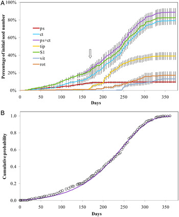
Germination time course of dormant dehulled red rice seeds. Data points are means ± SE of 14 dishes with 20 caryopses each. (A) Data are shown for pericarp splitting (ps), coat tearing (ct, which includes both seeds that promptly progressed to the seedling stage and seeds that lagged at this stage before growing), seeds with the embryo tip sticking out of the pierced caryopsis (tip, which is included in ct), ps+ct, the first growth stage (S1, i.e. rootlet or coleoptile were actively growing to ≥1 mm), seeds showing some vitrification (vit, recorded at S1), rotted seeds (rot, at any stage before S1). The vertical arrow indicates the time at which most of the seeds (about 95%) showed an evident annular bulge. (B) Datapoints for ps+ct (expressed as proportions of the final value) are fitted by a cumulative Gompertz distribution, G(t, k, α) = 1 − exp( − k(e αt − 1)/α), with k = 0.000350, α = 0.015421 and R2 adj = 0.999.

In many imbibed dormant caryopses, a first sign of the incumbent anomalous germination was the gradual formation of an unusually large annular bulge around the embryo base (Fig. 2C, D, E), apparently caused by the swelling of the scutellum. It developed gradually and at different times for the diverse seeds. Anyway, almost all the ungerminated seeds showed a more or less prominent annular bulge after about 6 months of incubation (this trait is not exactly definite, though, and therefore its accomplishment is a little subjective). Thence, after a few weeks to some months, the seed coat/pericarp covering the embryo of these caryopses was torn apart by the bulging collar (Fig. 2F), and the embryo tip stuck out of the pierced caryopsis coat (Fig. 2G). The tearing of the seed coat/pericarp always started above the lateral scales and the epiblast that, therefore, was the first part of the white embryo to appear through the fissuring of the red caryopsis coat, whereas the coleoptile was the first to jut out (Fig. 2H). This stage, characterized by some coat tearing and, commonly, by the embryo tip that sticks out of the pierced caryopsis coat but does not show subsequent growth, could persist for a few days to some weeks (for example, the seed in Fig. 2G has remained at this very stage for at least 41 d). The maximum recorded persistence in this stage was 53 d. Noteworthy, the presence of seeds with the embryo tip that sticks out of the pierced caryopsis coat became detectable (Fig. 3A) only when this stage had persisted for some time; otherwise, in normal germination, this is a very ephemeral condition, too short-lived to be observed systematically. Almost at the same time, the curves for the rupture of the caryopsis coat (by either pericarp splitting or coat tearing) and growth stage S1, diverged (plots for ps+ct and S1 in Fig. 3A), further indicating that these two stages became temporally separated at this time. In some instances, the tearing of the caryopsis coat extended along one side of the embryo toward the coleorhiza (full lateral tearing), still without it being immediately followed by seedling growth (Fig. 2F, I). Differences in the extent the tip protrudes from the caryopsis are fixed at the moment immediately after the collapse of the caryopsis coat covering the embryo and practically no growth occurs during this resting stage. So, the small expansion growth consequent to the sudden relief of the embryo compression and the transfer of the full turgor stress from the torn caryopsis coat to the embryo cell walls, does not show any follow up; that is, no further creeping of the cell walls and extension of the embryo tip occur subsequently (compare Fig. 2J and 2K), at least until germination growth is abruptly reactivated. Indeed, after some time, the embryo collar suddenly started swelling and, in 1 or at most 2 d, it was so prominent that the caryopsis coat was lifted from the side (Fig. 2L, M, N) like the lid of an opened can (Fig. 2O, P). Thus, in all cases the tearing of the caryopsis coat ultimately extended along one side of the embryo to the area above the coleorhiza (full lateral tearing) and this was followed by seedling growth (recorded in Fig. 3A as stage S1, that is, rootlet or coleoptile ≥1 mm; with the caveat that they, specifically the coleoptile, were actively growing, which was added for consistency with the findings of the present study). From there on, the sluggish germination gradually speeded up. No, or only a few, embryo collar rhizoids developed (Fig. 2P), and then the epiblast and the lateral scales remained clearly visible (Fig. 2Q). As normal in aerobic conditions, the seminal radicle promptly emerged through the coleorhiza (Fig. 2R, S), and then the collar further expanded and the coleoptile started to elongate (Fig. 2T). The latter grew (Fig. 2U) until the plumule emerged and new radicles eventually developed (Fig. 2V). In many cases, the seminal radicle soon stopped growing (Fig. 2V), but in some cases it grew normally.
Figure 3 shows that dormant dehulled red rice caryopses that do not show normal germination during the first 2–4 weeks of incubation in water (i.e. during the usual time for testing dormancy), can very gradually germinate in the subsequent months. Since these caryopses undergo anomalous germination, the rationale for limiting tests of germination and dormancy to 2–4 weeks, when anomalous germination is negligible, is confirmed. Notably, the overall time course of the anomalous germination extended for almost 13 months (when, finally, all the seeds in the experiment had either germinated or rotted) and showed an asymmetric sigmoid shape (Fig. 3A). The cumulative percentage of seeds that germinated by tearing away from the side the coat covering the embryo (Fig. 2F–Q) overtook the cumulative percentage of seeds that germinated by opening the coat along the ventral line of junction, i.e. pericarp splitting (Fig. 1A–H), after about 3 months of incubation (Fig. 3A). The number of newly germinating seeds that showed the (normal) opening of the coat along the ventral line of junction dropped close to zero after about 6 months of incubation (Fig. 3A). It is worth noting that, already, a very few caryopses germinated by tearing away the coat covering the embryo soon after the first 2–3 weeks of incubation (during which time very few seeds germinate because of their poor dormancy level, and pericarp splitting is the usual mode of starting germination); that is, well before the appearance of the other peculiar aspects of the anomalous germination described above (Fig. 3A). During the first months of incubation the subsequent growth of the eventually germinating seeds was normal, independently of whether they germinated by pericarp splitting or by coat tearing. The fully distinctive traits of the anomalous germination (specifically, the time lag between the initial rupture of the caryopsis coat and the start of seedling growth) were observed solely after several months of incubation, at least six in the case of the red rice genotype studied here, and only after about 9 months of incubation were all these traits shown by almost all the germinating seeds (Fig. 3A). Incidentally, besides seeds treated with bicarbonate solution, over long incubations all red rice caryopses gradually turned darker, but this effect was quite variable among Petri dishes (not shown) and such variability did not appear to be linked to the anomalous germination. Indeed, many months commonly passed between the anomalous germination of the first and last seeds within each dish (not shown).
The percentage of seeds that germinated by tearing of the caryopsis coat (the final value of which was 79.3% and included seeds whose coats were initially ruptured by either the emerging embryo tip or directly by full lateral tearing) closely approached the percentage of seeds that reached the minimal visible seedling growth (rootlet or coleoptile actively growing to ≥1 mm – that is, growth stage S1 – the final value of which was 82.9%), even though a portion of seeds germinated by pericarp splitting during early incubation (Fig. 3A). Thus, the overall percentage of seeds showing a rupture of the caryopsis coat (i.e. the sum of seeds germinated by pericarp splitting plus those germinated by coat tearing, whose final value was 88.8%) is higher than the number of seeds that reached the minimal visible seedling growth (Fig. 3A), and this is essentially due to a significant mortality of seeds resting at a stage at which the protection offered by the caryopsis coat is disrupted, as can be seen by the steep increment of rotted seeds following the rise in the percentage of seeds resting with the embryo tip sticking out (Fig. 3A; the final percentage of seeds that persisted in this stage for enough time to be detected was 39.1%). Evidently, the lag, sometimes a long one, between the initial rupture of the caryopsis coat (with the eventual protrusion of the embryo tip) and the start of seedling growth renders these seeds more susceptible to infection by microorganisms. Throughout the experiment, mortality (i.e. the percentage of rotted seeds) totalled 17.1%, of which 5.9% occurred after pericarp splitting or coat tearing but prior to growth stage S1, whereas the remaining 11.2% was due to seeds rotting before the caryopsis coat was ruptured either way.
The time course of the anomalous germination followed an asymmetric sigmoid curve (Fig. 3A), consistent with a Gompertz cumulative distribution G(t, k, α) = 1 − exp(−k(e αt − 1)/α) with k, α > 0 and t ≥ 0, where k and α are parameters and t is time (Johnson et al., Reference Johnson, Kotz and Balakrishnan1995). The shape of the distribution is given by parameter k, whereas α is a scale parameter that only affects the time scale of the process (Johnson et al., Reference Johnson, Kotz and Balakrishnan1995). This distribution gives the cumulative probability of failure, that is, of not attaining time t, and as such it has value G(t, k, α) = 0 at time t = 0 and it tends to G(t, k, α) = 1 for an infinite time. For distribution-fitting, data were expressed as a fraction of their end value (as distributions assume maximum values of 1, and once all the seeds have either germinated or rotted, end values represent maximum values), and fitting was performed on the probability density distribution (see the supplementary statistical notes), which is the derivative function of the cumulative distribution. The obtained function parameters were used to plot the cumulative distribution function, which is displayed in Fig. 3B to show its fitting to cumulative data. From Fig. 3A it can be noted that both ct, ps+ct and S1 show very similar curves, and all three fit remarkably well a Gompertz distribution (see the supplementary statistical notes). The overall percentage of seeds showing a rupture of the caryopsis coat (ps+ct in Fig. 3A) was, however, chosen (additional details are given in the supplementary statistical notes) to be shown in Fig. 3B. It is worth noting that the main assumption underlying this choice is that the observed pericarp splitting could be due to a failure of the ventral junction consequent to the embryo thrust rather than to a programmed weakening of this specialized tissue, as occurs in normal germination. In fact, if, in the anomalous germination, failure can occur at the caryopsis coat around the embryo, it can occur at the ventral junction as well, since the latter is just the predetermined breaking site for germination, and can therefore be expected to fail first.
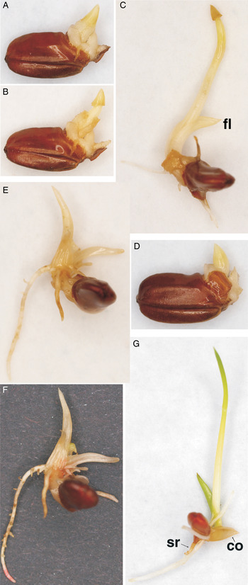
In the case of seeds that germinated after more than 8 months of incubation, sometimes the collar and also the coleoptile were swollen (Fig. 4A, D), and in some instances the seminal radicle, the first leaf, or even some other leaves, appeared vitrified (Fig. 4B, C, E). Nevertheless, often the seedling subsequently developed into a normal plant. Vitrification, or hyperhydricity, is the phenomenon by which leaves appear glassy, translucent, thick, rigid and brittle, with a turgid aspect (Ziv, Reference Ziv, Debergh and Zimmerman1991; Debergh et al., Reference Debergh, Aitken-Christie, Cohen, Grout, Von Arnold, Zimmerman and Ziv1992), and will not turn green when exposed to light (Fig. 4F). In the anomalous germination, however, most often the seedlings were only partially vitrified and subsequently developed normal greening leaves (Fig. 4G). Typically, vitrification is observed in vitro for tissues that are differentiated from cultured embryos or calluses (Ziv, Reference Ziv, Debergh and Zimmerman1991). The present observations show that a more or less intense vitrification can occur in germinating seeds too, though only after a long incubation (Fig. 3A). This suggests some similarity between the development of the embryo following anomalous germination and its culture in vitro. It also highlights that in the anomalous germination the metabolism of the seed might differ from that of normal germination, and in a very few cases the way to normality is permanently lost.
Some seedlings produced by anomalous germination show varying degrees of vitrification. (A, B, C) Development of a partially vitrified seedling: (A) both the embryo collar and the coleoptile are exceedingly swollen; (B) the coleoptile appears glassy and brittle, and its tip has been severed away by the growing plumule rather than being split open longitudinally as usual; (C) the first leaf (fl) is not swollen but resembles a coleoptile, while the plumule grows with a relatively normal aspect (but it still carries the detached coleoptile tip). (D, E, F) Development of a completely vitrified seedling: (D) as above, both the embryo collar and the coleoptile are swollen; (E) in the following 13 d the plumule has produced a few leaves that appear short, translucent and glassy; (F) growth has stopped and, even though the seedling has been transferred to conditions of 16 h of light per day for 10 d, the leaves have not turned green. Under the same conditions, (G) a seedling with vitrification limited to the coleoptile (co) and the seminal radicle (sr) shows greening leaves.

One problem with studying anomalous germination is that some data should be collected on every individual seed over a very long time span. For example, the duration of a given stage can be assessed only by measuring it for a number of individual seeds. This has been done on a restricted number of seeds, particularly toward the end of the experiment, when the single seeds were more easily identifiable, or on seeds that, earlier in the incubation time-course, were acknowledged to pass quickly through a given stage. However, this means that the reported times are indicative of just the most extreme times, that is, they represent the range of actual times. In addition, the slow germination increases the probability of infection by microorganisms, and this further complicates the study of anomalous germination.
Discussion
What is the relationship between dormancy and the anomalous germination?
In the soil, some dispersal units of red rice can survive for at least 10 years (Goss and Brown, Reference Goss and Brown1940; Delouche et al., Reference Delouche, Burgos, Gealy, de San Martin, Labrada, Larinde and Rosell2007), although most of them last only for a few months to a few years (Goss and Brown, Reference Goss and Brown1939; Vidotto and Ferrero, Reference Vidotto and Ferrero2000; Noldin et al., Reference Noldin, Chandler and McCauley2006). In Petri dishes, whereas almost all the viable red rice spikelets that do not germinate at optimal temperatures within 1–2 weeks of incubation persist in a dormant state for months or years (Fig. 2A; Gianinetti and Cohn, Reference Gianinetti and Cohn2008), dormant dehulled caryopses can very gradually germinate following the initial germination flush (Fig. 3) but, then, their germination assumes anomalous features (Fig. 2C–S).
The biological relevance of the anomalous germination is unclear, as the naturally germinating dispersal unit is not the dehulled caryopsis but the spikelet, and although the latter can show anomalous germination as well (not shown), this occurs later than for dehulled caryopses (apparently, about 8 months later, but only a few observations were available and no data are therefore shown), thus it is still more difficult to study. It is even unknown whether the anomalous germination occurs in the field, as specific observations are lacking. Anyway, the anomalous germination of caryopses in Petri dishes has a relevant physiological significance, since it reveals aspects of the dormancy/germination relationship that are not normally acknowledgeable.
Already in recently imbibed dehulled dormant seeds the scutellum shows a very slight tendency to bulge (Fig. 2B), indicating that it puts the caryopsis coat under tension. As a rule, expansion of the embryonic tissues is opposed by the restraint of the tissues enclosing them (Bewley et al., Reference Bewley, Bradford, Hilhorst and Nonogaki2013). In fact, the covering layers increase the yield threshold that must be exceeded to enable extension of the walls of the embryonic tissues (Schopfer, Reference Schopfer2006). Stress relaxation of the covering layers and consequent expansion of the internal tissues have a primary role in many instances of plant cell extension growth (Schopfer, Reference Schopfer2006). Presumably, as for primary walls, the caryopsis coat also shows a time-dependent extension (‘creep’) under load, and a time-dependent stress relaxation after stretching (Schopfer, Reference Schopfer2006), although such extension must be very limited, since the caryopsis coat is a quite rigid structure. In other words, the caryopsis coat is subject to wear and tear with time. Thus, the anomalous germination would occur because the turgid embryo stretches the covering layers until they fail and the protruding embryo can then continue to grow unrestricted by external forces.
Wide variations in the times and modes of germination (pericarp splitting, tearing of the caryopsis coat, and full anomalous germination) of dormant dehulled seeds might well be due to their different levels of dormancy, which correspond to a wide distribution of water potential thresholds for germination (Gianinetti and Cohn, Reference Gianinetti and Cohn2007), i.e. to diverse intensities of germination potential (as defined by Schopfer, Reference Schopfer2006) among seeds incubated in water. Germination requires this potential to be positive in the presence of an intact seed coat, whereas a negative germination potential signifies dormancy. This means that seeds with stronger dormancy exert a lower embryo thrust on the caryopsis coat, and therefore the degree of dormancy of each seed is expected to determine the time of coat breaking and germination. Thus, the most deeply dormant seeds, which have the weakest embryo thrust, would be the last to tear the caryopsis coat and those that show a long time-lag between the rupture of the caryopsis coat and the start of seedling growth. Clearly, besides the water potential threshold for germination, the readiness of each seed to germinate also depends on its level of dormancy, and this can be inferred here from the fact that non-dormant seeds, as well as poorly dormant seeds germinating in the first weeks of incubation, do not break the caryopsis coat randomly, but rather they show the splitting of the coat along the ventral junction as occurs in a programmed event of germination for fully afterripened seeds.
It is noteworthy that, if, after 9 months of incubation in water, dehulled caryopses (which did not show prior signs of coat tearing, but had an annular bulge, like every seed at this time point) are transferred to a solution containing fluridone, their dormancy is broken and they promptly germinate, but the coat covering the embryo is torn away from the side (not shown). This suggests that the surge in the embryo thrust consequent to the fluridone treatment precedes, and in these seeds prevents, any visible effect on the fissuring of the ventral junction (pericarp splitting), as is instead normally observed in more recently imbibed seeds (Gianinetti and Vernieri, Reference Gianinetti and Vernieri2007). The tearing of the coat covering the embryo even in caryopses induced to germinate by fluridone might well be due to an already relevant weakening of their caryopsis coat, an event that can indeed be expected after 9 months of incubation. Seedling growth follows promptly. Such a finding is interpreted as a confirmation that the metabolic blockage of germination after a long incubation is essentially still the same as that which can be removed by fluridone as observed (by Gianinetti and Vernieri, Reference Gianinetti and Vernieri2007) in the first weeks of incubation.
Anatomo-functional considerations regarding the anomalous germination
The fact that the collar completely envelops the embryo axis suggests that the former has a role in the early germination events in the anomalous as well as in the normal germination, by: (1) developing a positive embryo expansion potential (as defined by Schopfer, Reference Schopfer2006), and (2) controlling the enzymatic opening of the caryopsis coat along the ventral junction. Indeed, even in other species, it has been shown that germination first involves an increase in size of the cells near to the radicle–hypocotyl borderline and then further up the hypocotyl, whereas radicle emergence occurs only after these events (Musatenko et al., Reference Musatenko, Gašparíková, Martyn, Sytnik, Brouwer, Gašparíková, Kolek and Loughman1981; Sliwinska et al., Reference Sliwinska, Bassel and Bewley2009).
The fact that, in the anomalous germination, the local failure of the caryopsis coat most commonly begins above the lateral scales, suggests either that the covering layers are weaker or that the thrust is stronger in this area. Indeed, radicle extension is also constrained by the collar, which has no gap in the coleorhizal area. As many seeds remain blocked for several days at a stage characterized by some coat tearing, and eventually by the embryo tip that sticks out of the pierced caryopsis, without germinating or even showing any creeping (i.e. any slow expansion growth), and they commence to grow only at a later time, when the laceration of the caryopsis coat extends to the area above the coleorhiza, it seems that it is the expansion of the living tissues in the latter area that is effectively associated with the start of the active growth of the embryo. Accordingly, it has been proposed that, in grasses, the coleorhiza is functionally linked to dormancy by acting as a barrier to root elongation, thereby playing a major role in preventing germination (Barrero et al., Reference Barrero, Talbot, White, Jacobsen and Gubler2009). Since, differently from the acrospire, the radicle has to pierce through the collar to emerge, it is possible that it is precisely this action that elicits seedling growth. In fact, the displacement of the caryopsis coat covering the embryo is actuated by the emerging radicle, and this always precedes visible seedling growth. Indeed, severing, pricking or wounding the living tissues of the seeds is an effective way to break dormancy and induce germination (Cohn and Hughes, Reference Cohn and Hughes1981; Doherty and Cohn, Reference Doherty and Cohn2000; Gianinetti et al., Reference Gianinetti, Laarhoven, Persijn, Harren and Petruzzelli2007). Thus, it can be hypothesized that the failure of the caryopsis coat above the coleorhiza would leave the collar as the only restraint to the thrust of the radicle, which is then expected to push, stretch and pierce the collar itself. Ultimately, this would cause some failure even in the collar, which, differently from the caryopsis coat, is a living tissue, and can therefore activate a wounding response that, in turn, triggers seedling growth. Thus, once the radicle has emerged, germination (in the physiological sense) is a matter of fact.
The anomalous germination defies the traditional boundary between dormancy and germination
The fact that the caryopsis coat covering the embryo is stripped away from the side, rather than becoming unstuck along the ventral line of the junction, and the lack of development of embryo collar rhizoids, indicate that the anomalous germination is a much less coordinated process than normal germination. Interestingly, cells destined to become trichomes must enter an endoreduplication cycle (Ishida et al., Reference Ishida, Kurata, Okada and Wada2008) and, specifically, the elongation of the cells of collet hairs (trichomes) during early seedling growth is coupled with endoreduplication (Sliwinska et al., Reference Sliwinska, Mathur and Bewley2012). In arabidopsis, even completion of germination (i.e. radicle protrusion) is the result of cell expansion within the lower hypocotyl and the hypocotyl–radicle transition zone, consequent to endoreduplication (Sliwinska et al., Reference Sliwinska, Bassel and Bewley2009). So, the almost complete absence of embryo collar rhizoids in the anomalous germination of red rice caryopses (Fig. 2P–S) and the lack of radicle protrusion in seeds with the embryo tip sticking out of the pierced caryopsis coat (Fig. 2G–K), suggest that, initially, DNA replication does not accompany expansion as usually happens in normal aerobic germination. This is somewhat baffling, as DNA replication has been proposed to be essential for progression into germination (Vázquez-Ramos and Sánchez, Reference Vázquez-Ramos and Sánchez2003). On the other hand, it can be hypothesized that the seeds resting at a stage wherein the caryopsis coat is ruptured but the radicle does not emerge, have not actually germinated but, rather, are still dormant. This intriguing aspect of the anomalous germination can be approached from a more general point of view, as follows.
Unlike most other angiosperm species, in the grasses the primordia of some foliage leaves are already present in the embryo (specifically, three in the case of rice; Itoh et al., Reference Itoh, Nonomura, Ikeda, Yamaki, Inukai, Yamagishi, Kitano and Nagato2005). Indeed, the very advanced development of the embryo in the seed, with a well-differentiated radicle, and young leaves visible inside the coleoptile, is a character unique to the Poaceae (Tillich, Reference Tillich2007). Actually, both embryogenesis and part of the juvenile vegetative phase occur during seed development (Asai et al., Reference Asai, Satoh, Sasaki, Satoh and Nagato2002; Itoh et al., Reference Itoh, Nonomura, Ikeda, Yamaki, Inukai, Yamagishi, Kitano and Nagato2005). Subsequently, maturation and dormancy processes take place. This means that early vegetative stages elapse during seed development and are incorporated into embryos before dormancy takes place (Asai et al., Reference Asai, Satoh, Sasaki, Satoh and Nagato2002), thus the embryo in a ripe caryopsis resembles a resting seedling (Tillich, Reference Tillich2007). There is therefore a temporal reversion of embryo dormancy and early vegetative stages, which suggests that grasses can be regarded as heterochronic species; that is, they show an altered sequence of development, with a shift of the dormant period from the end of embryogenesis to the end of the juvenile phase (Asai et al. Reference Asai, Satoh, Sasaki, Satoh and Nagato2002; Itoh et al. Reference Itoh, Nonomura, Ikeda, Yamaki, Inukai, Yamagishi, Kitano and Nagato2005). Thus, the transition from a resting phase (dormancy) to a growth one (germination) is more a physiological switch than a sharp change of the morphological state (i.e. of the degree of development); although, typically, these two aspects (physiological and morphological) are closely linked, at least in a normal developmental programme, wherein the piercing of the caryopsis coat is a reliable morphological sign of the physiological transition from a developmental arrest to germination. When, instead, the two aspects are decoupled, the switching point has to be established on the basis of what the seed actually does: grow or rest. Hence, the seeds attaining the piercing of the caryopsis coat and the protrusion of the embryo tip, but resting at that point, appear to be just on the verge of the transition. However, the transition is not complete until their embryo axes really start to grow. If this is true, then, unlike normal germination, in the anomalous germination process the tearing of the caryopsis coat is not, by itself, a sign of germination. This is in agreement with the hypothesis made above that the seeds resting at this stage are still dormant.
Although this conclusion may appear puzzling at first, because it defies the traditional view on how to distinguish a dormant seed from a germinating one, it is not at odds with our general understanding of dormancy and germination. A gradual, reversible transition from dormancy to germination (Rajjou et al., Reference Rajjou, Duval, Gallardo, Catusse, Bally, Job and Job2012) is consistent with studies showing a concurrence between germination and dormancy induction in red rice seeds that are prevented from germinating (Gianinetti and Cohn, Reference Gianinetti and Cohn2008), and with a model that assumes the speed of germination of partially dormant seeds to depend upon the level of residual dormancy of the seeds (Gianinetti and Cohn, Reference Gianinetti and Cohn2007). In both cases, a trade-off between dormancy and germination occurs, rather than a sharp cut-off. Although between dormancy and germination there is a developmental switch, such a switch does not need to be sharply apparent, as some physiological overlapping is indeed common. Thus, the rupture of the caryopsis coat by the protruding embryo can take place prior to the physiological switch from dormancy to germination (though this is not what normally happens).
Ultimately, the unprogrammed yielding of the caryopsis coat drives the seed to enter germination before the physiological switch has occurred naturally; that is, compelling the switch to happen. This latter aspect is the most remarkable feature of the anomalous germination.
A Gompertzian time-course for the anomalous germination
We can turn now to examine the quantitative expression of the anomalous germination as a function of the time of incubation in water. The time course of three traits recorded for the anomalous germination (namely, the coat tearing, the sum of pericarp splitting and coat tearing, and the attainment of growth stage S1; Fig. 3A) shows an asymmetric sigmoid shape that fits a Gompertz distribution (Fig. 3B). The cumulative Gompertz distribution is the super-exponential function: G(t, k, α) = 1 − exp(−k(e αt − 1)/α), where k, α > 0 and t ≥ 0 (Johnson et al., Reference Johnson, Kotz and Balakrishnan1995). The shape of the distribution is given by parameter k, whereas α is a scale parameter that only affects the time scale of the process (Johnson et al., Reference Johnson, Kotz and Balakrishnan1995). The Gompertz distribution is typically used for the analysis of survival and is characterized by a failure rate that increases exponentially over time; that is, the logarithm of the failure rate grows linearly (Kirkwood, Reference Kirkwood2015). This distribution gives the cumulative probability of failure, that is, of not attaining time t, and as such it has value G(t, k, α) = 0 at time t = 0 and it tends to G(t, k, α) = 1 for an infinite time. The basic rationale for observing this distribution is that failure of a given system, or function, is dependent on the wearing-out of constituent elements that are irreplaceable and highly redundant (Abernethy, Reference Abernethy1979; Gavrilov and Gavrilova, Reference Gavrilov and Gavrilova2001; Kirkwood, Reference Kirkwood2015). Milne (Reference Milne2008) made explicit a series of logical assumptions able to explain the failure pattern described by the Gompertz function. The supplementary file ‘Theoretical considerations on the Gompertzian time-course of the anomalous germination’ explains that these assumptions can be easily adapted to the features of the anomalous germination. This supports the theoretical consistency of the Gompertz distribution with the presently described phenomenon and, therefore, a causal connection between the features of the anomalous germination and the fact that it turns out to have a Gompertzian time-course.
From a physiological point of view, the interesting feature is that these assumptions are entirely based on the mechanical properties of the caryopsis coat, and specifically upon the wearing-out of its capability to withstand embryo turgor. This quantitative model analysis corroborates anatomical observations and supports the idea that the factor that actually causes and shapes the anomalous germination of red rice dormant seeds is indeed the mechanical failure of the coats. Although other physiological controls must be in force too, a structural role of the caryopsis coat in dormancy maintenance is evident.
The role of the caryopsis coat
There are at least two locations for dormancy mechanisms in warm-season grass seeds with primary dormancy (Adkins et al., Reference Adkins, Bellairs and Loch2002): mechanisms based within the embryo-covering structures and mechanisms based within the embryo. The role of the caryopsis coat in maintaining seed dormancy cannot be tested directly by removing it, because the living tissues under it (i.e. either the aleurone layer or the embryo) could be very easily damaged by a mechanical intervention to remove the coat, thereby triggering a wounding response. However, just because there is an accelerated, unprogrammed loosening of the caryopsis coat, the anomalous germination allows us to probe the effect of the caryopsis coat on dormancy. This means that the two mechanisms envisaged by Adkins et al. (Reference Adkins, Bellairs and Loch2002) can be told apart.
The experiment with fluridone confirms that, as first noted by Esashi and Leopold (Reference Esashi and Leopold1968) in Xanthium, the thrust developed by non-dormant seeds is adequate to pierce the coat, and that developed by the dormant seeds is not; with this difference being due principally to the active enlargement of the embryo axis. In this sense, a population-based modelling approach (Gianinetti and Cohn, Reference Gianinetti and Cohn2007) showed that when red rice dormancy is relieved by dry-afterripening the water potential thresholds for germination of the seed population, which roughly (i.e. apart from the effect of weakening the covering structures, as discussed below) correspond to the germination potentials as defined by Schopfer (Reference Schopfer2006) with a change of sign, are shifted from values unsuitable for germination to values allowing germination. Accordingly, albeit in a reverse approach, studies with testa mutants of arabidopsis (Debeaujon and Koornneef, Reference Debeaujon and Koornneef2000) showed that when the restraint of the seed envelopes is weakened by mutations, the growth potential threshold required for germination is lowered. In accordance with the latter study, the present findings prove that if the yield threshold that must be exceeded for enabling germination is lowered (by a failure of the caryopsis coat, in this case), many seeds that were dormant when freshly incubated in water can then germinate because their germination potentials (as defined by Schopfer, Reference Schopfer2006) are suddenly shifted from negative to positive values. So, the envelopes are certainly involved in the maintenance and intensity of seed dormancy (Debeaujon and Koornneef, Reference Debeaujon and Koornneef2000; Schopfer, Reference Schopfer2006). However, the present study also suggests that seeds with strong dormancy can still maintain germination potentials below zero, that is, they do not germinate (actually, they do not show the growth typically associated with germination), even though the constraints of the hull and of the caryopsis coat are removed and a small expansion has occurred: the full turgor stress is transferred from the caryopsis coat to the embryo cell walls (which are then stretched as they now bear the full hydrostatic pressure of the embryo, and this allows for the small expansion growth observed), but evidently their actual yield threshold is not exceeded.
Although any tissue that exerts a mechanical restraint to the expansion of the embryo is expected to enforce dormancy (Debeaujon and Koornneef, Reference Debeaujon and Koornneef2000; Bewley et al., Reference Bewley, Bradford, Hilhorst and Nonogaki2013), the covering layers have a pre-established, passive role, acting as an external mechanical constraint to embryo expansion (Schopfer, Reference Schopfer2006). Thus, in normal germination (which means that dormancy has previously been relieved), it is the proactive action of the embryo tissues, such as the embryo collar, that is responsible for making the way of the acrospire and radicle through the caryopsis coat, and the siliceous lemma as well (Fig. 1K, L) (Xu et al. Reference Xu, Xu and Han1983). Typically, the mechanical constraint provided by the covering layers is overcome by a coordinated release of hydrolytic enzymes, which determines a regulated tissue weakening and rupture at predetermined breaking points (Finch-Savage and Leubner-Metzger, Reference Finch-Savage and Leubner-Metzger2006). In accordance, it has been shown that, in muskmelon, the testa does not appear to present a significant mechanical restraint to radicle emergence as the halves of the testa often split open slightly at the tip during imbibition, and should not directly restrict radicle growth (Welbaum and Bradford, Reference Welbaum and Bradford1990). This regulated weakening and rupture at a predetermined tissue is also observed in red rice, in which normal germination occurs by splitting the caryopsis coat, not by tearing it away. So, even though the thrust developed by caryopses whose dormancy has been broken by fluridone is adequate to push the coat apart, in more common conditions the increase in germination potential of imbibed non-dormant seeds is typically accompanied by the weakening and fissuring along the ventral junction. A similar programmed weakening is expected to work for the hull too, which during germination is split apart at a pre-fixed site above the ventral junction (Fig. 1K).
Therefore it is the mechanisms based within the embryo that rule the timing of the exit from dormancy, through both an increase of the thrust developed by the embryo (i.e. its germination potential) as well as an acquired ability to weaken the tissues that constrain it. Hence, there is a conceptual difference between the depth of dormancy as measured in terms, for example, of the time of dry-afterripening required to fully overcome dormancy (which is expected to measure the level of dormancy within the embryo) and the enforcement of dormancy in dormant seeds by the covering structures. The latter process helps to substantially reduce the chances of anomalous germination (it seems to delay it for several months at least), but it has almost no effect on the capability of the non-dormant seed to germinate once the physiological dormancy has been fully broken. This is why the afterripening time required to overcome dormancy is quite similar whether assessed on spikelets or dehulled caryopses, even though a relevant effect of the hull on the observed level of germination of a seed population is displayed at intermediate levels of dormancy (Roberts, Reference Roberts1961; Delouche and Nguyen, Reference Delouche and Nguyen1964; Cohn and Hughes, Reference Cohn and Hughes1981), probably because of an incomplete acquisition of the capability to weaken the covering structures in seeds whose dormancy is not yet fully released.
Ultimately, the anomalous germination evidences that the contribution of the caryopsis coat to seed dormancy is ancillary to the mechanisms based within the embryo, in the sense that the seed-covering structures enforce the depth of dormancy as measured in terms of germination potential (as defined by Schopfer, Reference Schopfer2006), but they do not affect the still unidentified physiological modulators that determine dormancy (Gianinetti and Vernieri, Reference Gianinetti and Vernieri2007) and eventually the time to full afterripening.
Conclusions
The main conclusions of this study are that: (1) the seed coverings contribute passively to restrain the growth (expansion) of the embryo and, therefore, germination; (2) embryo growth (by cell expansion) can be triggered by the failure of the caryopsis coat even if seed dormancy has not been previously relieved, and thus germination is started and the removal of dormancy is forced to occur; (3) this finally occurs also in seeds with strong dormancy, which apparently maintain a germination potential below zero even after the failure of the caryopsis coat and therefore show coat tearing, with an embryo tip that often sticks out of the pierced caryopsis coat, but do not progress immediately to normal growth.
To explain these features of anomalous germination, and especially the latter finding, the following mechanism is proposed: (a) seed dormancy limits the thrust of the embryo; that is, together with the covering coats, it prevents the initial cell expansion that usually starts seedling growth; in addition, (b) dormancy blocks other programmed steps of germination, such as pericarp splitting and cell division (this latter assures continuation of seedling growth); however, (c) after long incubations, in dormant dehulled seeds, some expansion growth can occur anyway because of the failure of the caryopsis coat (which gives a Gompertzian time-course to the anomalous germination), starting from the epiblast region (since the seed can rest at this stage before entering into active growth, it is suggested that the initial expansion growth is not necessarily a sign of germination); when (d) the failure of the caryopsis coat extends to the area above the coleorhiza, the radicle would also achieve some extension growth, thus stretching and piercing the coleorhiza; then, (e) after some time (probably varying with the depth of dormancy of each seed, since at this point seeds would still be dormant), this injury would become sufficient to induce a wounding response that suppresses dormancy and starts germination. Such a mechanism (the last two points of which are conjectural) can account for the most remarkable feature of the anomalous germination, i.e. the time lag, sometimes a long one, between the rupture of the caryopsis coat and the start of seedling growth, and therefore it provides an interpretative model to test in future studies.
Supplementary material
To view the supplementary material for this article, please visit http://dx.doi.org/10.1017/S0960258516000076
Supplementary statistical notes. These provide detailed considerations on the curve-fitting procedure adopted to build up Fig. 3B and additional comments about its interpretation.
Supplementary video. This shows the development of collar rhizoids during fully aerobic germination of a fully afterripened red rice caryopsis (lateral view).
Supplementary file: Theoretical considerations on the Gompertzian time-course of the anomalous germination. The theoretical assumptions entailed by the failure pattern described by the Gompertz function fit very well to the anomalous germination.
Acknowledgements
I thank Renzo Alberici for assistance with the photography.
Conflicts of Interest
None.