The bidirectional cavopulmonary shunt (modified Glenn shunt) provides increased systemic arterial oxygen saturation in patients with cyanotic cardiac disease without an increase in the ventricular workload or pulmonary vascular resistance. Long-term follow-up studies have shown that both the classic Glenn shuntReference Kopf, Laks, Stansel, Hellenbrand, Kleinman and Talner1 and the bidirectional Glenn shuntReference Lamberti, Spicer and Waldman2 provide excellent palliation with low mortality. However, late deterioration of the shunt may occur. Among the most common causes of a decrease in pulmonary blood flow years after palliation is the development of large venous collateral vessels from the superior caval vein to the inferior caval vein allowing desaturated blood to bypass the pulmonary bed.Reference Bargeron, Karp, Barcia, Kirklin, Hunt and Deverall3 A variety of obstructive lesions can form the substrate for the development of a host of possible decompressing channels. Such collateral vessels may develop in the absence of mechanical obstruction, and represent a “run-off” from the higher pressure caval venous system to the lower pressure atrium or hepatic vein. This results in increased cyanosis because of either a reduced effective pulmonary blood flow or an increased admixture of pulmonary venous return. Clinically, small collaterals will not lead to significant systemic desaturation. Large collaterals, however, result in clinically unacceptable desaturation and require intervention.Reference Magee, McCrindle, Mawson, Benson, Williams and Freedom4
Case report
We present an unusual case of progressive cyanosis in a child appearing several years after successful palliation of cyanotic cardiac disease with a bidirectional cavopulmonary shunt. The cause of the cyanosis was determined to be a large collateral decompressing the bidirectional Glenn shunt from the superior caval vein. We closed the collateral percutaneously with excellent results.
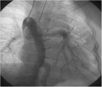
The patient was an 11-year-old child with complex cyanotic congenital cardiac disease with tricuspid atresia with pulmonary stenosis and had received a modified Blalock–Taussig shunt in infancy that was later revised to a bidirectional Glenn shunt at the age of 5 years. His growth after this shunt was excellent with significant clinical improvement. He, however, started noticing worsening cyanosis over the past year. The patient’s arterial saturation had fallen to 75% on room air and the haemoglobin level had risen to 19.5 grams per decilitre. Doppler echocardiography was suggestive of a vascular channel on the left side of the chest with flow in a caudal direction away from the heart. Cardiac catheterisation revealed that this vessel was a very large collateral connecting the superior caval vein to the tributaries of the inferior caval vein. The superior caval vein angiogram showed a large collateral arising from the superior caval vein and connecting to the supra-renal tributaries of the inferior caval vein with poor opacification of the pulmonary arteries (Fig 1). The haemodynamic effect of this collateral was to decompress the superior cavopulmonary shunt allowing desaturated blood to bypass the lungs and enter the inferior caval vein. The patient’s mean pulmonary artery pressure was 9 millimetres of mercury at the time of this catheterisation. The patient was taken up for percutaneous closure of the collateral.
Venous collateral connecting the superior caval vein to the inferior caval vein with poor opacification of the pulmonary arteries.

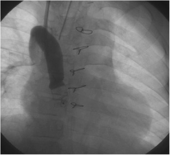
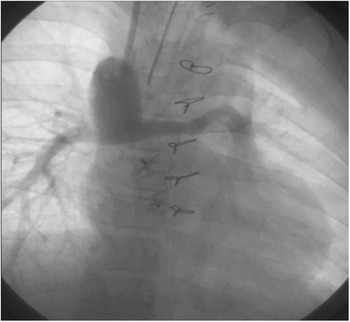
Under aseptic precautions, a right internal jugular vein access was taken with the Seldinger technique. A 035″ hydrophilic guide wire was passed into the superior caval vein through the right internal jugular vein. The wire was manoeuvred into the inferior caval vein through the collateral. A balloon flotation catheter (Swan Ganz) was tracked over the wire and positioned in the collateral. Test occlusion of the collateral with the inflated balloon showed significant improvement in systemic saturation and no haemodynamic instability, suggesting that the closure of the collateral would be beneficial. The size of the collateral was measured as 12 millimetres on fluoroscopy. The balloon was deflated and exchanged for an 8 French Cook’s Mullin sheath (Cook Incorporated, Bloomington, Indiana, United States of America). The Mullin sheath was positioned at the distal end of the collateral. A 14-millimetre Amplatzer vascular plug (AGA Medical Corporation, Plymouth, Minnesota, United States of America) was tracked through the sheath and positioned at the distal end of the collateral after withdrawing the sheath. Contrast injection through the sheath showed complete abolition of the shunt (Fig 2) and the vascular plug was released. Systemic saturation improved to 96%. The superior caval vein angiogram showed good opacification of the pulmonary arteries and no flow in the collateral (Fig 3).
Superior caval vein angiogram before device release showing complete occlusion of the collateral.

Superior caval vein angiogram after device deployment showing good opacification of the pulmonary arteries and complete occlusion of the collateral.

Discussion
With careful angiography, systemic venous anomalies can be found frequently after bidirectional cavopulmonary anastomosis. The incidence of venous collaterals after a Glenn shunt varies from 17% in one seriesReference Gross, Jonas, Castaneda, Hanley, Mayer and Bridges5 to as high as 31%, when they were actively sought. Large collaterals resulting in unacceptable desaturation require intervention. Collaterals between the superior caval vein and inferior caval vein can be safely left without intervention as this would be negated after completion of the Fontan circuit, unless they are so large as to significantly reduce effective pulmonary blood flow in the short term, as in the present case, in which a large venous collateral was decompressing the superior caval vein resulting in systemic desaturation and inadequate growth of pulmonary arteries. These collaterals can be managed surgically or percutaneously. Surgical intervention in the form of completion of Fontan was ruled out as it is known that the factors leading to systemic venous collateral channels may be indicative of a patient with suboptimal haemodynamic data for Fontan circulation and that pulmonary arteries had not grown adequately, and experience has shown that proper selection is probably one of the most important determinants of Fontan outcome. Coil occlusion of the venous collaterals has been performed successfully.Reference McElhinney, Reddy, Hanley and Moore6 A vascular plug has been used successfully to close a fistula between the superior caval vein and the left atrium.Reference Groote, Verhaaren, Suys and Wolf7 The collateral in the present case was too large to be occluded by a coil, and therefore we successfully closed the collateral with a vascular plug, as it does not have a retention disc like a device that may impinge on the walls of the collateral and erode it.
Conclusion
Systemic venous collaterals are frequently found after bidirectional cavopulmonary anastomosis. Progressive cyanosis is a clinical hallmark of this problem and requires investigation. These collaterals should be actively sought in a patient with systemic desaturation and worsening cyanosis, which can be closed percutaneously. The percutaneous procedure is safe and effective.