What is Known:
-
Previous studies conducted on adults have found that the likelihood of the patients diagnosed with arrhythmias requiring antiarrhythmic treatment increases in cases where palpitations persist for more than an hour, occur in isolation, or are ongoing at the time of emergency department admission.
What is New:
-
Since we are a specialised arrhythmia centre, the proportion of patients diagnosed with arrhythmias requiring antiarrhythmic treatment was found to be higher compared to other studies.
-
Our study highlights the importance of considering atrial tachycardia and atrial flutter, which can be a cause of tachycardia-induced cardiomyopathy in infants they present with symptoms such as fussiness or feeding difficulties, as they are unable to express their discomfort.
-
Our study is unique in providing a comprehensive report on emergency intervention, maintenance therapy, cardiac electrophysiological studies, and ablation outcomes for paediatric patients who present to the emergency department with palpitations and require treatment for arrhythmias.
Introduction
Palpitations, characterised by irregular, rapid, or strong pulsation in the chest, are a common complaint in emergency departments and can be a troubling symptom for both children and their caretakers. Reference Probst, Mower, Kanzaria, Hoffman, Buch and Sun1 Palpitations are usually benign, but they can indicate a cardiac or non-cardiac condition that requires careful evaluation. Reference Rivera, Chambers and Ceresnak2 Non-cardiac causes are more prevalent and may include physiological responses to stress, anxiety, dehydration, fever, or exercise. Reference Rivera, Chambers and Ceresnak2,Reference Doniger and Sharieff3 Cardiac causes include heart structure defects, arrhythmias, and conditions such as myocarditis or cardiomyopathy. Reference Sacchetti, Moyer, Baricella, Cameron and Moakes4 Arrhythmias, such as supraventricular tachycardia and Wolff-Parkinson-White syndrome, are among the most common cardiac arrhythmias causes in children. Reference Doniger and Sharieff3,Reference Abbot5 Clinicians to identify the cause inquire about the child’s medical history, physical examination, and associated symptoms such as the duration and frequency of episodes, chest pain, syncope, dyspnoea, or fatigue. Reference Geggel6 Since some conditions may have a genetic basis, a family history of sudden death should be investigated. Reference Gaita, Giustetto and Bianchi7 Diagnostic tests, such as electrocardiography, are crucial for detecting arrhythmias, prolonged QT interval, or pre-excitation syndromes. Reference Raviele, Giada and Bergfeldt8,Reference Clausen, Theophilos, Jackno and Babl9 Holter electrocardiography may be required to capture intermittent arrhythmias. In addition, echocardiography is essential when structural heart disease is suspected. Since our centre is the arrhythmia centre of our region, we receive a significant number of paediatric patients who present to the emergency department with palpitations.
Our study aims to demonstrate that a thorough medical history and electrocardiography evaluation can most likely lead to the diagnosis of arrhythmia in children presenting with palpitations. We believe that by sharing data on interventions for arrhythmia requiring emergency treatment, maintenance therapies, and cardiac electrophysiology and ablation procedures performed on necessary patients, we will make a valuable contribution to the literature.
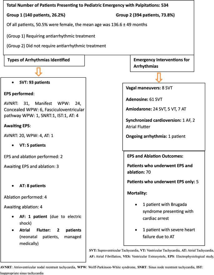
Diagram summaries outcomes of the study.

Materials and method
In this study, we examined all patients aged 0–18 who presented to the paediatric emergency department with complaints of palpitations between 1 October 2020 and 1 October 2024. A total of 534 patients were included, encompassing those managed in collaboration with other regional emergency departments. Of these, 22% (117 patients) initially presented to hospitals outside our centre, where their emergency evaluations and interventions were conducted in coordination with physicians from our institution. All of these patients were subsequently referred to our hospital either urgently or as outpatients for further evaluation or management of the underlying cause of palpitations.
Ethical approval was obtained from our hospital’s ethics committee with the approval date and reference number (20.11.2024/92).
Inclusion criteria
Patients aged between 0 and 18 years who presented with palpitations or were noted by their parents to have palpitations were included in the study.
Exclusion criteria
Children with a previously diagnosed arrhythmia and/or known chronic diseases were excluded from the study.
We collected demographic data such as age and gender, and we recorded patients’ vital parameters, physical examination findings, diagnostic tests including electrocardiography, echocardiography, Holter electrocardiography, and laboratory tests, potential infections, medications, and procedures. In addition, we reviewed hospital discharge records to capture final diagnoses. The electrocardiograms of patients were reviewed, and arrhythmia diagnoses were established. The isolated ventricular ectopics which were less than 10% and the isolated supraventricular episodes which were less than 10% were regarded as insignificant arrhythmias. Reference Paudel and Paudel10
Appropriate medical intervention applied for all patients who diagnosed with arrhythmia in paediatric emergency department, patients who exhibited arrhythmia despite medical therapy, had a diagnosis of Wolff-Parkinson-White syndrome, experienced adverse drug effects, or showed clinical deterioration due to arrhythmia underwent cardiac electrophysiological study and ablation therapy following informed consent from their families.
The procedures for electrophysiological study and ablation therapy were conducted under general anaesthesia using midazolam, propofol, fentanyl, and sevoflurane. All antiarrhythmic medications were discontinued for a period equivalent to five half-lives before the procedure. A multipolar electrode catheter (5 to 7 French) was introduced through the right and/or left femoral veins, while an ablation catheter was additionally employed for ablation-requiring cases. In some patients with an accessory pathway, a catheter was inserted into the right femoral artery and advanced towards the aorta to facilitate mapping of the mitral annulus. For some other patients with left-sided accessory pathways, the antegrade approach was used (transseptal approach). Before ablation, all patients underwent an electrophysiological study in the same session. During the electrophysiological study, intracavitary electrogram recordings were obtained, and supraventricular and ventricular stimulation was performed. If arrhythmia was not induced, intravenous isoproterenol or atropine was administered. Fluoroscopy was utilised in all cases to guide catheter placement within the cardiac chambers. The radiofrequency energy output, application duration, and temperature were individually adjusted by the responsible electrophysiologist. Cryoablation was employed for arrhythmias located in the parahisian region. Reference Paudel and Paudel10
For patients requiring ablation in the left accessory pathway or systemic circulation (left heart chambers), heparin therapy was administered. In addition, aspirin at a dosage of 5 mg/kg/day (maximum 100 mg/day) was prescribed for three months. Post-procedural follow-up was conducted in the paediatric cardiology unit. Reference Lee, Dorian and Downar11
Statistical analysis
Statistical analyses were performed using SPSS version 25.0.0. The normality of data distribution was assessed using the Shapiro–Wilk test. Continuous variables following a normal distribution were compared between two groups using the independent Student’s t-test, while the Mann–Whitney U test was applied for non-normally distributed variables. Correlations between variables were analysed using Pearson’s correlation test. Categorical variables were evaluated using Pearson’s chi-square test or Fisher’s exact test, as appropriate. Measurable parameters were expressed as mean ± standard deviation, while categorical variables were presented as frequency (n) and percentage (%). A p-value of <0.05 was considered statistically significant.
Results
A total of 534 patients presenting to the paediatric emergency department with complaints of palpitations were evaluated. Of these patients, 50.5% were female, with a mean age of 136.6 ± 49 months (Figure 1). Palpitations occurred at rest in 87.2% of cases, whereas 12.8% of cases were associated with physical or psychological stress. Sinus tachycardia was detected in 10% of the patients, while fever was present in 1.2%. Stress and anxiety were identified as the most common causes of sinus tachycardia.
When comparing the group requiring antiarrhythmic treatment (Group 1, 140 patients, 26.2%) with those who did not require such treatment (Group 2), no statistically significant differences were found in haemoglobin levels, thyroid function tests, electrolyte levels, and echocardiographic findings. No cases of sudden cardiac death were identified in the family histories of the patients.
The Holter electrocardiography recordings of 366 patients included in the study were obtained. All patients in Group 1 had available Holter recordings. In Group 1, atrial tachycardia was detected in 2 patients, supraventricular tachycardia in 3 patients, Wolff-Parkinson-White syndrome in 25 patients, less than 10% ventricular ectopic beats in 26 patients, less than 10% supraventricular ectopic beats in 34 patients, and normal Holter electrocardiography findings in 50 patients. In Group 2, Holter electrocardiography recordings were available for 226 patients (57.3%). Among these, 28 patients exhibited less than 10% supraventricular ectopic beat while 10 patients had less than 10% ventricular ectopic beats and normal Holter electrocardiography findings in 188 patients.
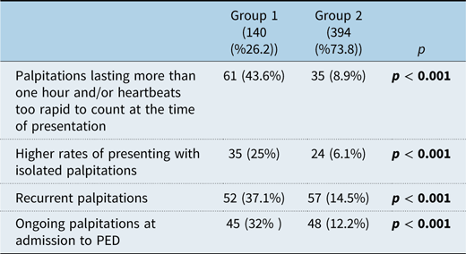
The patients presenting with isolated palpitations (absence of additional symptoms with no recurrent episodes of palpitation) were significantly more in Group 1 (25%) compared to Group 2 (6.1%) (p < 0.001). In Group 1, 61 patients (43.6%) described their palpitations at the time of presentation as lasting more than one hour and/or being too rapid to count (47 had too rapid to count heartbeats, 8 had palpitations lasting longer than one hour, and in 6 patients, both complaints were present together), whereas this was reported by only 35 patients (8.9%) in Group 2 (30 had too rapid to count heartbeats, 4 had palpitations lasting longer than one hour, and in 1 patient, both complaints were present together). The occurrence of these symptoms was significantly higher in group 1 (p < 0.001). The patients presenting with recurrent palpitations were significantly more in Group 1 (37.1%) compared to Group 2 (14.5%) (p < 0.001). Ongoing palpitations at the time of admission to the paediatric emergency department were observed in 45 patients (32%) in Group 1, whereas this was observed in 48 patients (12.2%) in Group 2. The presence of ongoing palpitations at admission was significantly higher in Group 1 (p < 0.001) (Table 1).
Comparing of the groups

n: Estimated cases, requiring antiarrhythmic treatment (Group 1), did not require such treatment (Group 2), PED Paediatric Emergency Department.
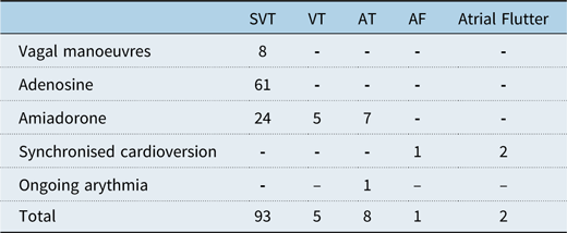
Among the 93 patients diagnosed with supraventricular tachycardia upon admission to the paediatric emergency department, 8 patients achieved sinus rhythm through vagal manoeuvers, 61 patients with adenosine, and 24 patients with amiodarone. All patients were initiated on beta-blockers as maintenance therapy.
Among the 8 patients diagnosed with atrial tachycardia, none responded to adenosine; 7 patients converted to sinus rhythm with amiodarone (Table 2), while 1 patient exhibited persistent atrial tachycardia despite treatment with amiodarone, esmolol, and flecainide. Although this patient achieved sinus rhythm following ablation, atrial tachycardia recurred later. This patient’s ejection fraction was 20%, the patient subsequently experienced fatal cardiac arrest following symptomatic palpitations.
Interventions for documented arrhythmias in the emergency department

SVT = Supraventricular Tachycardia; VT = Ventricular Tachycardia; AT = Atrial Tachycardia, AF = Atrial Fibrillation; VES = Ventricular Extrasystole. (A total of 140 patients). 12 patients were diagnosed with VES not requiring emergency treatment, 16 patients with WPW syndrome, 1 patient with Brugada syndrome who was died, and 2 patients with long QT syndrome.
The remaining 7 patients with atrial tachycardia were prescribed a combination of beta-blockers and propafenone for maintenance therapy. In 3 of these patients, intermittent atrial tachycardia persisted (intermittent atrial tachycardia was detected with Holter electrocardiography), accompanied by borderline ejection fraction (55–60%). According to maternal reports, these 3 infants exhibited symptoms of palpitations and fussiness. Consequently, propafenone was discontinued, and flecainide was introduced. Following this adjustment, all 3 patients successfully converted to sinus rhythm, and their ejection fractions normalised. Electrophysiological studies and ablation were performed in 4 patients with atrial tachycardia, all of whom achieved sinus rhythm. The remaining 3 patients are currently being followed in a stable condition, awaiting an appropriate age for electrophysiological studies and ablation. Except for 4 patients with atrial tachycardia, all patients had normal ejection fractions upon presentation to the paediatric emergency department. Apart from 1 patient diagnosed with Brugada syndrome who presented with cardiopulmonary arrest and another patient with refractory atrial tachycardia and heart failure, all other patients were hemodynamically stable at the time of admission. Synchronised cardioversion was performed on 1 patient who developed atrial fibrillation following an electrical injury and 2 neonates who developed atrial flutter, successfully restoring sinus rhythm.
In addition, 2 of our patients were diagnosed with long QT syndrome and are currently being monitored in a stable condition under beta-blocker therapy. Among the 5 patients who presented with ventricular tachycardia, sinus rhythm was successfully restored with intravenous amiodarone loading in all cases. Of these, 2 patients were diagnosed with posterior fascicular ventricular tachycardia and were initiated on verapamil for maintenance therapy. Subsequently, 2 patients underwent successful ablation. At present, 2 patients who have not yet undergone electrophysiological studies and ablation are being followed with beta-blocker therapy, while 1 patient is being managed with a combination of flecainide and a beta-blocker, with electrophysiological studies and ablation planned for an appropriate time.
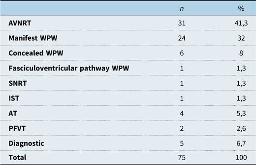
A total of 70 patients underwent both electrophysiological studies and ablation, whereas 5 patients underwent only electrophysiological studies. (Table 3). The locations of the Wolf-Parkinson-White patterns observed during electrophysiological studies were as follows: 1 in the middle cardiac vein, 3 midseptal, 3 parahisian, 5 right posteroseptal, 11 left lateral, and 7 left posterolateral. Fasciculoventricular pathway Wolff-Parkinson-White was diagnosed in 1 patient.
Cardiac electrophysiological study results (EPS)

n = Estimated Cases; AVNRT = Atrioventricular nodal reentrant tachycardia; WPW = Wolff-Parkinson-White syndrome; SNRT = Sinus node reentrant tachycardia; IST = Inappropriate sinus tachycardia; AT = Atrial tachycardia; PFVT = Posterior Fascicular Ventricular Tachycardia.
Atrioventricular nodal re-entrant tachycardia was identified in 31 patients, sinus node re-entrant tachycardia in 1 patient, unspecified supraventricular tachycardia in 1 patient, atrial tachycardia in 4 patients, and posterior fascicular ventricular tachycardia in 2 patients. A total of 65 patients currently remain under follow-up with antiarrhythmic therapy while awaiting electrophysiological studies and ablation at an appropriate time.
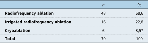
Of the patients who underwent ablation, 48 (68.6%) underwent radiofrequency ablation, 16 (22.8%) underwent irrigated radiofrequency ablation, and 6 (8.57%) underwent cryoablation (Table 4). In Group 1, among the 30 patients diagnosed with Wolff-Parkinson-White, 24 underwent ablation via the retrograde approach, and 6 via the antegrade approach. Since our centre has better experience with the retrograde technique, it was preferred more frequently, and no complications occurred in any of our patients.
Cardiac ablation results

n = Estimated Cases.
During radiofrequency ablation, 2 patients developed a transient first-degree atrioventricular block. Recurrence occurred in 4 of the patients who underwent radiofrequency ablation. Among these, 2 had atrioventricular nodal re-entrant tachycardia, 1 had AVRT localised to the middle cardiac vein, and 1 had atrial tachycardia (the patient had an ejection fraction of 20% and continued to experience arrhythmia despite triple antiarrhythmic therapy, ultimately passed away). In addition, 1 patient, despite initially undergoing successful ablation for atrioventricular nodal re-entrant tachycardia, later presented with atrial tachycardia. The patients who underwent ablation were followed up regularly, and no complications developed in the other patients.
Discussion
Palpitations are characterised by the awareness of irregular heartbeats, a common reason for paediatric emergency department visits. Studies on arrhythmias associated with palpitations in children remain limited. Palpitations were the primary cause of 5.8 per 1,000 emergency department visits for both children and adults. Reference Probst, Mower, Kanzaria, Hoffman, Buch and Sun1 The incidence of arrhythmia in children varies, with approximately 55.1 cases per 100,000 paediatric emergency department visits. Reference Sacchetti, Moyer, Baricella, Cameron and Moakes4 A retrospective study reported arrhythmia in 13.5% of paediatric patients presenting with palpitations. Reference Bobbo, Amoroso and Tamaro12 In our study, the prevalence of arrhythmia requiring treatment among paediatric patients presenting with palpitations was found to be 26.2%, which is higher than in other studies, this may be attributed to our hospital being a regional referral centre for critical arrhythmia cases. Bobbo et al. reported that 52.1% of patients presenting to the paediatric emergency department were female, with a mean age of 152.4 months. Reference Bobbo, Amoroso and Tamaro12 In our study, 50.5% of patients were female, with a mean age of 136.6 ± 49 months. Regarding physical examination, a study reported that 82.3% of patients had normal vital signs, while 17.7% exhibited age-related tachycardia, with 4.1% occurring during fever. The most common accompanying symptoms were chest pain (28.1%), dyspnoea (25%), and dizziness (14.6%), while 42.7% of cases presented with isolated palpitations, no patients or parents reported a history of syncope. Reference Bobbo, Amoroso and Tamaro12 In our study, 10% of patients had age-related sinus tachycardia, and 1.2% had fever. The most common cause of sinus tachycardia was stress and anxiety. The most frequently associated symptoms were chest pain (33.7%) and dyspnoea (12.3%). No statistically significant difference was observed between the group requiring antiarrhythmic treatment (Group 1) and the group not requiring antiarrhythmic treatment (Group 2) in terms of chest pain and dyspnoea. However, isolated palpitations were significantly more common in Group 1 (25%, 35 patients) compared to Group 2 (6.1%, 24 patients) (p < 0.001). In our study, 109 patients (20.4%) were diagnosed with supraventricular tachycardia, 12 (2.24%) with ventricular extrasystole, 8 (1.5%) with atrial tachycardia, 5 (0.94%) with ventricular tachycardia, 2 (0.37%) with atrial flutter, 2 (0.37%) with long QT syndrome, 1 (0.18%) with atrial fibrillation, and 1 (0.18%) with Brugada syndrome. Bobbo et al. reported 11.4% supraventricular tachycardia and 2.1% ventricular extrasystole. Reference Bobbo, Amoroso and Tamaro12 In a study by Probst et al., sinus tachycardia was the most frequently reported arrhythmia, accounting for nearly 50% of cases, followed by paroxysmal supraventricular tachycardia (13%), non-specific arrhythmia (10.6%), bradycardia (6%), and atrial fibrillation (4.6%). Reference Probst, Mower, Kanzaria, Hoffman, Buch and Sun1
In a study conducted by Leng et al., it was suggested that ambulatory cardiac monitoring using Holter monitors may provide valuable diagnostic information for most low-risk paediatric patients presenting to the emergency department with suspected cardiac arrhythmias. The study emphasised that, if the symptoms are shown not to be related to arrhythmia, this can offer reassurance for both patients and caregivers. In addition, this method of monitoring has the potential to reveal a subgroup of patients with clinically silent (asymptomatic) but potentially significant arrhythmias. Reference Leng, Aldalati and Homme13
In our study, no arrhythmias requiring treatment were detected on Holter electrocardiography in patients within Group 2. However, in the group requiring antiarrhythmic treatment (Group 1), Holter electrocardiography identified 1 patient with intermittent Wolff-Parkinson-White syndrome, 1 patient with atrial tachycardia, none of which were detected on the initial electrocardiography at presentation. Although the number of patients diagnosed exclusively by Holter electrocardiography was small, we believe that Holter monitoring should be performed in all patients with suspected arrhythmia.
In our study, 43.6% (61 patients) of Group 1 described their palpitations at the time of admission as “lasting more than an hour and/or being too rapid to count,” compared to only 8.9% (35 patients) in Group 2 (p < 0.001). This suggests that prolonged and extremely rapid palpitations are significant indicators of arrhythmia requiring treatment. Consistently, studies also found that palpitations lasting longer than one hour were significantly associated with arrhythmia diagnosis. Reference Bobbo, Amoroso and Tamaro12 At the time of admission, 32% (45 patients) of Group 1 and 12.2% (48 patients) of Group 2 had ongoing palpitations, a statistically significant finding (p < 0.001). Similarly, a study reported that the strongest predictor of arrhythmia diagnosis was the presence of tachycardia at the time of admission. Reference Bobbo, Amoroso and Tamaro12
The first-line treatment for paediatric arrhythmias remains antiarrhythmic medications, although vagal manoeuvres should be attempted before pharmacological intervention when appropriate. Reference Clausen, Theophilos, Jackno and Babl9 The goal of treatment is to terminate acute episodes and prevent recurrence. Reference Clausen, Theophilos, Jackno and Babl9 Vagal manoeuvres and adenosine are first-line therapies for terminating acute supraventricular tachycardia by slowing atrioventricular conduction. Reference Clausen, Theophilos, Jackno and Babl9 Amiodarone is effective in treating most atrial, re-entrant, and ventricular arrhythmias. Reference Clausen, Theophilos, Jackno and Babl9 In our study, among 93 patients diagnosed with supraventricular tachycardia, 8 successfully reverted to sinus rhythm with vagal manoeuvres, 61 with adenosine, and 24 with amiodarone. All 5 patients with ventricular tachycardia and 7 of the 8 patients with atrial tachycardia were successfully treated with amiodarone. In 2 cases of atrial flutter and 1 case of atrial fibrillation, successful treatment was achieved with synchronised cardioversion. One major drawback of maintenance therapy for supraventricular tachycardia is the frequent side effects of antiarrhythmic drugs. In low-risk patients, therapy should be discontinued after a few years to assess whether the arrhythmia persists or changes in character, as symptoms may resolve as children grow. Reference Kafalı and Ergül14 Beta-blockers are widely used in paediatric re-entrant supraventricular tachycardia and in preventing ventricular arrhythmias associated with congenital long QT syndrome. Reference Clausen, Theophilos, Jackno and Babl9 Amiodarone and flecainide are alternative antiarrhythmic agents. Flecainide, a class Ic sodium channel blocker, is effective for various atrial and re-entrant arrhythmias but should be carefully used in patients with structural heart disease or ventricular dysfunction. Due to its potential long-term side effects, it is typically reserved for resistant arrhythmias. Reference Clausen, Theophilos, Jackno and Babl9 In our study, all supraventricular tachycardia patients received beta-blockers as maintenance therapy, and 64 patients underwent electrophysiological study and ablation. Ventricular tachycardia is a potentially life-threatening arrhythmia that can lead to sudden death in both adult and paediatric patients. It is rare in children, accounting for approximately 6% of patients monitored for tachycardia. Beta-blockers may be used for maintenance therapy. Reference Rhodes, Wernovsky and Keane15,Reference Hanash and Crosson16 Among 5 patients who presented with ventricular tachycardia, all successfully reverted to sinus rhythm with intravenous amiodarone. Of these, 2 patients were diagnosed with posterior fascicular ventricular tachycardia and started maintenance therapy with verapamil. Both subsequently underwent successful ablation. Currently, 2 of the remaining patients are being managed with beta-blockers, while 1 patient is receiving a combination of flecainide and beta-blockers. Ablation therapy is planned at an appropriate time for these 3 patients.
Ectopic atrial tachycardia is a rare condition, accounting for approximately 10% of supraventricular tachycardias in children. Reference Walsh, Berul, Triedman, Keane, Lock and Fyler17 It is the most common cause of tachycardia-induced cardiomyopathy and is associated with improved cardiac function following successful treatment. Reference Hanash and Crosson16 Ectopic atrial tachycardia exhibits a poor response to adenosine and synchronised cardioversion. For symptomatic patients, amiodarone is the first-line treatment. In patients with normal cardiac function, beta-blockers may be beneficial by slowing atrioventricular node conduction, reducing ventricular rate, and alleviating symptoms, more aggressive treatment options include flecainide, amiodarone, and/or sotalol. Reference Hanash and Crosson16 In cases where medical therapy fails, catheter ablation is performed. Younger children (under 3 years of age) have a higher likelihood of spontaneous resolution with medical therapy, whereas older children often require radiofrequency ablation due to persistent tachycardia despite optimal medical management. Reference Hanash and Crosson16 In our study, the emergency interventions, maintenance treatments, and electrophysiological study-ablation outcomes of the 8 patients diagnosed with atrial tachycardia were found to be consistent with the literature.
Paediatric electrophysiological study and radiofrequency ablation are increasingly utilised, but many clinicians lack sufficient knowledge regarding their management due to challenges unique to paediatric patients, including limited vascular access, smaller cardiac anatomy, and CHD variations. Radiofrequency ablation is the first-line treatment for paediatric supraventricular tachycardia, the most common type of arrhythmia in children. Reference Lee, Hwang and Chen18,Reference Simão, Rios and Leiria19 The primary indications include AVRT, atrioventricular nodal re-entrant tachycardia, and atrial tachycardia. Given its rarity, radiofrequency ablation is infrequently indicated for ventricular tachycardia in children. Reference Lee, Hwang and Chen18 Catheter ablation is a highly successful and low-complication method for treating common paediatric arrhythmias. It is recommended for children weighing over 25 kg but can be performed in smaller children if clinically necessary. Reference Clausen, Theophilos, Jackno and Babl9 Radiofrequency ablation eliminates arrhythmogenic tissue by heating the catheter tip, with a success rate of 95%. Cryoablation, which eliminates arrhythmogenic substrates by freezing, has a success rate of 85–90%, with a slightly higher recurrence rate compared to radiofrequency ablation. However, cryoablation is preferred for accessory pathways near the atrioventricular node due to its safety profile. Reference Clausen, Theophilos, Jackno and Babl9 Catheter placement and arrhythmia mapping are typically performed under fluoroscopic guidance. However, with advancements in three-dimensional electroanatomic mapping and intracardiac echocardiography, the entire procedure can be conducted without the use of X-ray radiation.
Van Hare et al. reported a 95.7% success rate for radiofrequency ablation in paediatric patients with supraventricular tachycardia due to accessory pathways and atrioventricular nodal re-entrant tachycardia. Reference Van Hare, Javitz and Carmelli20 Similarly, Tanel et al. documented a 90% success rate for radiofrequency ablation in paediatric arrhythmias. Reference Tanel, Walsh, Triedman, Epstein, Bergau and Saul21 In another study, ablation procedures achieved a success rate of 91.7% for atrioventricular nodal re-entrant tachycardia and 83.5% for accessory pathways. The relatively lower success rate for accessory pathways may be attributed to the absence of transseptal puncture in these cases and the higher prevalence of complex CHD (12%) in this study population. The complication rate in this study was 1.4%, while the literature reports a range of 1.2% to 8.7%. Reference Simão, Rios and Leiria19 In our study, transient first-degree atrioventricular block developed in 2 patients during radiofrequency ablation, resulting in a complication rate of 2.8%.
In our study, radiofrequency ablation was performed in 68.6% (48 patients), irrigated radiofrequency ablation in 22.8% (16 patients), and cryoablation in 8.57% (6 patients). Recurrence was observed in 4 cases. The success rate for atrioventricular nodal re-entrant tachycardia ablation was 93.6%, for accessory pathways 96.8%, for radiofrequency ablation 95.4%, and for cryoablation 83.4%.
Only 2 patients with arrhythmia diagnoses died, 1 due to cardiac arrest secondary to Brugada syndrome and the other due to atrial tachycardia with an ejection fraction of 20%.
Conclusion
Although palpitations are a common reason for admission to paediatric emergency departments, arrhythmias are rarely detected in these patients. However, the likelihood of identifying arrhythmias is higher in patients referred to specialised arrhythmia centres, such as ours. As observed in our study, vagal manoeuvres are not adequately performed. Vagal manoeuvres should be applied appropriately in eligible patients before medical intervention. Arrhythmia diagnosis is particularly important in cases where palpitations persist for more than an hour, occur at an uncountable rapid rate, present as isolated or recurrent episodes, or continue at the time of admission. This underscores the significance of performing an electrocardiogram during episodes of palpitations. In addition, in infants presenting with symptoms such as irritability and feeding difficulties, atrial tachycardia, and atrial flutter should be considered. Performing an electrocardiography is especially crucial in patients with a family history of sudden cardiac death, as genetic conditions such as Brugada syndrome, long QT syndrome, hypertrophic cardiomyopathy, and various other rhythm disorders can either be diagnosed or suspected based on electrocardiography findings. Given the lack of studies in the literature that provide detailed data on paediatric arrhythmias similar to our research, we believe that our study will make a significant contribution to the field by aiding in the early detection of life-threatening arrhythmias, guiding initial management and follow-up, and providing insights into the outcomes of cardiac ablation for arrhythmias.
Limitations
The primary limitation of this study is its retrospective design. This aspect has hindered the ability to obtain a complete medical history in some cases and to determine whether electrocardiography was performed during symptomatic periods for all patients. Another limitation is that not all patients in the group without detected arrhythmia underwent a comprehensive evaluation with 24-hour Holter electrocardiography monitoring, which suggests that some diagnoses may have been missed. To the best of our knowledge, no other study includes a similarly large cohort consisting exclusively of paediatric patients, which limits our ability to compare our findings with those of other studies.
Financial support
There is no funding source. Data availability No datasets were generated or analysed during the current study.
Ethical standard
This article does not contain any studies with human participants or animals performed by any of the authors. The study was conducted in accordance to the Declaration of Helsinki, and the study protocol was approved by Dicle University institutional review board and ethical committee (20.11.2024/92). Informed consent was waived because of the retrospective nature of the study and that no personal identifiable data will be processed or published.
Declarations
None.