Introduction
The success of assisted reproduction techniques (ARTs) depends on the availability of competent oocytes that are able to develop into healthy embryos and allow the establishment of pregnancy.
When oocytes are removed from the follicular environment, they automatically resume meiosis without completing cytoplasmic maturation. Therefore, those that have not achieved total competence will not develop into viable embryos (Gilchrist et al., Reference Gilchrist, Lane and Thompson2008). Oocytes used for in vitro maturation (IVM) are usually recovered from smaller follicles and form a heterogeneous population with various degrees of competence. Consequently, when IVM is applied in ART it lowers embryo development. A possible strategy to improve the outcome of IVM is to keep oocytes meiotically arrested in vitro for a longer period of time rather than allowing them to undergo germinal vesicle breakdown (GVBD) as soon as they are retrieved from follicles (Dode & Adona, Reference Dode and Adona2001; Vanhoutte et al., Reference Vanhoutte, Sutter, Nogueira, Gerris, Dhont and Van der Elst2007; Bilodeau-Goeseels, Reference Bilodeau-Goeseels2011; Guemra et al., Reference Guemra, da Silva Santo, Zanin, Monzani, Sovernigo, Ohashi, Leal and Adona2014).
Thus, a pre-maturation culture (PMC) would give the oocytes additional time to undergo cytoplasmic changes and to acquire total competence before they are submitted to IVM. Moreover, PMC would also enhance synchronization of the nucleus and cytoplasm to provide a more homogenous population of immature oocytes (Anderiesz et al., Reference Anderiesz, Fong, Bongso and Trounson2000; Dieleman et al., Reference Dieleman, Hendriksen, Viuff, Thomsen, Hyttel, Knijn, Wrenzycki, Kruip, Niemann and Gadella2002; Luciano et al., Reference Luciano, Modina, Vassena, Milanesi, Lauria and Gandolfi2004; Nogueira et al., Reference Nogueira, Cortvrindt, Everaerdt and Smitz2005).
Various physiological and pharmacological methods have been used to inhibit the resumption of meiosis in bovine oocytes. Physiological methods, such as culture in follicular fluid (Carolan et al., Reference Carolan, Lonergan, Monget, Monniaux and Mermillod1996) and in hemi-sections of follicles (Sirard & Coenen, Reference Sirard and Coenen1993; Oliveira e Silva et al., Reference Oliveira e Silva, Vasconcelos, Caetano, Gulart, Camargo, Bao and Rosa e Silva2011), generally are of shorter duration and are less efficient than pharmacological methods. Substances that increase the levels of cAMP, such as inhibitors of phosphodiesterase (PDE) and activators of adenylate cyclase (AC), have been shown to be efficient in inhibiting resumption of spontaneous meiosis in mice (Nogueira et al., Reference Nogueira, Cortvrindt, Everaerdt and Smitz2005), cattle (Aktas et al., Reference Aktas, Leibfried-Rutledge and First2003; Luciano et al., Reference Luciano, Franciosi, Modina and Lodde2011) and humans (Nogueira et al., Reference Nogueira, Cortvrindt, Everaerdt and Smitz2005). Other drugs, such as 6-dimethylaminopurine (6-DMAP) that acts on meiosis-promoting factor (MPF) and specific inhibitors of cyclin-dependent kinases (CDKs), such as butyrolactone-I and roscovitine, have also been used successfully to maintain oocytes in the GV stage (Dode & Adona, Reference Dode and Adona2001; Adona & Leal, Reference Adona and Leal2004, Reference Adona and Leal2006; Barretto et al., Reference Barretto, Caiado Castro, Garcia and Mingoti2011). Recently, inhibitors of phosphodiesterase type 3A (PDE-3A), which is specific to oocytes (Sasseville et al., Reference Sasseville, Albuz, Cote, Guillemette, Gilchrist and Richard2009) and is responsible for hydrolyzing cAMP, have been used (Luciano et al., Reference Luciano, Franciosi, Modina and Lodde2011; Dieci et al., Reference Dieci, Lodde, Franciosi, Lagutina, Tessaro, Modina, Albertini, Lazzari, Galli and Luciano2013). The advantage of using specific inhibitors of PDE-3A is that they maintain elevated cAMP levels, which retain the oocyte in the GV (Conti et al., Reference Conti, Andersen, Richard, Mehats, Chun, Horner, Jin and Tsafriri2002; Thomas et al., Reference Thomas, Armstrong and Gilchrist2002) without affecting the cumulus cells (Sasseville et al., Reference Sasseville, Albuz, Cote, Guillemette, Gilchrist and Richard2009). Among those agents, cilostamide has been used in humans (Vanhoutte et al., Reference Vanhoutte, Sutter, Nogueira, Gerris, Dhont and Van der Elst2007; Shu et al., Reference Shu, Zeng, Ren, Zhuang, Liang, Shen, Yao, Ke and Wang2008), mice (Nogueira et al., Reference Nogueira, Cortvrindt, Everaerdt and Smitz2005; Yeo et al., Reference Yeo, Gilchrist and Lane2009), sheep (Gharibi et al., Reference Gharibi, Hajian, Ostadhosseini, Hosseini, Forouzanfar and Nasr-Esfahani2013; Rose et al., Reference Rose, Gilchrist, Kelly, Thompson and Sutton-McDowall2013) and pigs (Dieci et al., Reference Dieci, Lodde, Franciosi, Lagutina, Tessaro, Modina, Albertini, Lazzari, Galli and Luciano2013). However, there are few reports of its use in cattle (Mayes & Sirard, Reference Mayes and Sirard2002; Albuz et al., Reference Albuz, Sasseville, Lane, Armstrong, Thompson and Gilchrist2010; Luciano et al., Reference Luciano, Franciosi, Modina and Lodde2011; Ulloa et al., Reference Ulloa, Heinzmann, Herrmann, Timmermann, Baulain, Grossfeld, Diederich, Lucas-Hahn and Niemann2014).
Although many studies have shown that these substances successfully inhibit the resumption of meiosis and keep the oocytes at the germinal vesicle (GV) stage for a certain period (Kubelka et al., Reference Kubelka, Motlík, Schultz and Pavlok2000; Dode & Adona, Reference Dode and Adona2001; Adona & Leal, Reference Adona and Leal2004), the results show no improvement in embryo production. We hypothesized that if beneficial factors are added to the pre-maturation medium, they can prevent possible toxic effects of the inhibitor and provide a more suitable environment for the oocytes, allowing them to be better prepared for fertilization and development. Therefore, in this study, we tested an alternative method of using meiotic arrest to improve embryo development. PMC was performed by 8 and 24 h using cilostamide to inhibit PDE-3A and retain meiosis and the combination of insulin, transferrin and selenium (ITS) to promote cell survival and to protect against toxic damage during the PMC period.
Materials and methods
Unless otherwise indicated, all chemicals were purchased from Sigma-Aldrich (St. Louis, MO, USA).
Oocyte recovery
Ovaries from crossbred cows (Bos indicus × Bos taurus) were collected at local abattoirs immediately after slaughter and were transported to the laboratory in saline solution (0.9% NaCl) supplemented with streptomycin sulfate (100 μg/ml) and penicillin G (100 UI/ml) at 35°C. Cumulus–oocyte complexes (COCs) were aspirated from 3–8-mm-diameter follicles with an 18-gauge needle and pooled in a 15-ml conical tube. After sedimentation, COCs were recovered and selected using a stereomicroscope. Follicular fluid was centrifuged for 5 min and used for searching and selection. Only COCs presenting homogenous cytoplasm and at least three layers of cumulus cells were used.
Pre-maturation culture (PMC)
Selected COCs were incubated for 8 or 24 h in the presence of cilostamide, a specific PDE-3 A inhibitor, at a final concentration of 20 μM in TCM-199 (Gibco® Invitrogen, Carlsbad, CA, USA) supplemented with 10% fetal bovine serum (FBS, Invitrogen®, Carlsbad, CA, USA), 0.1 mg/ml l-glutamine and an antibiotic (0.075 mg/ml, amikacin). Depending on the treatment group, a combination of ITS at a final concentration of insulin 10 mg/l, transferrin 5.5 mg/l, selenium 5 μg/l, was also added into the pre-maturation medium. Droplets were covered with silicone oil and cultured at 39°C in an atmosphere of 5% CO2 in air.
In vitro maturation (IVM)
After selection, the COCs were washed and transferred in groups of 20 to 30–200 μl droplets of maturation medium under silicone oil and incubated for 22 h at 39°C and 5% CO2 in air. Maturation medium consisted of TCM-199 supplemented with 10% FBS, 0.01 IU/ml follicle stimulating hormone (FSH), 0.1 mg/ml l-glutamine and antibiotic (amikacin, 0.075 mg/ml).
In vitro fertilization (IVF) and embryo culture (IVC)
Following maturation, COCs (groups of 25 to 30) were transferred to a 200-μl droplet of fertilization medium, which consisted of TALP (Parrish et al., Reference Parrish, Krogenaes and Susko-Parrish1995) supplemented with penicillamine (2 mM), hypotaurine (1 mM), epinephrine (250 mM) and heparin (10 μg/ml−1). Frozen semen from a Nellore bull, which was previously tested in our laboratory, was used for IVF. Motile spermatozoa were obtained by the Percoll (GE® Healthcare, Piscataway, NJ, USA) gradient method in microtubes (Machado et al., Reference Machado, Carvalho, Filho, Caixeta, Franco, Rumpf and Dode2009) and were added into the fertilization droplets containing the COCs at a final concentration of 1 × 10⁶ spermatozoa ml−1. The spermatozoa and oocytes were co-incubated for 18 h at 39°C in 5% CO2 in air, and the day of in vitro insemination was considered as day 0 (D0).
Eighteen hours post insemination (pi), presumptive zygotes were washed and transferred to 200-μl droplets of synthetic oviduct fluid medium (SOFaaci) (Holm et al., Reference Holm, Shukri, Vajta, Booth, Bendixen and Callesen1998) supplemented with 2.77 mM of myo-inositol and 5% FBS and cultured at 39°C in 5% CO2 in air for 7 days. The embryos were evaluated for cleavage on day 2 pi, and the blastocyst rates were determined on days 6, 7 and 8.
Assessment of nuclear maturation
For meiotic progression evaluation, oocytes were removed from the PMC and/or maturation medium at 0, 8, 18 and 24 h. Then, they were denuded and fixed for at least 48 h in fixing solution consisting of glacial acetic acid and ethanol at a concentration of (3:1). On the day of the evaluation, the oocytes were placed on a slide, covered with a coverslip and stained with 1% lacmoid in 45% glacial acetic acid. The maturational stage of each oocyte was determined using phase contrast microscopy (Nikon Eclipse E200, ×1000). Oocytes were classified as follows: germinal vesicle (GV), germinal vesicle break down (GVBD), metaphase I (MI), anaphase I (AI), telophase I (TI) or metaphase II (MII). Any oocytes that had diffuse or undefined chromatin or had some chromosomal aberration were considered abnormal/degenerate.
Determination of embryo total cell number
On day 7 (D7), blastocysts were measured using Motic Image Plus 2.0, (Moticam®, Xiamen, China) and were classified into three categories according to their diameter: 120–140, 140–160 or >160 μm. The embryos with a diameter ≥160 μm were used to determine the cell number. Embryos were exposed to Hoechst 33342 staining solution at a concentration of 1 μg/ml in phosphate-buffered saline (PBS) for 5 min and then placed on a slide and covered with a coverslip. The cell number was determined under an epifluorescence microscope (Zeiss Axiophot, Germany®; filter 24 with a wavelength of 330–365 nm excitation/emission) (×100).
Cumulus cell viability
Cumulus cells from 10 COCs were obtained by repeatedly pipetting in 100 μL of PBS to form a cell suspension. A 10-μl sample of this suspension was removed and transferred to a tube containing 10 μl trypan blue stain. Afterward, 10 μl was used in a Neubauer chamber to count viable (unstained cells) and non-viable cells (stained cells), and the percentage of viable cells was determined.
Experimental design
Experiment 1: Effect of cilostamide on the nuclear maturation of bovine oocytes
Initially, we evaluated the efficacy of cilostamide at maintaining meiotic arrest for 8 and 24 h of culture. In total, 489 COCs was used, and in each replica, oocytes were cultured in pre-maturation medium containing 20 μM cilostamide for 8 and 24 h. Samples of oocytes were removed at 0, 8 and 24 h of culture to determine the stage of meiosis.
Subsequently, we assessed whether the meiotic arrest at different periods of time would affect meiosis progression post blockage. This information was needed to determine the most appropriate time to perform the IVF on the pre-matured oocytes. Because our main objective was to add beneficial factors to the medium during PMC to increase embryonic production, at this phase of the experiment, we had supplemented the pre-maturation medium with ITS.
After PMC for 8 and 24 h, oocytes were transferred to maturation medium and matured for 18 or 24 h. Oocytes from the control group were matured for 18 and 24 h as well. At the end of the maturation period, oocytes from both groups were stained and evaluated to determine the stage of meiosis.
Experiment 2: Effect of PMC for 8 or 24 h in the presence of cilostamide and ITS on embryo production and quality
In this experiment, COCs were submitted to PMC for 8 and 24 h and then matured for 18 h, which was when IVF was performed. In the control groups, oocytes were matured for 18 or 24 h. After IVF, the embryos were assessed for cleavage on day 2 (D2) and for blastocysts on day 6 (D6) and day 7 (D7). Blastocysts on D7 were measured, and those with a diameter ≥160μm were stained with Hoechst 33342 to determine the total cell number. The percentage of embryos with a diameter greater than 160 μm and the total number of cells of those embryos were used as embryo-quality parameters.
Experiment 3: Effect of reducing the cilostamide concentration during PMC in the presence of ITS on embryo production and quality
To verify if cilostamide had a deleterious effect on COCs, the same protocol used in the previous experiments was carried out, except that the concentration of cilostamide was reduced by half. Then, the viability of the cumulus cells after meiotic arrest and maturation and the embryo production and quality were evaluated. COCs were distributed into six treatments as follows: (i) T1 control 18: oocytes matured for 18 h; (ii) T2 control 24: oocytes matured for 24 h; (iii) T3 PMC 8 [20] + IVM 18 h: oocytes pre-matured for 8 h in the presence of 20 μM cilostamide and ITS and matured for 18 h; (iv) T4 PMC 8 [10] + IVM 18 h: oocytes pre-matured for 8 h in the presence of 10 μM cilostamide and ITS and matured for 18 h; (v) PMC 24 [20] + IVM 18 h: oocytes pre-matured for 24 h in the presence of 20 μM cilostamide and ITS and matured for 18 h; and (vi) PMC 24 [10] + IVM 18 h: oocytes pre-matured for 24 h in the presence of 10 μM cilostamide and ITS and matured oocytes for 18 h.
After IVM, a group of COCs was denuded, and the viability of the cumulus cells was determined by trypan blue staining. The other group was subjected to IVF. The cleavage rate, blastocyst rate, percentage of blastocysts with diameter ≥160 μm and the total number of cells were evaluated using the same procedures as described above.
Experiment 4. Effect of a PMC in the absence of ITS on embryo production and quality
Finally, to ensure that the effect of cilostamide was not influenced by the presence of ITS, oocytes were pre-matured with cilostamide but in the absence of ITS. COCs were distributed into the following treatment groups: (i) T1 Control 18 h: oocytes were matured for 18 h; (ii) T2 Control 24 h: oocytes were matured for 24 h; (iii) T3 PM8 + IVM 18 h: oocytes were pre-matured for 8 h in the presence of 10 μm cilostamide and matured for 18 h; and (iv) T4 PM24 + IVM 18 h: oocytes were pre-matured for 24 h in the presence of 10 μm cilostamide and matured for 18 h.
After IVF, the embryos were assessed for cleavage at day 2 (D2) and for blastocysts on day 6 (D6) and day 7 (D7). On D7, the embryos were measured, and those with diameters ≥160 μm were stained with Hoechst 33342 to determine the total number of cells.
Statistical analysis
The maturation rate and embryo development data were analyzed using the chi-squared test (P < 0.05). Data comparing embryo diameter and the total cell number were compared using the Kruskal–Wallis test. All statistical analyses were performed using the Prophet program, version 5.0 (BBN Systems and Technologies, 1996).
Results
Experiment 1: Effect of cilostamide on the nuclear maturation of bovine oocytes
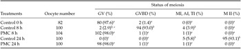
We assessed the ability of cilostamide to maintain oocyte meiotic arrest for 8 and 24 h. At 0 h, before being placed in culture, the majority of oocytes were at GV. In the groups exposed to cilostamide for either 8 or 24 h, the resumption of meiosis was inhibited, while in the control group, the majority of oocytes resumed and completed meiosis after 24 h of culture (Table 1).
Assessment of nuclear maturation of bovine oocytes submitted or not to pre-maturation culture (PMC) in the presence of cilostamide (20 μM) for 0, 8 and 24 h

a, b, c Different letters within the same column indicate significant differences by χ2 (P < 0.05).
AI: anaphase I; GV: germinal vesicle; GVBD: germinal vesicle break down; MI: metaphase I; MII: metaphase II; TI: telophase I.
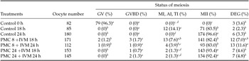
When COCs were submitted to different arresting periods and subsequently matured for 18 and 24 h, it was observed that after PMC for either 8 or 24 h, the percentage of oocytes that reached the metaphase II stage was similar for those matured for 18 and 24 h. In contrast, oocytes from the control group that were matured for 24 h showed a higher maturation rate than those matured for 18 h (Table 2).
Assessment of the nuclear maturation of bovine oocytes submitted to pre-maturation culture (PMC) in the presence of cilostamide (20 μM) and insulin, transferrin and selenium (ITS) for 8 and 24 h and subsequently in vitro matured (IVM) for 18 or 24 h

a, b, c, d Different letters within the same column indicate significant differences by χ2 (P < 0.05).
AI: anaphase I; DEG: degenerated; GV: germinal vesicle; GVBD: germinal vesicle break down; MI: metaphase I; MII: metaphase II; TI: telophase I.
Experiment 2: Effect of PMC for 8 and 24 h in the presence of cilostamide and ITS on embryo production and quality
Because at 18 and 24 h, there was no difference in the maturation rates of the oocytes that had been inhibited for 8 or 24 h, we chose to use the 18-h period of IVM to evaluate embryonic development. The cleavage rate was similar (P > 0.05) among groups, except for the group that was pre-matured for 24 h, which showed a smaller rate (P < 0.05) than the others (Table 3). Although the cleavage rate was similar for the oocytes from the control groups, those matured for 24 h showed the highest (P < 0.05) blastocyst rate on D7. On the other hand, pre-maturation had a detrimental effect on embryo development, i.e., the longer the period of meiotic arrest, the greater the effect (Table 3).
Embryonic development of bovine oocytes submitted to pre-maturation culture (PMC) in the presence of cilostamide (20 μM) and insulin, transferrin and selenium (ITS) for 8 or 24 h and subsequently in vitro matured (IVM) for 18 h

a, b, c Different letters within the same column indicate significant differences by χ2 (P < 0.05).
Embryos from the control group that had matured for 24 h had a greater number of cells and were larger than those of other groups. However, the group pre-matured for 24 h followed by matured for 18 h had embryos with the lowest cell number and that were also the smallest (Table 4).
Percentage, total cell number and size of the D7 embryos with diameter ≥160 μm obtained from oocytes submitted to pre-maturation culture (PMC) in the presence of cilostamide (20 μM) and insulin, transferrin and selenium (ITS) for 8 or 24 h and subsequently in vitro matured (IVM) for 18 h

a,b,c Different letters within the same column indicate significant differences (P < 0.05).
SD, standard deviation.
Experiment 3: Effect of reducing the cilostamide concentration during PMC in the presence of ITS on embryo production and quality
To assess whether the negative effect of pre-maturation on oocyte quality was due to cilostamide, we tested if a lower concentration of cilostamide would change the response. It was observed that the concentration and time of exposure to cilostamide drastically affected the blastocyst rate. The best embryonic development between the treated groups was observed in the group pre-matured for 8 h in the presence of half the concentration of cilostamide (Table 5).
Embryonic development of oocytes submitted to pre-maturation culture (PMC) in the presence of 20 μM [20] or 10 μM [10] of cilostamide and insulin, transferrin and selenium (ITS) for 8 or 24 h and subsequently in vitro matured (IVM) for 18 h

a,b,c,d Different letters within the same column indicate significant differences by χ2 (P < 0.05).
Furthermore, embryo quality was assessed (Table 6). The pre-matured groups, regardless of time, showed an improvement in embryo production and quality when the cilostamide concentration was reduced by half, i.e., embryos from the group pre-matured for 8 h were similar to those from the control group (Table 6).
Percentage, total cell number and size of the D7 embryos with diameter ≥160 μm obtained from oocytes submitted to pre-maturation culture (PMC) for 8 or 24 h in the presence of 20 μM [20] or 10 μM [10] of cilostamide and insulin, transferrin and selenium (ITS) and subsequently in vitro matured (IVM) for 18 h

a,b,c,d Different letters within the same column indicate significant differences (P < 0.05).
SD, standard deviation.
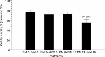
Because the oocytes pre-matured for 24 h always showed lower embryos, even after reduction of the cilostamide concentration, we evaluated the cell viability of cumulus cells from those oocytes. In the control group, the percentage of viable cells was similar before (0 h) and after IVM (24 h), with averages of 89.2 and 75.1%, respectively. When the cells from COCs submitted to pre-maturation for 8 and 24 h were evaluated, no differences were observed at the end of the PMC. However, a decrease in cell viability (P < 0.01) was observed after maturation only in the group pre-matured for 24 h (Figure 1).
Viability of cumulus cells after pre-maturation (PM) for 8 (PM 8) or 24 h (PM 24) in medium supplemented with cilostamide (10 μM) and ITS (0.05 mg/ml) after 0 and 18 h of in vitro maturation (IVM).

Experiment 4. Effect of a PMC in the absence of ITS on embryo production and quality
To eliminate a possible adverse effect of ITS on the reduction of embryo production in the pre-matured oocytes, the same system was used without ITS. It could be observed (Table 7) that the cleavage and blastocyst rates were similar (P > 0.05) between the control group and the group pre-matured for 8 h. Similar to the previous experiment, a lower production of embryos was observed for the group in which oocytes were pre-matured for 24 h (Table 7).
Embryonic development of oocytes submitted to pre-maturation culture (PMC) for 8 or 24 h in the presence of cilostamide (10 μM) and subsequently in vitro matured (IVM) for 18 h

a, b Different letters in the same column indicate significant differences by χ2 (P < 0.05).
Regarding embryo quality (Table 8), the results were similar to the results of embryo production, in which the control and 8 h pre-matured groups had similar percentages of embryos ≥160 μm and similar cell numbers. The group pre-matured for 24 h showed fewer and lower-quality embryos.
Percentage, total cell number and size of D7 embryos with diameter ≥160 μm obtained from oocytes submitted to pre-maturation culture (PMC) for 8 or 24 h in the presence of cilostamide (10 μM) and subsequently in vitro matured (IVM) for 18 h

a,b Different letters in the same column indicate significant differences (P < 0.05).
SD, standard deviation.
Discussion
Meiotic arrest in oocytes immediately after they are removed from follicles has been studied for many years with the aim to improve the shortcomings of conventional IVM of bovine oocytes. A variety of inhibitory substances has been tested, however no advances have been achieved in embryo production (Adona & Leal, Reference Adona and Leal2006; Gharibi et al., Reference Gharibi, Hajian, Ostadhosseini, Hosseini, Forouzanfar and Nasr-Esfahani2013; Rose et al., Reference Rose, Gilchrist, Kelly, Thompson and Sutton-McDowall2013; Ulloa et al., Reference Ulloa, Heinzmann, Herrmann, Timmermann, Baulain, Grossfeld, Diederich, Lucas-Hahn and Niemann2014). Unlike other inhibitors, cilostamide, which inhibits PDE-3A, specifically acts in the oocyte to inhibit cAMP degradation and therefore retains meiotic progression (Sasseville et al., Reference Sasseville, Albuz, Cote, Guillemette, Gilchrist and Richard2009). Despite few reports showing a beneficial effect of cilostamide on bovine oocytes (Albuz et al., Reference Albuz, Sasseville, Lane, Armstrong, Thompson and Gilchrist2010; Luciano et al., Reference Luciano, Franciosi, Modina and Lodde2011), its use as an inhibitor in that species is not well established.
Thus, we initially evaluated the ability of cilostamide to maintain meiotic arrest in our system. The utilized concentration was based on previous studies (Jee et al., Reference Jee, Chen and Chian2009; Albuz et al., Reference Albuz, Sasseville, Lane, Armstrong, Thompson and Gilchrist2010; Gharibi et al., Reference Gharibi, Hajian, Ostadhosseini, Hosseini, Forouzanfar and Nasr-Esfahani2013; Rose et al., Reference Rose, Gilchrist, Kelly, Thompson and Sutton-McDowall2013; Ulloa et al., Reference Ulloa, Heinzmann, Herrmann, Timmermann, Baulain, Grossfeld, Diederich, Lucas-Hahn and Niemann2014). Our results showed that cilostamide was able to maintain meiotic arrest for 8 or 24 h in nearly 100% of the oocytes. Subsequently, we assessed whether the pre-maturation culture affected the kinetics of meiosis progression. This study was performed to determine the most appropriate time for fertilization. Thus, COCs were submitted to PMC for 8 and 24 h in the presence of cilostamide and ITS. Because our main objective was to test if the addition of ITS during the PMC would have beneficial effects on the oocytes, we had already supplemented the pre-maturation medium. As expected, in the control group, an increase in the nuclear maturation rate was observed when oocytes remained in culture for 24 h compared with 18 h. However, when the oocytes were arrested for 24 h and subsequently underwent maturation, the percentage of oocytes at the metaphase II stage was similar to those matured for 18 or 24 h. These results suggest that the kinetics of meiosis accelerated after the oocytes were retained, and at 18 h of maturation, most of the oocytes had reached MII. Other authors have also reported an acceleration of meiosis after meiotic inhibition using butyrolactone-I (Adona & Leal, Reference Adona and Leal2004, Reference Adona and Leal2006; Barretto et al., Reference Barretto, Caiado Castro, Garcia and Mingoti2011). It seems that this acceleration is due to the arrest per se because it has also been observed when other drugs were used (Adona & Leal, Reference Adona and Leal2004, Reference Adona and Leal2006). However, this type of behaviour on nuclear maturation kinetics using cilostamide has not been reported (Mayes & Sirard, Reference Mayes and Sirard2002; Jee et al., Reference Jee, Chen and Chian2009; Sasseville et al., Reference Sasseville, Albuz, Cote, Guillemette, Gilchrist and Richard2009; Luciano et al., Reference Luciano, Franciosi, Modina and Lodde2011). This information is critical for IVF to be performed at the right time to prevent the fertilization of aged oocytes, which would impair embryonic development. Based on these results, we performed IVF in oocytes that had been submitted to PMC after 18 h of maturation.
In contrast with our expectation, oocytes pre-matured in the presence of cilostamide and ITS showed a markedly reduced blastocyst rate as well as reduced blastocyst quality. It was also observed that the higher the PMC, the greater deleterious effect of treatment on embryo development. PMC in the presence of inhibitors is an attempt to provide the oocytes extra time to undergo additional changes that could increase their developmental potential (Adona & Leal, Reference Adona and Leal2006; Gilchrist et al., Reference Gilchrist, Lane and Thompson2008; Bilodeau-Goeseels, Reference Bilodeau-Goeseels2012). Several studies have used oocyte meiotic arrest for this purpose; however, an increase in embryo production has not been observed, and deleterious effects have not been reported (Dode & Adona, Reference Dode and Adona2001; Mayes & Sirard, Reference Mayes and Sirard2002; Adona & Leal, Reference Adona and Leal2004; Nogueira et al., Reference Nogueira, Cortvrindt, Everaerdt and Smitz2005). Based on those results, we hypothesized that supplementation of pre-maturation medium with additional factors would improve embryo development. We chose to add ITS because of its action in cells; insulin exerts mitogenic and antiapoptotic actions (Spicer & Echternkamp, Reference Spicer and Echternkamp1995; Lee et al., Reference Lee, Kang, Lee and Hwang2005), selenium acts as a stimulator of glutathione peroxidase synthesis (GSH; Raghu et al., Reference Raghu, Reddy and Nandi2002), and transferrin is a chelating, radical hydroxyl and a transporter of metals (Córdova et al., Reference Córdova, Morató, Izquierdo, Paramio and Mogas2010). ITS has been routinely used in IVM systems for oocytes of several species, such as mice, bovine (De La Fuente et al., Reference De La Fuente, O'Brien and Eppig1999; De Bem et al., Reference De Bem, Chiaratti, Rochetti, Bressan, Sangalli, Miranda, Pires, Schwartz, Sampaio, Fantinato-Neto, Pimentel, Perecin, Smith, Meirelles, Adona and Leal2011) and pigs (Jeong et al., Reference Jeong, Hossein, Bhandari, Kim, Kim, Park, Lee, Park, Jeong, Lee, Kim and Hwang2008) and in pre-antral follicle culture systems (Hammami et al., Reference Hammami, Morató, Romaguera, Roura, Catalá, Paramio, Mogas and Izquierdo2013; Huanmin & Yong, Reference Huanmin and Yong2000). Therefore, based on its beneficial effects, we expected that ITS would protect the oocytes during PMC. However, we could not detect any beneficial effect of ITS in our study. In addition, to rule out the possibility that supplementation of pre-maturation medium with ITS was not involved in the deleterious effects of cilostamide, we removed it from the pre-maturation medium. As expected, the embryonic development and embryo-quality results were the same as the previous experiment. Therefore, the presence of ITS in the pre-maturation medium did not affect the results, which did not confirm our hypothesis.
Because we found that the deleterious effect of pre-maturation was more pronounced as the time of meiotic arrest increased, we wondered if it could be due to the concentration of cilostamide and be associated with the period of exposure to the inhibitor. To verify this effect, half of the concentration of cilostamide was used. The results demonstrated that, by reducing the concentration of cilostamide to 10 μM, it was possible to increase blastocyst development in both groups submitted to pre-maturation. Nevertheless, the difference in embryo development between 8-h and 24-h inhibition was still present, suggesting that the time of exposure and concentration of cilostamide are important factors in the oocyte response. A study comparing different concentrations of cilostamide on mouse oocytes for 6 or 24 h showed that time of exposure showed a more deleterious effect (Jee et al., Reference Jee, Chen and Chian2009). Previous experiments conducted to evaluate the effect of the cilostamide concentration showed that lower doses are effective in retaining meiosis and do not interfere with embryonic development. Concentrations such as 1 μM in sheep (Gharibi et al., Reference Gharibi, Hajian, Ostadhosseini, Hosseini, Forouzanfar and Nasr-Esfahani2013; Rose et al., Reference Rose, Gilchrist, Kelly, Thompson and Sutton-McDowall2013), 10 μM in bovine (Luciano et al., Reference Luciano, Franciosi, Modina and Lodde2011), 0.1 μM in mice (Vanhoutte et al., Reference Vanhoutte, Nogueira, Gerris, Dhont and De Sutter2008) and 1 μM in humans (Vanhoutte et al., Reference Vanhoutte, Sutter, Nogueira, Gerris, Dhont and Van der Elst2007) have been used with success. The reason why the present study showed the opposite effect is not clear but could include a factor, such as the inhibitor agent used such as Org 9935 (Nogueira et al., Reference Nogueira, Cortvrindt, De Matos, Vanhoutte and Smitz2003, Reference Nogueira, Cortvrindt, Everaerdt and Smitz2005; Romero & Smitz, Reference Romero and Smitz2010) or milrinone (Thomas et al., Reference Thomas, Armstrong and Gilchrist2004; Grupen et al., Reference Grupen, Fung and Armstrong2006; Naruse et al., Reference Naruse, Iga, Shimizu, Takenouchi, Akagi, Somfai and Hirao2012), the species/subspecies, the donor follicular stimulation treatment previously to oocyte retrieval, and the dose and composition of the medium in which cilostamide was added. It is interesting to note that most of the reports that showed a positive effect of cilostamide also have used a low concentration of FSH in the PMC (Shu et al., Reference Shu, Zeng, Ren, Zhuang, Liang, Shen, Yao, Ke and Wang2008; Luciano et al., Reference Luciano, Franciosi, Modina and Lodde2011; Lodde et al., Reference Lodde, Franciosi, Tessaro, Modina and Luciano2013). In bovine, it has been shown that the maintenance of communication between cumulus cells and oocytes during pre-maturation can be attained using IBMX and low concentrations of FSH (Lodde et al., Reference Lodde, Franciosi, Tessaro, Modina and Luciano2013). Similar increases in developmental capacity were obtained in mouse and human oocytes when cilostamide was used in the maturation medium in the presence of FSH and LH (Vanhoutte et al., Reference Vanhoutte, Nogueira, Gerris, Dhont and De Sutter2008; Vanhoutte et al., Reference Vanhoutte, Nogueira, Dumortier and De Sutter2009) as well as when a PMC was applied in oocytes collected from super stimulated ovaries. It seems that inhibitors in the presence of FSH keeps the oocytes in meiotic arrest and extends the coupling between cumulus cells and oocyte. It is possible that in our study, where pre-maturation occurred in the absence of FSH, the gap junctional communication was not maintained during the entire PMC, which affected maturation after meiotic inhibition. PMC for only 8 h produced better blastocyst development rates than those for 24-h culture.
When we evaluated the cell viability of cumulus cells in the control group, the percentages of viable cells were similar before (0 h) and after IVM (24 h). The cells from COCs submitted to PMC for 8 or 24 h were evaluated, but no differences were observed at the end of the meiotic arrest period. However, after IVM, the oocytes that were blocked for 24 h showed a decrease in CC viability, confirming that the time of exposure to cilostamide had a detrimental effect. The decrease in cumulus cell viability indicated that, possibly at the end of pre-maturation, the gap junctional communication could already be lost its integrity. These findings seem to be in accordance with results showing that a PDE3A inhibitor extended gap junctional communication between oocytes and cumulus cells, but only during the first 9 h (Thomas et al., Reference Thomas, Armstrong and Gilchrist2004). It is possible that the presence of FSH during pre-maturation, can maintain the gap junctional crosstalk between the germinal and somatic compartment longer (Webb et al., Reference Webb, Marshall, Swann and Carroll2002). Putting all the results together, we can conclude that, under our conditions, temporary nuclear arrest for 8 or 24 h by the specific PDE3-A inhibitor cilostamide did not improve embryo development or quality and in fact had a deleterious effect. In addition, ITS had no effect on the oocytes during the PMC and was not involved in their poor response to the meiotic arrest prior to IVM. Adjustment of the pre-maturation conditions to sustain oocyte–granulosa cell interactions up to and during IVM might improve this outcome. Therefore, additional studies are needed that evaluate the duration-dependent and dose-dependent effects of cilostamide as well as the addition of other substances in the PMC to improve oocyte competence.
Acknowledgements
Thanks to EMBRAPA and CAPES for financial support and the Qualimax slaughterhouse (Luziania-GO) for providing biological material to perform these experiments. The authors also acknowledge Mateus Nunes Diógenes for assistance on statistical analysis.