Introduction
Toxoplasma lymphadenitis is a zoonotic infectious disease in humans caused by the protozoan intracellular parasite Toxoplasma gondii. Transmission of the disease mainly occurs through ingestion of food or water contaminated with oocysts. Felines such as domestic cats serve as the definite hosts, while humans and other mammals are intermediate hosts. T gondii transmission to a fetus can occur when a woman acquires a primary infection during pregnancy. The prevalence of congenital infection ranges from 0.1 to 0.3 per 1000 live births.Reference Kieffer and Wallon1 Congenital toxoplasmosis occurs through transplacental passage of the parasite from the mother to the fetus. Most toxoplasmosis cases in immunocompetent hosts do not cause serious illness, while severe symptoms including encephalitis, chorioretinitis, pneumonia and myocarditis occur in immunocompromised hosts or congenitally infected newborns. A diagnosis of toxoplasmosis cannot usually be established by either direct detection of the parasite or serological methods alone. Here, we report a case of toxoplasmosis in an immunocompromised male who had recurrent acute myelogenous leukaemia with cervical lymphadenopathy. He was diagnosed with toxoplasma lymphadenitis via a rare detection of toxoplasma cysts by fine-needle aspiration.
Case report
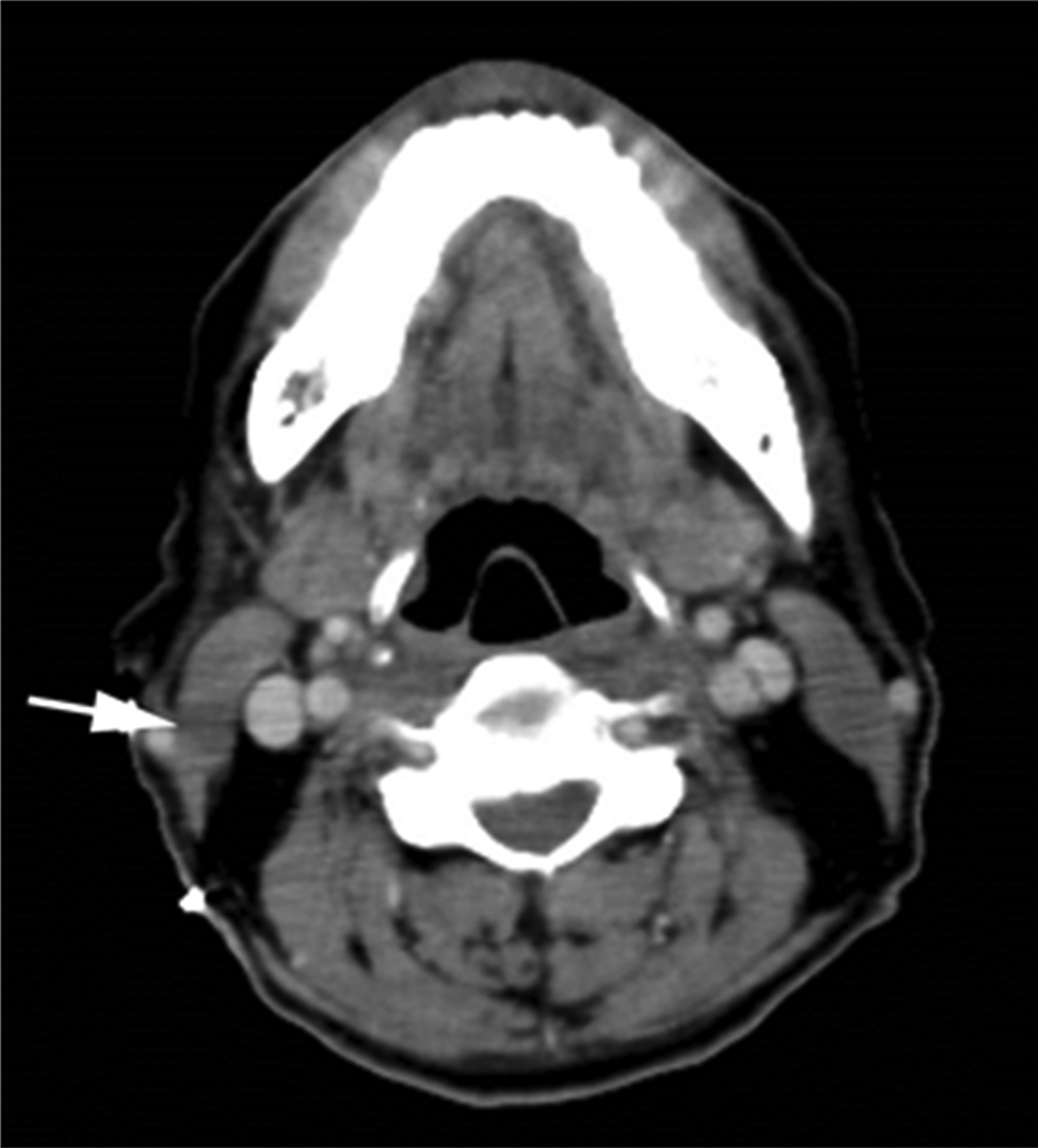
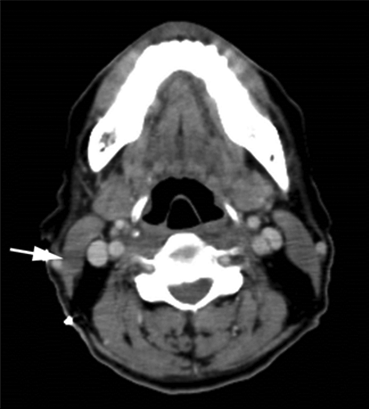
A 57-year-old man was referred to the Department of Otolaryngology from the Department of Medicine of our institution. The patient had a history of a gradually enlarging, painless neck swelling and had experienced a fever of over 38°C for about a week before presenting at our hospital. Eight months prior to this event, the patient had received three cycles of chemotherapy for acute myelogenous leukaemia, one of which was given for recurrent disease. Enhanced computed tomography of the patient's neck showed a right postero-lateral mass within the sternocleidomastoid muscle, measuring about 1.5 cm in diameter (Figure 1).
Enhanced computed tomography scan of the neck showing a right post-lateral neck mass measuring 1.5 cm in diameter (arrow)
Physical examination revealed a soft, elastic, enlarged lymph node, which was palpable in the middle portion of the right posterior triangle and measured 3 cm in diameter. The patient had no other constitutional symptoms, and physical examination of the ears, nose and throat was otherwise unremarkable.
A fine-needle aspiration biopsy was taken using a 21-gauge needle and a 20 ml syringe attached to a Chiba aspiration gun (Nagai Tokurindo, Tokyo, Japan). Cytological spreads and cytocentrifuge preparations were fixed in 95 per cent ethanol and stained with Papanicolaou stain, or air-dried and stained with Wright–Giemsa stain.
Cytology
Cytology revealed an increased number of inflammatory cells, including neutrophils and mononuclear cells, in the striated muscle of the sternocleidomastoid. The Papanicolaou-stained smear showed a large, round, dark-stained cyst measuring 50 µm in diameter. The interior of the cyst was difficult to visualise; however, after destaining with an aqueous hydrochloric acid solution, it exhibited features typical of a toxoplasma cyst and contained numerous tachyzoites (Figure 2a). The rest of the smear contained reactive tachyzoites with larger dispersed histiocytes, which were easier to identify by Wright–Giemsa staining. Dark-stained, elongated particles measuring 3–5 µm in diameter and consistent with T gondii tachyzoites were occasionally observed in the background of these preparations. These particles were also present in a few histiocytes that formed a pseudocyst (Figure 2b).
Photomicrographs showing (a) Papanicolaou staining of a toxoplasma cyst containing tachyzoites (arrow heads) and (b) Wright-Giemsa staining of toxoplasma cysts containing tachyzoites (arrow heads) (×400)

Serology
The biopsy specimen showed a cytological structure characteristic of toxoplasmosis infection. To confirm the cytological diagnosis, immunoglobulin (Ig) G and IgM enzyme-linked immunosorbent assays were performed on the serum specimen. The titres of IgG and IgM antibodies specific to T gondii were found to be 17 IU/ml (normal, less than 5) and 0.1 IU/ml (normal, less than 0.5), respectively. These serological findings provide evidence of an inactive or previous infection. An open biopsy provided material for a histological diagnosis.
Histology
Histological studies on lymph nodes within the sternocleidomastoid muscle showed follicular hyperplasia associated with monocytoid cell hyperplasia, clusters of epithelioid histiocytes and loosely formed non-necrotising granulomas. Histological analysis also revealed toxoplasma cysts. Further, tachyzoites were visualised by Periodic acid-Schiff, haematoxylin and eosin (H&E), and Grocott's staining followed by light microscopy. The sample showed hyperplastic structural features of lymph nodes with well-defined pseudocyst areas (Figure 3). Immunohistochemical analysis using an antibody recognising T gondii primarily showed strong cytoplasmic staining of large histiocytes. A few smaller cells, including small lymphoid cells, showed positive staining, and some background granular staining was also present, including the occasional tachyzoite. The cyst showed strong positive staining. However, we were unable to identify any other cells with similar reactivity, although the histology of all was consistent with toxoplasma lymphadenitis (Figure 4).
Histological images of tachyzoites showing well-defined pseudocysts via (a) Periodic acid–Schiff, (b) H&E and (c) Grocott's staining. (×400)
Positive immunohistochemical staining of some smaller cells, with occasional background granular staining, including tachyzoites. The cyst showed strong positive staining. ((a) ×40, (b) ×400) (Photo by Y. Tsutsumi)
Once a diagnosis of lymphadenopathy was established, an immediate investigation was initiated and chemotherapy was scheduled. The chemotherapy regimen comprised pyrimethamine (50 mg/day) and sulfadiazine (4.0 g/day). However, after approximately three months of treatment (when the first course of chemotherapy for recurrent acute myelogenous leukaemia was complete), severe chemotherapy-related adverse effects developed, including pancytopenia, rash and high fever. As a result, chemotherapy was discontinued. The patient also exhibited worsening symptoms of pneumonia and increased numbers of leukaemic cells, coupled with a recurrent high fever. Further, he developed dyspnoea and, at the request of the patient and his family, only symptomatic therapy was given from then onwards. The patient died of respiratory dysfunction a month later.
Discussion
Toxoplasma infections are transmitted by a ubiquitous parasite.Reference Kieffer and Wallon1–Reference Markell, John and Krotoski3 The incidence of infection is very high in regions with warm, humid climates, where a significant percentage of the population has low antibody titres against this parasite. As much as 40 per cent of the global adult population is infected with T gondii,Reference Jayaram, Ramaprasad, Chethan and Sujay4 and it is thought that 15–20 per cent of unexplained lymphadenopathy cases, especially those affecting cervical nodes, are caused by toxoplasmosis.Reference Kieffer and Wallon1–Reference Jayaram, Ramaprasad, Chethan and Sujay4
T gondii, the intracellular parasite that causes toxoplasma, exists in three forms: oocysts, tachyzoites and bradyzoites (tissue cysts). Oocysts contain sporozoites and are produced by completion of the reproductive cycle in the intestine of definitive hosts (felines). The felines then excrete these oocysts in their faeces. Tachyzoites are the asexual invasive form, which can replicate in all nucleated cells. Bradyzoites can remain dormant in tissues for decades before becoming reactivated.Reference Markell, John and Krotoski3
A diagnosis of toxoplasmosis is based on a combination of direct visualisation by fine-needle aspiration cytology or open biopsy histology and serological assays to detect antibodies against the parasite. However, serological analysis of an immunocompromised patient can yield a false negative result.Reference Chen, Remotti, Tong, Gorczyca and Hamele2 In our patient, for example, the serum IgG antibody titre was elevated but the serum IgM antibody titre was not. Therefore, confirmation of active disease by other serological methods is always desirable. For this purpose, enzyme-linked immunosorbent assay and indirect immunofluorescence methods are available for titration of the parasite-specific IgM antibody.Reference Jayaram, Ramaprasad, Chethan and Sujay4–Reference Zaharopoulos6
There are few reports of cervical toxoplasma lymphadenitis cases being diagnosed exclusively by fine-needle aspiration cytology (with serology).Reference Pathan, Francis, Das, Mallik, Sheikh and Hira5, Reference Zaharopoulos6 Fine-needle aspiration cytology typically shows microgranulomas, consisting of small clusters of characteristic epithelioid histiocytes with only a few mature lymphocytes, and no evidence of necrosis, suppuration or giant cells, on a background of reactive lymphoid hyperplasia.Reference Viguer, Jiménez-Heffernan, López-Ferrer, González-Peramato and Vicandi7 Very few reports have defined the cytological criteria for a diagnosis of toxoplasma lymphadenitis.Reference Zaharopoulos6–Reference Argyle, Schumann, Kjeldsberg and Athens8 There is a similar lack of reports on histopathological analysis. The histological criteria for a diagnosis of toxoplasma lymphadenitis include follicular hyperplasia with preservation of lymph node architecture, focal proliferation of transformed monocytoid B lymphocytes and small, scattered clusters of large, epithelioid-like histiocytes (with larger aggregates of these cells defined as granulomas).Reference Pathan, Francis, Das, Mallik, Sheikh and Hira5, Reference Zaharopoulos6
• Toxoplasma lymphadenitis was successfully diagnosed by fine-needle aspiration cytology
• This method is useful for differentiating toxoplasma lymphadenitis from more serious causes of lymphadenopathy
• Toxoplasma lymphadenitis can be diagnosed by haematoxylin and eosin staining of tissue sections or immunohistochemical analysis of fine-needle aspirates
Tissue cysts are very rarely identified in tissue sections (less than 1 per cent of all cases), and hardly ever in cytological smears.Reference Viguer, Jiménez-Heffernan, López-Ferrer, González-Peramato and Vicandi7 In addition, the identification of tachyzoites or bradyzoites in histological preparations has rarely been reported, and only a few studies have described these structures in fine-needle aspiration cytology smears.Reference Zaharopoulos6–Reference Argyle, Schumann, Kjeldsberg and Athens8 Zaharopoulos reported pseudocysts (tachyzoites in histiocytes), with free tachyzoites observed in the background.Reference Zaharopoulos6 According to this author, Wright–Giemsa staining is useful for identifying tachyzoites in cytological preparations of toxoplasma lymphadenitis because detection with the Papanicolaou stain can be challenging.
Immunohistochemical staining with an antibody against T gondii is valuable for localising toxoplasma antigen in cellular organisms or in inflammatory cells that neutralise these organisms.Reference Al Adnani, Badawi, Thorburn and Rao9
The present case exhibited typical cytomorphological features of toxoplasma lymphadenitis, including high cellularity, reactive lymphoid cells, tingible body macrophages and groups of epithelioid cells. In addition, we observed a T gondii pseudocyst containing numerous tachyzoites.
Conclusion
Toxoplasma lymphadenitis is a common cause of lymph node enlargement. Fine-needle aspiration cytology is a useful method for diagnosing and differentiating toxoplasma lymphadenitis from more serious causes of lymphadeno-pathy, such as metastatic lymphadenopathy or lymphoma. Toxoplasma lymphadenitis can be indicated by histological analysis of H&E-stained paraffin-processed tissue sections or immunohistochemical analysis of fine-needle aspiration cytology smears. Serological analyses are essential to confirm the diagnosis of toxoplasmosis, especially when microscopic studies alone yield inconclusive results.
Acknowledgements
The authors thank Professor Yutaka Tsutsumi for his advice on immunohistochemical analysis and for helpful comments on this manuscript.