Introduction
Members of the Family Allocreadiidae include trematodes commonly found in the digestive tract of freshwater fishes (Caira & Bogéa, Reference Caira, Bogéa, Jones, Bray and Gibson2005); however, two species – Caudouterina rhyacotritoni and Bunoderella metteri – are parasites of salamanders or frogs, respectively (Schell, 1964; Martin, 1966). Species composition, validity of genera and taxonomic arrangement in subfamilies has fluctuated over time and the taxonomic history of the Family has been controversial, and its placement within the Trematoda was unstable (Looss, Reference Looss1902; Yamaguti, Reference Yamaguti1958, Reference Yamaguti1971) until recently (Choudhury et al., Reference Choudhury, Rosas-Valdez, Johnson, Hoffman and Pérez-Ponce de León2007; Pérez-Ponce de León & Hernández-Mena, Reference Pérez-Ponce de León and Nadler2019). Currently, the family is composed of 14 valid genera and approximately 130 species (unpublished data). Due to the morphological similarity of the larval and adult stages of the allocreadiids, in addition to the morphological study, it was also necessary to assess the systematic, ecological and biological analysis for correct taxonomic determination (Caira & Bogéa, Reference Caira, Bogéa, Jones, Bray and Gibson2005). However, in recent years, molecular phylogenetic studies have emerged as a fundamental approach to help the allocation of the species in their taxonomic groups (Pérez-Ponce de León & Choudhury, Reference Pérez-Ponce de León and Choudhury2010; Pérez-Ponce de León & Nadler, Reference Pérez-Ponce de León and Hernández-Mena2010).
Among teleosts, Characiformes possess a highly diverse parasite fauna with approximately 270 genera and 2258 species (Eschmeyer, Reference Eschmeyer2020). These fish are mainly distributed in freshwater environments across the Neotropical region (Baumgartner et al., Reference Baumgartner, Pavanelli, Baumgartner, Bifi, Debona and Frana2012). Astyanax spp., described as the most diverse genus (Ornelas-García et al., Reference Ornelas-García, Domínguez-Domínguez and Doadrio2008), with approximately 261 described species (Eschmeyer, Reference Eschmeyer2020), have feeding habits ranging from herbivores, insectivores (Uieda et al., Reference Uieda, Buzzato and Kikuchi1997) to omnivores (Silva et al., Reference Ronquist, Teslenko and Mark2012). Two species of the genus Astyanax Baird & Girard, 1854 were studied herein – Astyanax lacustris and Astyanax fasciatus – and, until this moment, only one species of the allocreadiid genus Wallinia Pearse, 1920 has been recorded parasitizing A. lacustris (Dias et al., Reference Dias, Mueller, Almeida, Silva, Azevedo, Pérez-Ponce de Leon and Abdallah2018). The genus Creptotrematina Yamaguti, 1954 was erected to include two species of allocreadiids originally described by Freitas (Reference Freitas1941) as belonging to the genus Creptotrema Travassos, Artigas & Pereira, 1928 from Brazil and Argentina, as parasites of characid fish: Creptotrematina dissimilis (Freitas, Reference Freitas1941) Yamaguti, 1954 in Tetragonopterus argenteus Cuvier, 1816 in Brazil, and in Astyanax bimaculatus (Linnaeus, 1758) and Astyanax eigenmanniorum (Cope, 1894) in Argentina; and Creptotrematina dispar (Freitas, 1941) Manter, 1962 in Triportheus paranensis (Günther, 1874) in Brazil and Argentina (see Kohn et al., Reference Kohn, Fernandes and Cohen2007). A third species was described by Jiménez-Guzmán, Creptotrematina aguirrepequenoi Jiménez-Guzmán, 1973, in Astyanax mexicanus (De Filippi, 1853) in north-eastern Mexico, and later found also in Costa Rica in Astyanax aeneus (Günther, 1860) (Curran et al., Reference Curran, Tkach and Overstreet2011). Species of Creptotrematina are characterized by the presence of an elongated body, ventral sucker larger than the oral sucker, large number of small eggs, uterus extending to the posterior end of body and by having an oral sucker with a pair of ventrolateral muscle lobes (Caira & Bogéa, Reference Caira, Bogéa, Jones, Bray and Gibson2005). Three species have been allocated in the genus. In this study, we describe a new species of Creptotrematina from the intestine of A. lacustris and A. fasciatus in Brazil. We used morphological and molecular data of internal transcribed spacers 2 (ITS2) and D1–D3 domains of the 28S ribosomal RNA (rRNA) gene, as well as host association and geographical distribution data to validate the finding, and to provide the description of the new species.
Materials and methods
Specimens of A. fasciatus and A. lacustris were collected between June 2013 and July 2014 in the Batalha River, State of Sao Paulo, Brazil. A total of 31 specimens of A. fasciatus and 75 specimens of A. lacustris were analysed and we found ten digenean specimens in the intestine of their hosts. The prevalence was 16.13% for A. fasciatus and 5.33% for A. lacustris. The mean intensities were 1.2 and 1.33 digeneans per infected host for A. fasciatus and A. lacustris, respectively. Specimens were fixed in 99% ethanol for DNA extraction, and in 70% ethanol for parasite morphological identification. All procedures were approved by the applicable committees (CEUA number 3353050417; SISBio number 40998-2; SisGen number A4901A5).
For the morphological study, samples were stained with Gomori's trichrome and mounted in Canada balsam. Motic Images Plus™ 2.0 software (Motic Asia, Hong Kong, China)was used to perform micrometre measurements. Drawings were made with the help of a stabilization tube mounted on the Hund Wetzlar H-600 phase-contrast microscope (Wetzlar, Hessen, Germany). Samples were deposited with the National Institute for Amazonian Research (INPA), Manaus, Amazonas, Brazil (holotype number INPA 811 and paratype number INPA 812)
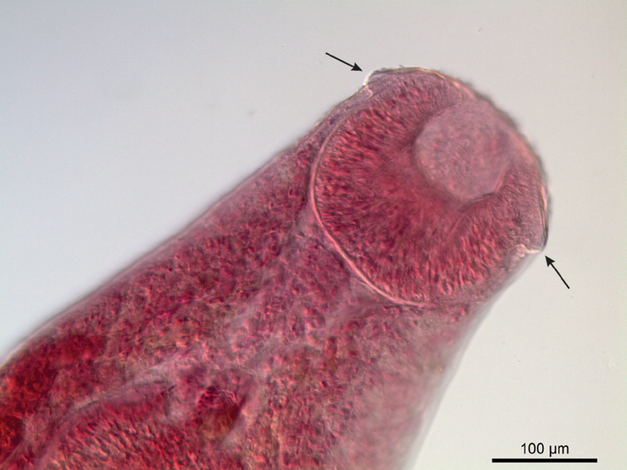
For the molecular characterization, the specimens were cut medially, and the anterior portion was stained with Gomori's trichrome and mounted in Canada balsam to record a molecular voucher for visualization of morphological characters (hologenophore sensu Astrin et al., Reference Astrin, Zhou and Misof2013). Genomic DNA was extracted from the posterior median portion following Qiagen DNeasy® Blood & Tissue kit (Germantown, Maryland, USA)protocols. Fragments from the D1–D3 domains of the 28S rRNA and ITS were amplified by polymerase chain reaction (PCR), following the procedures detailed by Razo-Mendivil et al. (Reference Razo-Mendivil, Vázquez-Domínguez, Rosas-Valdez, Pérez-Ponce de León and Nadler2014a, Reference Razo-Mendivil, Mendoza-Garfias, Pérez-Ponce de Leon and Rubio-Godoyb). The primers used for 28S rRNA amplification were 28dig12 (5′-AAGCATATCACTAAGCGG-3′; Curran et al., Reference Curran, Tkach and Overstreet2011) and reverse 1500R (5′-GCTATCCTGAGGGAAACTTCG-3′; Olson et al., Reference Olson, Cribb, Tkach, Bray and Littlewood2003) with a melting temperature of 54°C, and for ITS amplification, were forward Glyp1 (5′-GCTGAGAAGACGACCAAACTTGAT-3′; Razo-Mendivil et al., Reference Rambaut2010) and reverse BD2 (5′-TATGCTTAAATTCAGCGGGT-3′; Bowles et al., Reference Bowles, Blair and McManus1995) with a melting temperature of 55°C. PCR results were visualized on 1% agarose gel (Razo-Mendivil et al., Reference Razo-Mendivil, Vázquez-Domínguez, Rosas-Valdez, Pérez-Ponce de León and Nadler2014a, Reference Razo-Mendivil, Mendoza-Garfias, Pérez-Ponce de Leon and Rubio-Godoyb) and purified with the Qiagen purification kit before sequencing. The sequencing reaction was performed using the BigDye® Terminator v3.1 Cycle Sequencing Kit (Applied Biosystems, Foster City, California, USA) and ethanol/EDTA (Ethylenediaminetetraacetic acid)/sodium acetate precipitation, according to the manufacturer instructions. Automatic sequencing was performed by capillary electrophoresis on the ABI3730xl DNA Analyzer (Applied Biosystems, Waltham, MA, USA). After sequencing, electropherogram analysis was performed using the programs EMBOSS: merger (http://www.bioinformatics.nl/cgi-bin/emboss/merger), Chromas Lite (https://chromas-lite.software.informer.com) and Geneious 4.8.3 (https://geneious.com) to obtain the consensus sequence. The sequence was assembled and edited in Sequencher TM version 5.2.4 (Gene Codes, Ann Arbor, MI, USA) and was submitted to GenBank associated with voucher that was deposited with the INPA (voucher number INPA 813), Manaus, Amazonas, Brazil. The 28S rRNA gene sequences from 19 members of the Allocreadiidae were downloaded from GenBank for phylogenetic analysis (table 1). Prosthenhystera obesa (Diiesing, 1850) (AY222206) was used as an outgroup for rooting the trees. Callodistomidae are the sister group of Allocreadiidae (Pérez-Ponce de León et al., Reference Pérez-Ponce de León, Choudhury, Rosas-Valdez and Mejía-Madrid2015; Choudhury et al., Reference Choudhury, García-Varela and Pérez-Ponce de León2017). The final alignment was 1002 bp long to the 28S rRNA gene and 491 bp to ITS2. The sequences were aligned using the MUSCLE Software (Edgar, Reference Edgar2004), implemented within the Geneious Server database program, version 7.1.3 using the default settings (Kearse et al., Reference Kearse, Moir and Wilson2012) by cutting the ends of the alignment. To assess the occurrence of saturation substitution, the substitution saturation index was estimated using DAMBE5 (Xia ,Reference Xia2013). The number of base substitutions per site between sequences was calculated. Standard error estimates were obtained using the bootstrap procedure (1000 replicates). Analyses were performed using the Tamura–Nei model (Tamura & Nei, Reference Stamatakis1993) in the MEGA6 program (Tamura et al., Reference Tamura and Nei2013). The evolutionary model used to estimate the ultrametric tree was GTR + G. maximum likelihood analyses, using the RaxML version 8 program (Stamatakis, Reference Silva, Pessoa, Costa, Chellappa and Chellappa2014). The model parameter and bootstrap value (1000 repetitions) were also estimated using the RaxML program. Bayesian analyses were performed using MrBayes on Xsede software (Ronquist et al., Reference Razo-Mendivil, Pérez-Ponce de Leon and Rubio-Godoy2012). MrBayes and RaxML analyses were performed through an online computer site CIPRES (Miller et al., Reference Miller, Pfeiffer and Schwartz2010). Phylogenetic trees were generated and visualized in the program FigTree version 1.3.1 (Rambaut, Reference Petkeviciute, Stunzenas, Staneviciute and Sokolov2009).
Species, hosts, locality, access to GenBank number and references to parasite sequences used in phylogenetic analyses of 28S rDNA gene.

Results
Systematics
Allocreadiidae (Looss, 1902) Stossich, 1903
Creptotrematina Yamaguti, 1954
Creptotrematina batalhensis Dias and Abdallah n. sp.
Description
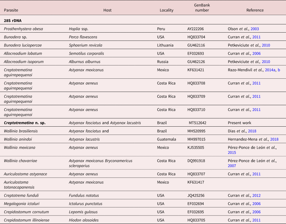
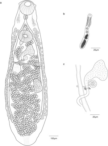
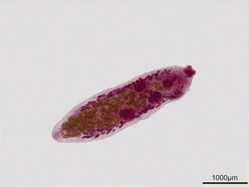
Based on four whole-mount specimens (figs 1 and 2). Body elongate, 2040–3590 (2810) long; maximum width 567–847 (707) in posterior third of body; anterior extremity bluntly rounded; body widening towards posterior end of body. Eyespots remnants present at pharynx level. Oral sucker subterminal, 169–221 (191) long, 174–223 (216) wide, with anteriorly directed oral opening bearing two muscular oral lobes on each side (fig 3). Ventral sucker rounded, 154–216 (189) long, 155–223 (196) wide. Ratio of oral sucker length to ventral sucker length 1:1.02–1.09 (1.05). Ratio of oral sucker width to ventral sucker width 1:1–1.12 (1.06). Prepharynx lacking. Pharynx muscular 60–75 (64) long, 57–105 (80) wide. Oesophagus narrow, relatively long, not sinuous, 70–111 (90) long. Gland cells forming lateral clusters on either side of pharynx, reaching anterior third of oesophagus in some specimens. Caecal bifurcation short, distance, anterior to ventral sucker; caeca terminating blindly near posterior end of body, 1530–2165 (1916) from posterior end. Ovary smooth, irregularly shaped, immediately posterior to and not overlapping ventral sucker, 156–290 (231) long, 114–274 (188) wide and ventrally situated on sinistral position; proximal region of ovary containing oocytes. Seminal receptacle rounded 64–102 (84) long, 57–101 (80) wide, with thick muscular wall immediately posterior to ovary. Vitelline receptacle between anterior testis and ovary. Vitellarium follicular, caecal and extracaecal in two ventrolateral fields extending from mid-level between genital pore and anterior end of ventral sucker to posterior end of body reaching the end of caeca. Eggs numerous, ovoid, embryonated, 38–57 (49) long, 20–39 (34) wide (supplementary videos S1–S3). Genital pore median, between caecal bifurcation and anterior border of ventral sucker. Testes two, ellipsoidal, oblique, intercaecal, with smooth margins; anterior testis 175–310 (251) long, 118–301 (206) wide; posterior testis 203–368 (299) long, 127–316 (220) wide; 771–1572 (1113) from posterior end of body; distance between testes 169–349 (275); distance between ovary and anterior testis 66–168 (123); distance between anterior testis and posterior extremity of ventral sucker 295–349 (326); distance between ovary and posterior extremity of ventral sucker 56–288 (139); cirrus sac elongated, median and dorsal, extending slightly posterior to margin of ventral sucker, with maximum width of 310–469 (398). Excretory vesicle I-shaped. Excretory pore terminal.
Creptotrematina n. sp. from Astyanax fasciatus and Astyanax lacustris. (a) Holotype: ventral view of whole-mount specimen from A. fasciatus. (b) Ventral view of the cirrus sac of a paratype from A. lacustris. (c) Female reproductive complex from an A. fasciatus holotype.

A ventral view of whole-mount specimen of Creptotrematina n. sp. from Astyanax lacustris and Astyanax fasciatus.

Oral sucker with a pair of ventrolateral muscle papillae, as highlighted by the arrows.

Taxonomic summary
Type material. Holotype (number available after article is accepted).
Type host. Astyanax fasciatus (Cuvier, 1819) (Actinopterygii: Characidae).
Other hosts. Astyanax lacustris Lucena & Soares, 2016 (Actinopterygii: Characidae).
Type locality. Batalha River, located on the border between the cities of Bauru and Piratininga, Sao Paulo, Brazil (22°24′46″S, 49°05′05″W).
Site of infection. Intestine.
Etymology. The specific epithet batalhensis refers to the name of the river where the parasite was discovered.
GenBank accession numbers. MT512641; MT512642; MT512643.
Remarks
The new species conforms with the diagnosis of the genus Creptotrematina as previously described. The specimens obtained in the present study morphologically resemble C. aguirrepequenoi in the posterior extension of the intestinal caeca, the extension of the vitellaria to the posterior end of the body and by having oblique testes. The species C. aguirrepequenoi was described from the Mexican tetra, A. mexicanus, in north-eastern Mexico. The new species is readily distinguished from C. aguirrepequenoi mostly by the presence of eyespot remnants at pharynx level, the elongated shape of vitelline follicles and the posterior extent of the caeca in the posterior end of hindbody. Additionally, the ovary size is almost similar compared to the testes in Creptotrematina batalhensis Dias and Abdallah, n. sp., while in C. aguirrepequenoi the ovary is much smaller than testes. Two species of Creptotrematina occur in South American characids – C. dissimilis and C. dispar. However, the new species can also be clearly distinguished by the testes position (oblique vs. symmetrical) and the extension of the vitellaria (lateral fields along most of the body vs. restricted to the anterior part, between ventral sucker and testes).
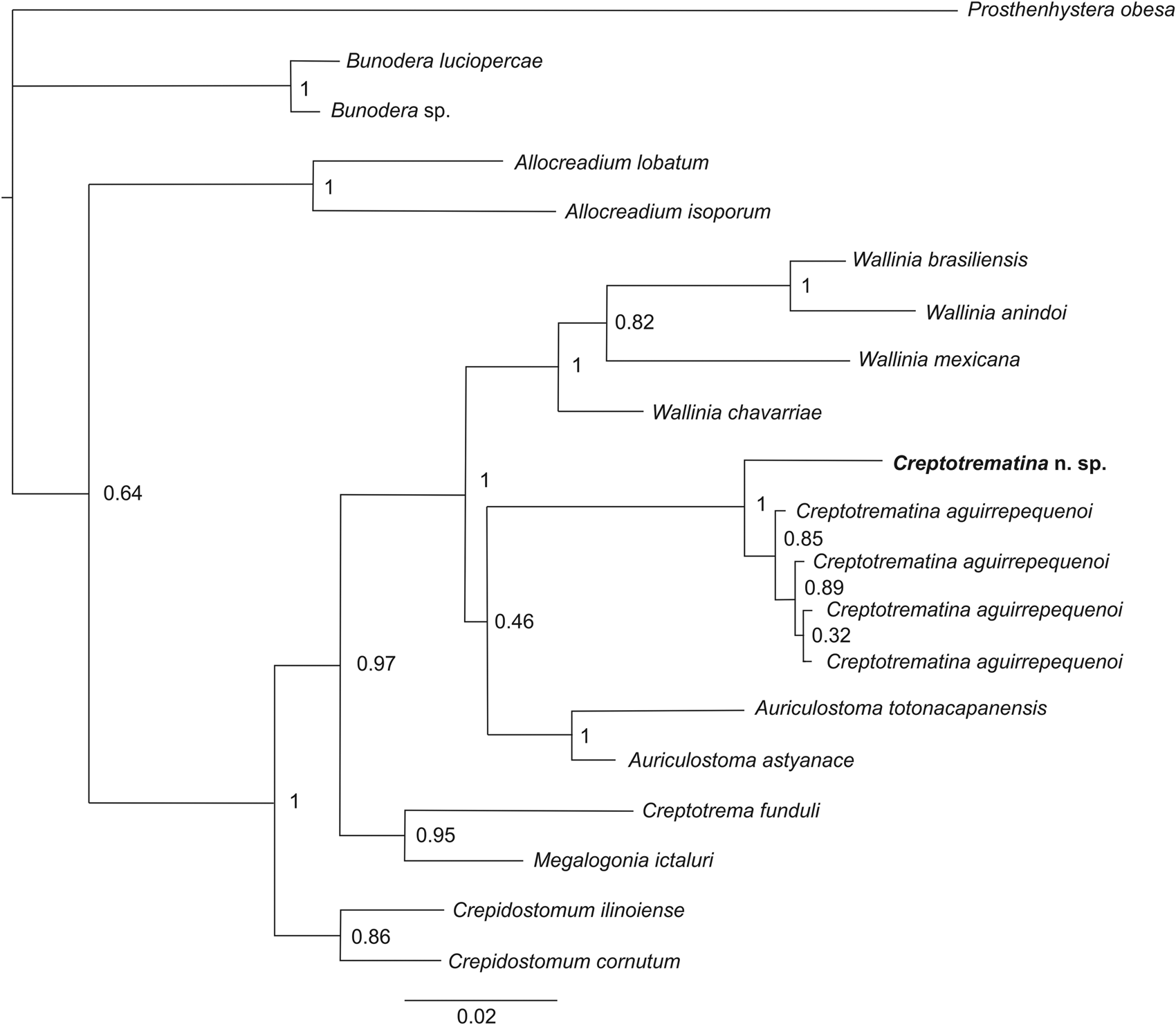
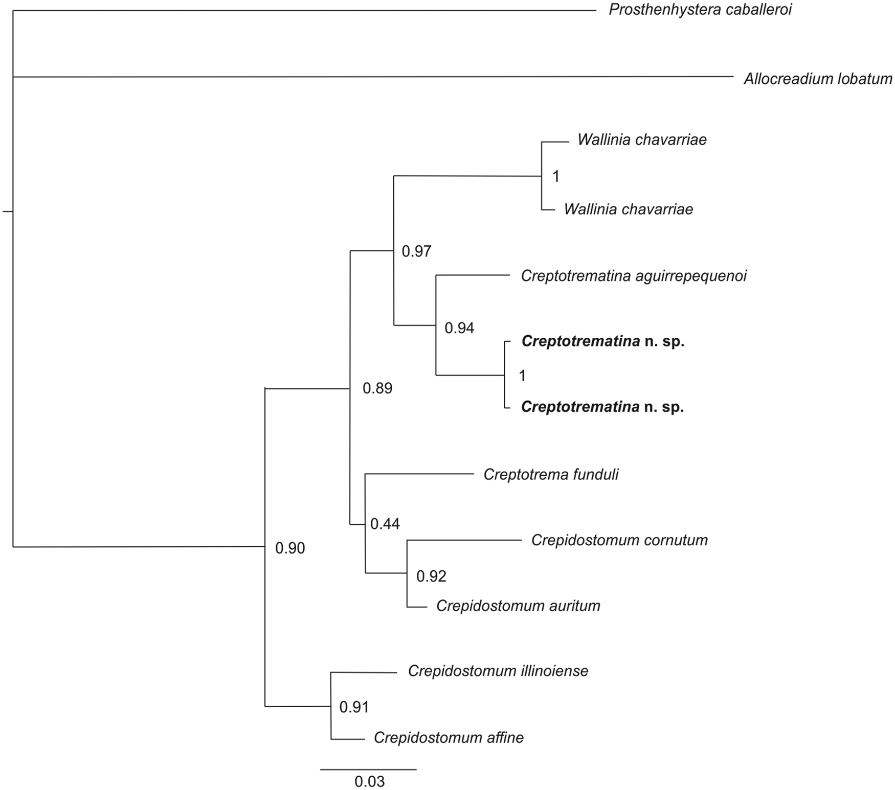
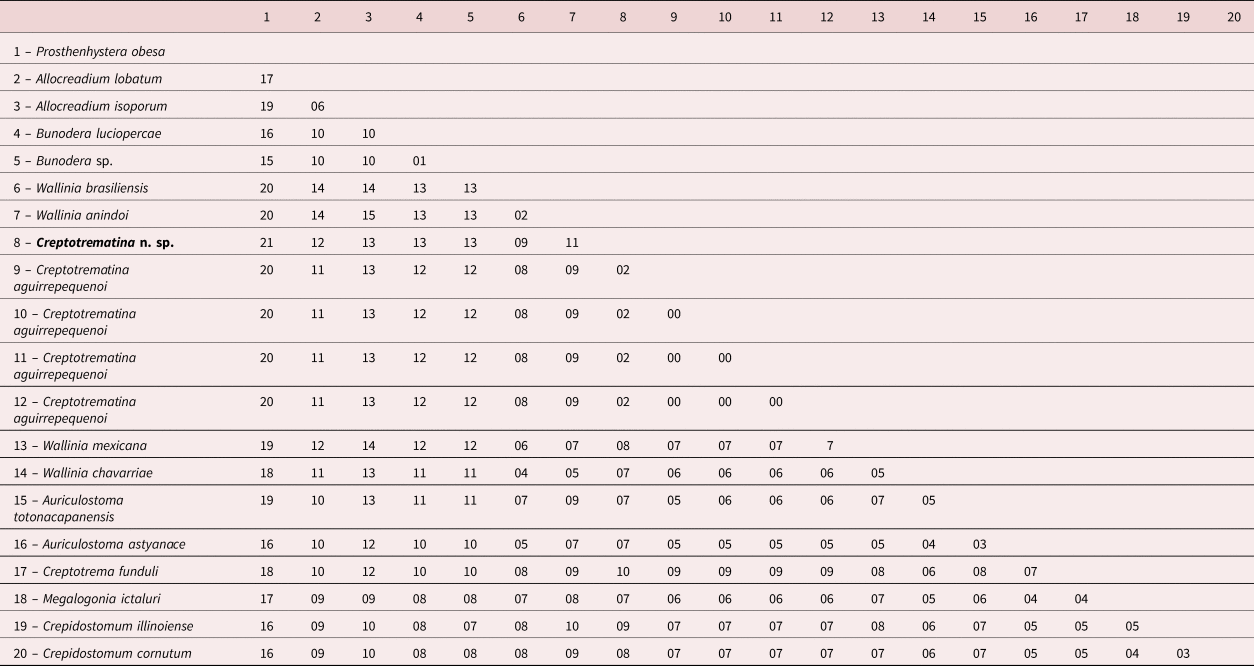
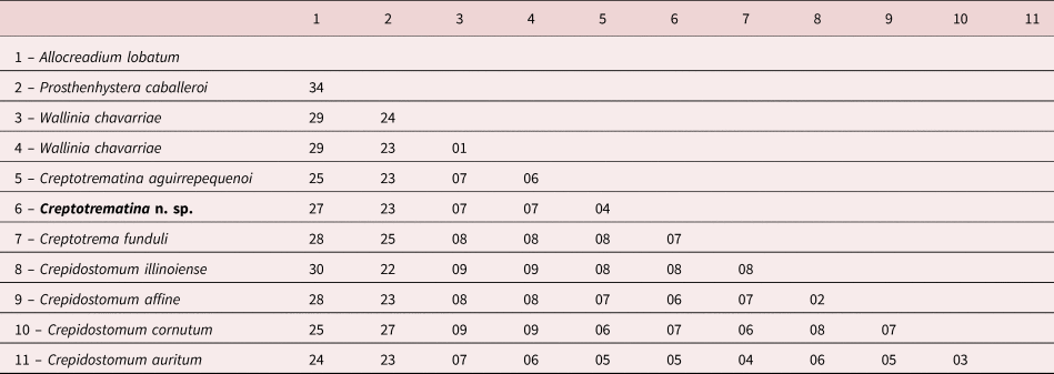
Molecular data further corroborated the distinction of the new species. The three isolates sequenced for two ribosomal markers show reciprocal monophyly. The new species is yielded as the sister taxa of C. aguirrepequenoi, the only congeneric species for which sequences are still available, with specimens sequenced from a geographical range that spans between Mexico and Costa Rica, in Central America (Curran et al., Reference Curran, Tkach and Overstreet2011; Razo-Mendivil et al., Reference Razo-Mendivil, Vázquez-Domínguez, Rosas-Valdez, Pérez-Ponce de León and Nadler2014a, Reference Razo-Mendivil, Mendoza-Garfias, Pérez-Ponce de Leon and Rubio-Godoyb) (figs 4 and 5). These relationships are well supported by high posterior probability and bootstrap values, except for the clade that includes the Auriculostoma and Creptotrematina species. The genetic distance for the 28S rRNA gene between the specimens of C. batalhensis Dias and Abdallah, n. sp. of Brazil and those of C. aguirrepequenoi from A. mexicanus and A. aeneus from Mexico and Costa Rica, respectively, was 2% (table 2), which is similar to what has been reported for other species pairs in allocreadiids. Our results show significant genetic divergence for species delimitation of members of Allocreadiidae; these molecular markers have been used for species differentiation in the group (Curran et al., Reference Curran, Tkach and Overstreet2006; Pérez-Ponce de Léon et al., Reference Pérez-Ponce de León, Choudhury, Rosas-Valdez and Mejía-Madrid2015, Reference Pérez-Ponce de León, Razo-Mendivil, Mendoza-Garfias, Rubio-Godoy and Choudhury2016; Dias et al., Reference Dias, Mueller, Almeida, Silva, Azevedo, Pérez-Ponce de Leon and Abdallah2018; Hernández-Mena et al., Reference Hernandez-Mena, Lynggaard, Mendoza-Garfias and Peréz-Ponce de Leon2018). For instance, Hernández-Mena et al. (Reference Hernandez-Mena, Lynggaard, Mendoza-Garfias and Peréz-Ponce de Leon2018) validated two new species of allocreadiids using information from the 28S gene. The genetic divergence between their new species Wallinia anindoi and its sister taxa Wallinia brasiliensis was 1.19%, and between Auriculostoma tica and its sister taxa Auriculostoma totonacapanensis was 2.11%. Therefore, the genetic differences corroborate the delimitation of the new species described in this study. In addition to 28S, the internal ITS2 was also used, since they have been considered relatively consistent for species differentiation among trematodes (Morgan & Blair, Reference Morgan and Blair1995; Jousson et al., Reference Jousson, Bartoli, Zaninetti and Pawlowski1998; Curran et al., Reference Curran, Tkach and Overstreet2006). The genetic divergence between the new species we described herein and C. aguirrepequenoi was 4% (table 3), which is also similar to the distance referred by the authors mentioned above, corroborating the species distinction.
Bayesian topology based on partial 28S ribosomal DNA sequences of some members of the Allocreadiidae. Support values are above the nodes of posterior probabilities for the Bayesian analyses. The Prosthenhystera obesa were used as an outgroup. Branch length scale bar indicates number of substitutions per site.

Bayesian topology based on ITS ribosomal DNA sequences of some members of the Allocreadiidae. Support values are above the nodes of posterior probabilities for the Bayesian analyses. The Prosthenhystera obesa were used as an outgroup. Branch length scale bar indicates number of substitutions per site.

Percentage of genetic diversity (Tamura–Nei) of 28S rDNA gene with Allocreadiidae family sequences taken from GenBank with Creptotrematina n. sp. Prosthenhystera obesa (Callostomidae) was used as outgroup.

Percentage genetic divergence (Tamura–Nei) of the ITS rDNA with sequences from the Allocreadiidae family taken from GenBank with Creptotrematina n. sp. Prosthenhystera obesa (Callostomidae) were used as an external group.

Discussion
Before the present study, only two species of Creptotrematina had been reported in Brazil: C. dissimilis from A. bimaculatus in the Paraná River, Paraná State, Brazil (Hamann, Reference Hamann1983) and in T. argenteus in the Miranda River, Mato Grosso, Brazil (Freitas, 1941); and C. dispar from T. paranensis in the Miranda River, Mato Grosso, Brazil (Freitas, 1941). Thus, the current work contributes with a new species associated with a new host and locality record of a congeneric species in Brazil. The genus now contains four species, three of them occur in South American characids, and apparently only one, C. aguirrepequenoi, in Middle America, where characids reach their most northern distributional range in the Americas. The 28S phylogenetic tree shows some level of variation among samples of C. aguirrepequenoi from different localities, although the variations is very low for this molecular marker. Still, the genetic variation between the new species and C. aguirrepequenoi warrants the proposal that it corresponds to a separate species and these results are also corroborated by ITS2 sequence data. A detailed study of C. aguirrepequenoi using more variable genes such as mitochondrial cox1 is still required to demonstrate that they might represent the species complex. The most northern distribution record of C. aguirrepequenoi is from Mexican tetra, A. mexicanus, where it was originally described, while the record from Costa Rica was made for the banded tetra, A. aeneus. Also, DNA sequences from the two remaining species, C. dispar and C. dissimilis, are required to fully understand the interrelationships among the species of Creptotrematina. With the available information, we can corroborate that the genus is widely distributed across the Americas, in close association with the historical biogeography of their characid hosts (see Ornelas-Garcia et al., Reference Ornelas-García, Domínguez-Domínguez and Doadrio2008; but see discussion in Choudhury et al., Reference Choudhury, García-Varela and Pérez-Ponce de León2017). Clearly, DNA analysis plays a major role in the definition of species limits and classification in allocreadiids, and thorough sampling is required in areas of Brazil and Argentina to collect specimens of trematodes to conduct DNA sequencing.
Supplementary material
To view supplementary material for this article, please visit https://doi.org/10.1017/S0022149X20000474
Financial support
This study was supported by the Brazilian National Research Council (CNPq; case number 158778/2013-8); Coordenação de Aperfeiçoamento de Pessoal de Nível Superior, Brazil (CAPES; finance code 001); the São Paulo Research Foundation (FAPESP; grant numbers 2016/21040-9, and 2017/16546-3); Programa Nacional de Pós-Doutorado (PNPD; grant number AUX-PE-PNPD 3005/2010); Young Researcher Program (PROPE-UNESP; grant number 02/2016); and CONACyT (G.P.P.L., grant number A1-S-21694).
Conflicts of interest
None.
Ethical standards
All applicable institutional, national and international guidelines for the care and use of animals were followed under license number N3295230615 CEUA and this article does not contain any studies with human participants performed by any of the authors.
Author contributions
All authors contributed to the study conception and design. Material preparation, data collection and analysis were performed by Karina Gabriele Alves Dias, Maria Isabel Muller and Aline de Almeida Camargo. The first draft of the manuscript was written by Karina Gabriele Alves Dias and all authors commented on previous versions of the manuscript. All authors read and approved the final manuscript. Conceptualization: Karina Gabriele Alves Dias and Vanessa Doro Abdallah; methodology: Karina Gabriele Alves Dias and Maria Isabel Muller; formal analysis and investigation: Karina Gabriele Alves Dias and Gerardo Pérez-Ponce de Léon; writing – original draft preparation: Karina Gabriele Alves Dias; writing – review and editing: Gerardo Peréz-Ponce de Léon; funding acquisition: Reinaldo José da Silva and Vanessa Doro Abdallah; resources: Reinaldo José da Silva and Rodney Kozlowiski de Azevedo; and supervision: Rodney Kozlowiski de Azevedo and Vanessa Doro Abdallah.