Introduction
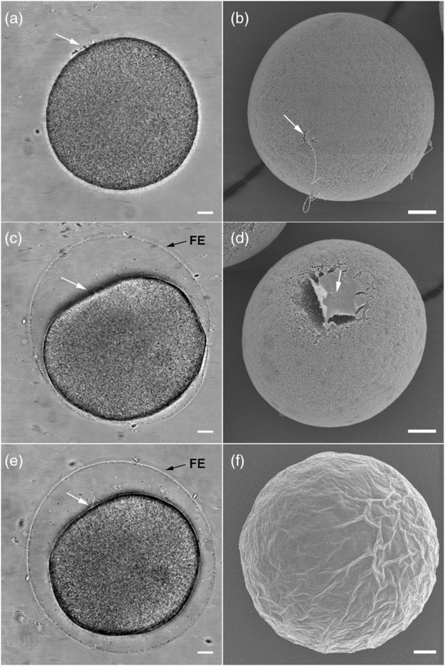
For more than a century, scientists have been fascinated by the fact that, under laboratory conditions, sea urchin eggs exposed to high concentrations of spermatozoa are still invariably monospermic (Just, Reference Just1939). Based on the observation that the fertilizing spermatozoon triggers a series of successive and overlapping physiological changes in the egg transforming it from a quiescent cell into a dynamic zygote, it was proposed that several of these activation events play a secondary role in prevention of supernumerary sperm entry. At fertilization, the main structural event of the activated sea urchin eggs, the cortical reaction, was described a century ago (Just, Reference Just1919) and involves the fusion of the cortical granules (CG) with the egg plasma membrane following Ca2+ increase in the egg, and the release of their contents into the space between the plasma membrane and the vitelline envelope. This exocytotic process leading to the formation of the fertilization envelope (FE) takes several minutes (Fig. 1), and has been suggested as a mechanical barrier to polyspermy. However, the process is too slow or inefficient to ensure monospermy, even when the FE is fully elevated (Santella et al., Reference Santella, Limatola, Chun, Sawada, Inoue and Iwano2014, Reference Santella, Limatola and Chun2016, Reference Santella, Limatola, Vasilev and Chun2018; Chun et al., Reference Chun, Limatola, Vasilev and Santella2014; Limatola et al., Reference Limatola, Vasilev, Chun and Santella2019). In line with this, it was suggested that:
…With membrane separation the eggs undergo some change and it is this change – not its result, membrane separation – which constitutes the block to the entrance of additional spermatozoa… (Just, Reference Just1939)
Cortical reaction in an intact P. lividus egg. Bright field view photomicrograph (left panel) and scanning electron micrograph (right panel) of the fertilized eggs at the key moments of the cortical reaction. (a, b) Contact of the sperm’s acrosomal process with the egg plasma membrane generates a dimple on the egg surface at which the fertilization envelope (FE) begins to elevate. (c, d) A few seconds later, the concavity becomes more pronounced, while the spermatozoon penetrates deeper into the egg with the formation of the fertilization cone (black arrow in e). After a few minutes, the spermatozoon completes its incorporation, and the FE is fully elevated over the entire surface of the egg (e, f). Note that once the cortical reaction has started, not many spermatozoa surround the egg and only the fertilizing spermatozoon remains attached to the egg surface. White arrows (a–e) indicate the site of sperm–egg interaction and penetration. Scale bars, 10 µm.

It was subsequently proposed that an additional fast, yet transient, change also exists, namely a fast block to polyspermy that complements the cortical reaction in preventing the entry of unwanted spermatozoa (Rothschild, Reference Rothschild1954). Two decades later, it was suggested that this fast polyspermy block was electrical in nature; the fertilizing spermatozoon depolarizes the egg plasma membrane, rendering it unresponsive to supernumerary spermatozoa (Jaffe, Reference Jaffe1976). Despite decades of contention based on experimentation (Dale, Reference Dale2014) and the fact that, under natural conditions, the sperm:egg ratio is very low and would not favour the evolution of polyspermy-preventing mechanisms (Dale, Reference Dale and Skinner2018), the concept that egg activation events play a role in achieving monospermy gained popularity and entered the general and specific scientific literature (Wong and Wessel, Reference Wong and Wessel2006; Gilbert, Reference Gilbert2014).
As the activation events overlap with each other both spatially and temporally, experimental design has been fraught with technical difficulties to dissect cause from effect. In this study, we have used a single procedure, a reduction in external Na+ concentration, to perturb early activation events such as membrane depolarization, Ca2+ signalling, and the cortical reaction, and have studied sperm–egg kinetics in the sea urchin using intact eggs under natural conditions. We show that, even when the eggs were inseminated with high concentrations of spermatozoa as in the earlier studies (Jaffe, Reference Jaffe1980), monospermy prevails, and that the changes in Ca2+ signalling and the cortical reaction in low Na+ seawater are the result of a perturbed cortical actin cytoskeleton. Furthermore, we show that the aberrant cleavage of eggs fertilized in low Na+ is due to the altered behaviour of cortical actin filaments (F-actin) following fertilization and it is not the result of the formation of multiple sperm asters.
Materials and Methods
Gamete preparation and sperm entry visualization
Specimens of the Mediterranean species of sea urchin (Paracentrotus lividus) were collected in the Gulf of Naples during the breeding season (from November to May) and maintained in circulating seawater (16°C). Spawning in adult animals was induced by intracoelomic injection of 0.5 M KCl, or by manual dissection. (We found no difference in the behaviour or quality of the eggs collected by these two methods.) Eggs were collected in filtered natural seawater (NSW) and washed three times before use, while dry sperm were collected with a pipette from the aboral surface of the animal. Fertilization was performed with the fresh sperm diluted at 1:1000 to 1:12,500 in artificial seawater (ASW), as previously reported (Jaffe, Reference Jaffe1980). To obtain different Na+ concentrations, ASW (575 mM NaCl, 10 mM KCl, 10 mM CaCl2, 55 mM MgSO4, 2 mM NaHCO3, pH 8.1) was mixed with Na+-free choline-substituted ASW (575 mM choline chloride, 10 mM KCl, 10 mM CaCl2, 55 mM MgSO4, 2 mM NaHCO3, pH 8.1) at various ratios (Schuel et al., Reference Schuel, Schuel, Dandekar, Boldt and Summers1982). To determine the number of spermatozoa incorporated into the egg, sperm nuclei were stained with 5 µM Hoechst 33342 (Sigma–Aldrich) for 15 s before fertilization. Sperm incorporation was monitored 5 min after egg insemination using a CCD camera (MicroMax, Princeton Instruments, Inc.) mounted on a Zeiss Axiovert 200 microscope with a Plan-Neofluar ×40/0.75 objective and a UV laser.
Microinjection, confocal microscopy, and video imaging
Microinjection of the eggs was performed with an air pressure transjector (Eppendorf, FemtoJet). The amount of injected substance was approximately 1% of the egg volume, therefore the final concentration was estimated to be 100-fold lower than that in the injection pipette. Fresh intact eggs (without pre-treatment to remove egg jelly) were microinjected by gently tapping the needle against the egg surface. The injection chamber was prepared by fixing two coverslips at an angle of about 45° with wax, into which the eggs in NSW were pipetted. This arrangement immobilized the eggs during the insertion of microinjection needle. To monitor Ca2+ and F-actin changes in living P. lividus eggs, 10 µM Alexa Fluor 568–phalloidin (Molecular Probes) was microinjected into the egg cytoplasm as previously described (Chun et al., Reference Chun, Limatola, Vasilev and Santella2014). The injected eggs were then placed into a space created by two pieces of coverslip glued together by double-sided adhesive tape in the imaging chamber. F-actin was visualized with a Leica TCS SP8 confocal laser scanning microscope equipped with a white light laser and hybrid detectors (Leica Microsystem, Srl). For Ca2+ imaging, 500 µM Calcium Green 488 conjugated to 10 kDa dextran (CG) was mixed with 35 µM Rhodamine Red (RR) (Molecular Probes) in the injection buffer (10 mM Hepes, 0.1 M potassium aspartate, pH 7.0). The relative Ca2+ changes were scored using the fluorescence of the Ca2+-sensitive dye CG normalized to that of the Ca2+-insensitive dye RR. Bright field views and epifluorescence images of cytosolic Ca2+ changes (acquisition time = 3 s) were collected using a cooled CCD camera (MicroMax, Princeton Instruments, Inc.) mounted on a Zeiss Axiovert 135TV microscope with a Plan-Neofluar ×40/0.75 objective, and analyzed with MetaMorph (Universal Imaging Corporation, PA, USA). The quantified relative Ca2+ signal at a given time point was normalized to the baseline fluorescence (F0) following the formula Frel = (F − F0)/F0, where F represents the average fluorescence level over the entire egg. The instantaneous incremental changes of the Ca2+ rise at a given time point (t) was analyzed by applying the formula Finst = [(Ft − Ft-1)/Ft-1]. This allowed visualization of the instantaneous Ca2+ release at a particular site. Therefore, Frel was expressed as relative fluorescence units (RFU) in plotting Ca2+ trajectories. To compensate for the batch-to-batch variability of Ca2+ signal intensities, the peak amplitudes of the Ca2+ wave and the cortical flash of each egg were normalized with the averages of the corresponding values in the control eggs from the same batch. Subsequently, the data presented in reference to the batch-matching control (taken as 100%) were pooled together and subjected to statistical analyses.
Statistical analysis
Numerical MetaMorph data were compiled and analyzed using MS Excel software, Microsoft Office 2010. The average and variation of the data were reported as ‘mean ± standard deviation (SD)’ in all cases in this manuscript. One-way analysis of variance (ANOVA) tests and the post hoc analyses were performed using VassarStats (http://vassarstats.net/anova1u.html), and P-values less than 0.05 (P < 0.05) were considered to be statistically significant.
Sample preparation for scanning electron microscopy
Intact sea urchin eggs before and after insemination were fixed in ASW and low sodium ASW containing 0.5% glutaraldehyde for 1 h at 20°C. The samples were washed several times in distilled water, post-fixed for 1 h in 1% osmium tetroxide, and subsequently dehydrated by dipping into a series of ethanol–water mixtures containing increasing concentrations of ethanol, ending at 100% ethanol. A Leica CPD300 critical point dryer and an EM ACE600 sputter coater were used, respectively, to dry and coat the specimens with conductive metal before imaging them using a JEOL 6700F scanning electron microscope.
Results
Cortical reaction in intact Paracentrotus lividus eggs
In sea urchin, the morphological hallmark of ‘egg activation’ by the fertilizing spermatozoon is the elevation of the FE, which results from Ca2+-induced exocytosis of the CG into the perivitelline space. The current opinion is that FE elevation serves as a mechanical barrier to prevent the entry of supernumerary spermatozoa and to protect the embryo during development. In P. lividus eggs, at ambient temperature, several minutes elapse from the initial attachment of a successful fertilizing spermatozoon to the final completion of FE elevation over the entire surface of the egg. However, as shown in Fig. 1, and in contrast with previous reports (Schatten and Mazia, Reference Schatten and Mazia1976), a few spermatozoa remain attached to the egg surface beyond the initiation of the cortical reaction. When the acrosome-reacted spermatozoon exposing adhesive protein binding interacts with the egg surface (Vacquier, Reference Vacquier2012), a dimple is generated on the egg surface (Fig. 1 d, arrow). At this depression, the plasma membranes of the spermatozoon and the egg begin to fuse (Green and Summers, Reference Green and Summers1980). Through a newly formed protrusion on the surface of the egg, namely the fertilization cone, the spermatozoon is progressively incorporated into the cytoplasm in a process mediated by its anchorage to the eggs’ actin filaments (Puppo et al., Reference Puppo, Chun, Gragnaniello, Garante and Santella2008). In the meantime, the FE starts to rise. Within 1–2 min, the spermatozoon completes its entry, and the FE becomes fully raised around the entire surface of the egg.
Decreased levels of sodium do not induce polyspermy, but do negatively affect embryonic development
Early experiments performed on the eggs of Arbacia punctulata, demonstrated that sodium cations were more effective than potassium or magnesium in causing polyspermic fertilization (Clark, Reference Clark1936). Low external Na+ reduces the amplitude of the membrane depolarization at fertilization (Dale and De Santis, Reference Dale and De Santis1981) and therefore should induce polyspermy, if the reigning hypothesis that the fast block to polyspermy is electrically mediated is correct. According to this hypothesis proposed by Jaffe (Reference Jaffe1976), when the spermatozoon contacts the surface of an egg of the sea urchin (Strongylocentrotus purpuratus), it triggers a rapid (1–3 s upon sperm addition) depolarization of the egg cell plasma membrane, whose resting potential is normally kept around −70 mV (negative inside, positive outside). This depolarization (to around +15 mV) prevents the entry of additional spermatozoa. When the membrane potential was artificially maintained at a positive value, however, fertilization could not occur. Conversely, when membrane potential was held at a more negative value, polyspermy sometimes resulted. Jaffe (Reference Jaffe1976) concluded that the consequence of sperm binding is a rapid shift in membrane potential towards a more positive value, and that it is this shift that prevents additional spermatozoa from binding and constitutes the fast block to polyspermy. Our results presented below, which show monospermic fertilization in the presence of lower-than-normal external Na+, argue against the idea that the block is electrically mediated.
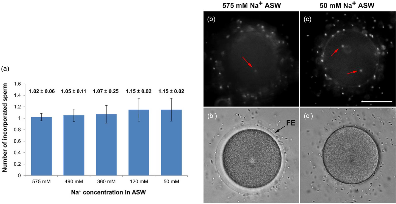
P. lividus eggs were incubated for 15 min in ASW containing different concentrations of Na+, and inseminated at high sperm density (final dilution 1:1000 = 1.15 × 10⁷ spermatozoa/ml) (Jaffe, Reference Jaffe1980). When examined under a fluorescence microscope to count the number of egg-incorporated spermatozoa (Fig. 2 a), only one spermatozoon successfully penetrated each egg in most cases, irrespective of the Na+ concentration (1.02 ± 0.065 in 575 mM Na+ ASW, representing NSW and used as a positive control; 1.05 ± 0.11 in 490 mM Na+ ASW; 1.07 ± 0.155 in 360 mM Na+ ASW; 1.15 ± 0.2 in 120 mM Na+ ASW; 1.15 ± 0.2 in 50 mM Na+ ASW; P = 0.06). Polyspermic fertilization took place only rarely in low Na+ medium and, even in that case, the number of the egg-incorporated sperm was limited to two in most cases (Fig. 2 c and Table S1). Interestingly, in the rare cases in which two spermatozoa penetrated the egg, they always entered at opposite poles of the egg (Fig. 2 c, arrows), arguing against the existence or effectiveness of a fast transmitting electrical mechanism that blocks polyspermy at the plasma membrane level. When fertilized at a lower sperm density (final sperm dilution 1:12,500 = 1.84 × 10⁶ spermatozoa/ml), these eggs were all monospermic regardless of Na+ concentration (n = 20 eggs tested at each Na+ concentrations, total eggs = 100). These results showed that lower external Na+ levels do not significantly induce supernumerary sperm entry.
Fertilization in low Na+ medium does not induce polyspermy. P. lividus eggs were transferred from natural seawater (NSW) to the artificial seawater (ASW) containing various amounts of NaCl: 575 mM, 490 mM, 360 mM, 120 mM, or 50 mM. To remove remnants of NSW, eggs were sequentially rinsed with the corresponding ASW in three Petri dishes. After a 15 min incubation, the eggs were fertilized using Hoechst 33342-stained sperm prepared in the same ASW. The number of incorporated spermatozoa was counted in the cooled CCD camera 5 min after fertilization. (a) Graphic summary of the intracellular sperm counts in the fertilized eggs. The average number of incorporated spermatozoa was calculated from the pooled data at the given concentration of Na+ in ASW. Mean ± SD are indicated above the corresponding bars. One-way analysis of variance (ANOVA) indicated no statistically significant differences among the conditions tested (P = 0.064). The number of eggs examined in each experiment was 60, and the experiment was repeated in three independent batches (n = 60 × 3 = 180 total eggs tested for each condition). (b, c) Incorporated spermatozoa visualized in epifluorescence microscopy. Red arrows indicate incorporated sperm pronuclei. (b′, c′) Elevation of the fertilization envelope (FE) is shown in the bright field view. Scale bar, 50 µm.

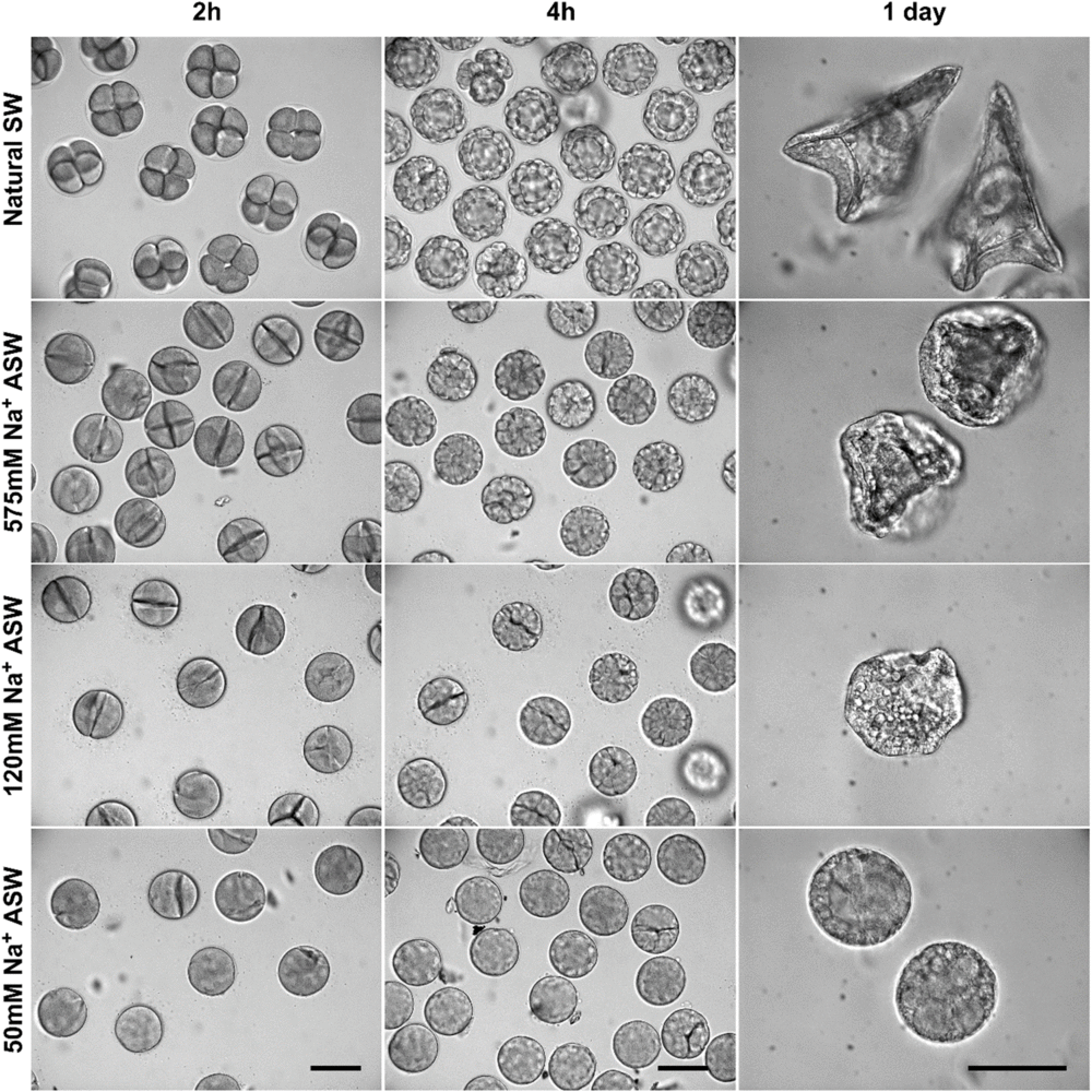
P. lividus eggs incubated and inseminated in ASW containing 50 mM Na+ failed to elevate the FE, as shown in bright field images (Fig. 2c′). Sodium deficiency also affected the formation of the FE in eggs inseminated in 120 mM Na+. As already reported, at this concentration, the FE has collapsed on the egg surface and was no longer visible 10 min after insemination (Schuel et al., Reference Schuel, Schuel, Dandekar, Boldt and Summers1982). This situation indicated that sodium deficiency may alter Ca2+ increase or the reorganization of the cortical actin cytoskeleton that normally takes place following fertilization, both of which are indicators that the cortical reaction has occurred in a normal way (Chun et al., Reference Chun, Limatola, Vasilev and Santella2014; Puppo et al., Reference Puppo, Chun, Gragnaniello, Garante and Santella2008; Chun et al., Reference Chun, Puppo, Vasilev, Gragnaniello, Garante and Santella2010; Chun et al., Reference Chun, Vasilev, Limatola, Santella, Kloc and Kubiak2018). In earlier studies, it was shown that sea urchin eggs fertilized in the presence of reduced concentrations of external Na+ cleaved abnormally (Jaffe, Reference Jaffe1980). This observation further supported the hypothesis of a fast electrical block to polyspermy, which was supposedly impaired due to the reduced Na+ entry and the weakened fertilization potential. Apparently, however, it was too readily assumed that this abnormal cleavage was due to the entry of multiple sperm centrosomes. Here we have shown that this is not the case: (i) polyspermy hardly occurred under the conditions of lower-than-normal external Na+; and (ii) the abnormal cleavage of the eggs fertilized in low Na+ (Fig. 3) may well be due to an alteration of cortical actin cytoskeleton, and not to meiotic defects following the formation of multipolar spindles by the supernumerary sperm asters (Just, Reference Just1939).
Embryonic development of monospermic eggs fertilized in low Na+ artificial sea water. Bright field view images of the embryos 2 h (first column), 4 h (second column) and 1 day (third column) after insemination. The number of fertilized eggs examined for each set of experiment is 100. Representative results from one of the three independent batches of experiments are presented. Scale bars, 100 µm.

Low concentrations of Na+ in the medium affect the Ca2+ response of fertilized sea urchin eggs
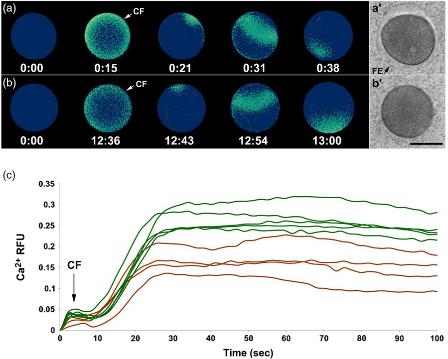
Ca2+ signalling at fertilization is considered to be the major activation event that transforms the quiescent egg into a dynamic zygote (Stricker, Reference Stricker1999; Runft et al., Reference Runft, Jaffe and Mehlmann2002; Santella et al., Reference Santella, Lim and Moccia2004, Reference Santella, Vasilev and Chun2012; Whitaker, Reference Whitaker2006; Ramos and Wessel, Reference Ramos and Wessel2013). In P. lividus eggs, the first detectable Ca2+ signalling event was an overall simultaneous increase in sub-cortical Ca2+. This so-called ‘cortical flash (CF)’ lasts around 8 s, and is known to be generated by the entry of external Ca2+ following the sperm-induced egg plasma membrane depolarization. As previously shown, low external Na+ reduces the amplitude of the membrane depolarization at fertilization (Chambers and De Armendi, Reference Chambers and De Armendi1979; Schuel and Schuel, Reference Schuel and Schuel1981), and here we show it also reduces the amplitude of the CF (Table S2). This situation suggests that both the Na+ and Ca2+ influxes may occur in sea urchin eggs through non-specific ion channels, perhaps via ‘the fertilization channels’ as first described (Dale et al., Reference Dale, De Felice and Taglietti1978) and later characterized in ascidians (Dale and De Felice, Reference Dale and De Felice1984). The CF is followed by a Ca2+ wave that starts at the sperm–egg interaction site and propagates to the opposite pole (Fig. 4). Low Na+ causes a delay in the onset of the sperm-induced Ca2+ signals of approximately 12 min in ASW containing 50 mM Na+ (Fig. 4 b), and also reduces the peak amplitude of the CF and Ca2+ wave at egg activation (brown lines in Fig. 4 c). Therefore, there is a significant effect of low Na+ on the time required for sperm to activate the eggs, i.e. the time lapse from the addition of spermatozoa to the medium to the generation of the CF (Table S2). But low Na+ does not significantly change the length of the latent period (the time delay between the CF and Ca2+ wave) or the speed of the Ca2+ wave as judged by the time it takes to traverse the egg from the site of fertilization to the antipode.
Effects of low Na+ on the intracellular Ca2+ increase in sea urchin eggs at fertilization. P. lividus eggs microinjected with calcium dyes were incubated in ASW containing 575 mM Na+ (a, control) or 50 mM Na+ (b) for 15 min. Eggs were then inseminated, and the changes in the intracellular Ca2+ levels were monitored (c) in the control eggs (green curves) and in the eggs fertilized in low Na+ ASW (brown curves). The pseudo-coloured relative fluorescence images at the top represent the sites of momentary Ca2+ increases at the key time points (min:s). In (a) and (b), to highlight the conspicuous delay of the sperm-induced Ca2+ response in the low Na+ eggs, the time of sperm addition in the medium was set as t = 0. In the graph presented in (c), the time frame immediately before the first detectable Ca2+ signal was set to time zero (t = 0) for the sake of comparison of the Ca2+ trajectories in each egg. (a’ and b’) Bright field view images of the eggs 15 min after fertilization. Scale bar, 50 µm.

Low Na + alters actin filaments dynamics at fertilization
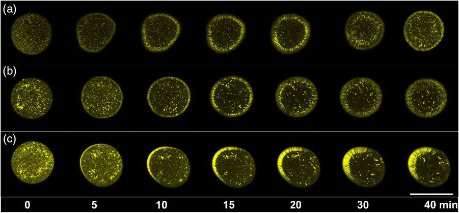
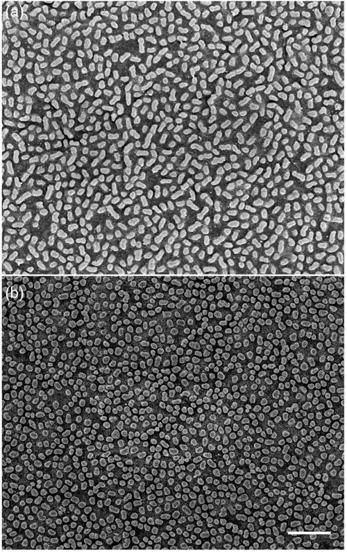
To observe F-actin in living eggs, fluorescent phalloidin, which binds F-actin (Terasaki, Reference Terasaki1996), was microinjected as described in Materials and Methods. The eggs were then transferred to ASW at various concentrations of Na+. After 15 min of incubation, spermatozoa were added and the actin filaments in the eggs were observed by confocal microscopy (Fig. 5). Under control conditions (575 mM Na+), 5 min after sperm addition, actin filaments become arranged perpendicularly to the egg cortex (Fig. 5 a). These filaments migrated progressively towards the centre of the egg within 30 min post-fertilization, and then a layer of filaments re-formed in the subplasmalemmal region by about 40 min (Fig. 5 a). When the eggs were incubated and fertilized in 120 mM Na+ ASW, however, such characteristic rearrangement of the cortical F-actin was significantly altered in that the late restoration of the subplasmalemmal F-actin layer is appreciably absent (Fig. 5 b). Curiously, in lower (50 mM) Na+ ASW, three out of five (60%) eggs tested manifested a massive actin polymerization only on one side of the zygote with no characteristic migration of actin fibres (Fig. 5 c), while the remaining 40% showed a disorganization in F-actin dynamics, very similar to that observed in the eggs incubated with 120 mM Na+ ASW. In addition to the altered functional dynamics of the actin filaments at fertilization, subtle changes in the fine structure of the actin filaments at the cell surface were observed in low Na+ ASW. On the external surface of the eggs, the microvilli of the eggs incubated for 15 min in ASW containing 50 mM Na+ were appreciably shortened as judged by the images of scanning electron microscopy (Fig. 6 b).
Alteration in the actin dynamics in sea urchin eggs fertilized in low Na+ ASW. P. lividus eggs microinjected with Alexa Fluor 568–phalloidin (10 µM in the pipette) were transferred into ASW containing various amounts of Na+: 575 mM (a), 120 mM (b), or 50 mM (c). After 15 min incubation, the eggs were fertilized, and the changes of the actin cytoskeleton were monitored with the confocal microscope on the equatorial plane. Images of representative eggs in each condition (n = 5 or 6) are shown. For each experiment, the moment immediately before sperm addition was taken as t = 0. Scale bar, 100 µm.

Ultrastructural changes at the surface of sea urchin eggs in low Na+ artificial sea water (ASW). P. lividus eggs were incubated for 15 min in ASW containing two different Na+ concentrations and subsequently fixed for scanning electron microscopy (SEM): 575 mM Na+ (a); 50 mM Na+ (b). Note that the microvilli appear shorter and flattened on the surface of the eggs incubated in ASW containing 50 mM Na+. Scale bar, 1 µm.

Discussion
In this study, we have used a single experimental variable, reduced external Na+ level, to perturb activation events in the sea urchin egg. As reduced external Na+ alters the fertilization-induced membrane depolarization of the egg (Jaffe, Reference Jaffe1980; Dale and De Santis, Reference Dale and De Santis1981), it was expected that lowering Na+ concentration in the fertilization medium would induce polyspermy, if the hypothesis of a fast electrical block to polyspermy is correct. We have shown that this is not the case. When intact sea urchin eggs were used without harsh treatments such as dejellying, electrostatic attachment to poly-l-lysine-covered microscope slides, or prolonged use of an intracellular microelectrode (as was done for the experiments described by Jaffe, Reference Jaffe1980), even in low Na+ and a high sperm density, the fertilized eggs were overwhelmingly monospermic. Therefore, while the original suggestion by E.E. Just (Reference Just1939) may be correct in its principle that ‘the block to polyspermy is most rapidly interposed’ in eggs fertilized in natural conditions, its precise mechanism is still obscure, although it is likely to be established before the membrane separation and to be dependent upon the state of excitability of the surface of eggs (Byrnes and Newman, Reference Byrnes and Newman2014). In line with the latter hypothesis, our previous studies suggested that starfish and sea urchin eggs are penetrated by multiple spermatozoa when the egg surface has been ‘injured’ by the experimental conditions affecting its structural organization (Puppo et al., Reference Puppo, Chun, Gragnaniello, Garante and Santella2008; Vasilev et al., Reference Vasilev, Chun, Gragnaniello, Garante and Santella2012, Reference Vasilev, Limatola, Chun and Santella2019; Chun et al., Reference Chun, Limatola, Vasilev and Santella2014). This finding is in keeping with the experimental observations of E.E. Just (Reference Just1939) who wrote:
…The eggs of Arbacia in my experience, if in best condition are never polyspermic…
and
…I have also made the initial insemination with the heaviest sperm-concentration procurable, i.e., ‘dry’ sperm as it exudes from the male, and have obtained only mono-spermic fertilization…
He added:
…Normally monospermic eggs can be rendered polyspermic by experimental treatment as staling (long residence in sea water), treatment with alkali, acids or poisons, subjection to low temperature, in short, by methods for inducing injury or weakness…
and
…polyspermy in them thus is a sign of weakness and hence pathological… (Just, Reference Just1939, p. 202).
Therefore, it is conceivable that under experimental conditions that induce perturbation of the structure of the actin cytoskeleton of the egg cortex, the damaged surface of starfish and sea urchin eggs is less rapid in responding to the attachment of the first spermatozoon. As a consequence, this ‘slow’ egg reaction may eventually lead to failure to block penetration of additional spermatozoa. These multiple sperm–egg interactions give rise to deregulated Ca2+ signalling events such as suppression or reduction of the cortical flash, initiation of multiple Ca2+ signals, and aberrant sperm entry (Kyozuka and Osanai, Reference Kyozuka and Osanai1988; Puppo et al., Reference Puppo, Chun, Gragnaniello, Garante and Santella2008). It is important to note that:
…[the cortical structural (ectoplasmic) changes] occurring at the very outset are most striking; they mark out the course, direct the way toward the final outcome of fertilization, the formation of the complex organism out of a single cell, the egg… (Just, Reference Just1939, p. 205)
Therefore, the occurrence of these rapid cortical structural changes exhibited by eggs fertilized in their optimum physiological state:
…constitutes an index of the condition of the egg and the quality of its later development. (Just, Reference Just1929).
During sea urchin fertilization, in addition to the membrane fusion and exocytosis of the CG (Wessel et al., Reference Wessel, Brooks, Green, Haley, Voronina, Wong, Zaydfudim and Conner2001), sperm–egg interaction brings about other dramatic F-actin linked structural reorganizations of the egg surface. A few minutes after sperm attachment, formation of a network of actin filaments in the egg cortex and fertilization cone correlates with the accumulation of the F-actin-bundling protein, alpha-actinin, which is a visible sign that the egg surface has been highly responsive to the first successful sperm attachment (Tilney and Jaffe, Reference Tilney and Jaffe1980; Hamaguchi and Mabuchi, Reference Hamaguchi and Mabuchi1986; Yonemura and Mabuchi, Reference Yonemura and Mabuchi1987). The cortical F-actin restructuring also involves two bursts of microvillus elongation and the formation of spikes containing actin in the perivitelline space (Burgess and Schroeder, Reference Burgess and Schroeder1977; Chun et al., Reference Chun, Puppo, Vasilev, Gragnaniello, Garante and Santella2010)concomitant with the equidistant separation of the vitelline envelope from the plasma membrane. Moreover, Just stated,
…membranes that separate incompletely and are not equidistant from the egg at all points and are slow in rate of separation mean eggs of poor fertilizability that subsequently develop abnormally… (Just, Reference Just1939)
Therefore, the importance of the actin in the egg surface cytoplasm (ectoplasm) was highlighted even before its discovery (Just, Reference Just1939). Finally, a translocation and redistribution of actin filaments from the surface to the centre of the fertilized egg mark the late phase of the fertilization process and might control the cleavage cycles of zygotes (Vasilev et al., Reference Vasilev, Chun, Gragnaniello, Garante and Santella2012).
Our results showed that low external Na+ induces alteration in microvilli and CG exocytosis (Figs. 2c′ and 6) and changes in the structure and dynamic functionality of the cortical actin filaments (Fig. 5). In this context, it is noteworthy that the amplitude of CF (or egg’s membrane depolarization step) and the Ca2+ wave in these eggs at fertilization were significantly reduced (Fig. 4 and Table S2). In line with the finding that Na+-induced Ca2+ signals changes may be due to the structural alterations of the actin cytoskeleton of the egg surface (Nusco et al., Reference Nusco, Chun, Ercolano, Lim, Gragnaniello, Kyozuka and Santella2006; Puppo et al., Reference Puppo, Chun, Gragnaniello, Garante and Santella2008; Kyozuka et al., Reference Kyozuka, Chun, Puppo, Gragnaniello, Garante and Santella2009; Chun et al., Reference Chun, Vasilev and Santella2013, Reference Chun, Limatola, Vasilev and Santella2014), modifications of the electrical properties of the plasma membrane of the maturing oocytes of starfish are linked to the shortening of microvilli, which contain F-actin bundles (Miyazaki et al., Reference Miyazaki, Ohmori and Sasaki1975; Moody and Lansman, Reference Moody and Lansman1983). We have previously shown that the retraction of microvilli in starfish oocytes matured in Ca2+-free sea water appears to be related to inhibition of the CF at fertilization (Limatola et al., Reference Limatola, Chun, Kyozuka and Santella2015; Santella et al., Reference Santella, Limatola and Chun2015, Reference Santella, Limatola, Vasilev and Chun2018). Conversely, alteration of CGs, which are closely associated with microvilli via F-actin, and are known to contain between 30 to 95 mM of total calcium (Gillot et al., Reference Gillot, Ciapa, Payan and Sardet1991) may explain the lower amplitude of the Ca2+ wave elicited in eggs inseminated in low Na+. This is in light of the finding that microvilli and acidic cortical vesicles (CG and yolk granules) significantly affect the sperm-induced Ca2+ signals in P. lividus eggs (Vasilev et al., Reference Vasilev, Limatola, Chun and Santella2019). It has been reported that successful exocytosis of CG in the fertilized starfish eggs requires exquisite regulation of the cortical actin cytoskeleton in addition to the sizeable Ca2+ increase (Puppo et al., Reference Puppo, Chun, Gragnaniello, Garante and Santella2008, Kyozuka et al., Reference Kyozuka, Chun, Puppo, Gragnaniello, Garante and Santella2009). Therefore, sea urchin eggs pretreated with actin drugs often fail to elevate FE at fertilization, but these eggs do not incorporate multiple spermatozoa (Chun et al., Reference Chun, Limatola, Vasilev and Santella2014, Reference Chun, Vasilev, Limatola, Santella, Kloc and Kubiak2018). In line with these findings, the results presented in the current study strongly suggest that cortical reaction does not serve as a mechanical block to polyspermy, as inhibiting the FE elevation does not necessarily lead to polyspermy (Figs. 2c′ and 5b′).
Low Na+ does induce abnormal cleavage, but here we have shown that this is not due to polyspermic fertilization and the consequent formation of multipolar spindles associated with supernumerary sperm centrosomes (Schatten et al., Reference Schatten, Schatten, Mazia, Balczon and Simerly1986); for a description of Boveri’s theory see Scheer, who states:
The centrosome is the true division organ of the cell, it mediates the nuclear and cellular division. (Scheer, Reference Scheer2014)
Our results, as originally suggested by Just (Reference Just1939), show that the observed aberrant development was due to sodium-induced alterations in actin filament structural dynamics of the egg surface, and that these changes play a definite role in induction of defective mitosis of the developing embryos. In fact, upon fertilization, F-actin dynamics was altered drastically in low Na+ sea water (Fig. 5), even leading to polarization of the egg surface F-actin by 50 min post-fertilization. This abnormal development, resulting from the aberrant low sodium-induced ectoplasmic behaviour, strongly supported the idea that the sperm-triggered reorganization of the cortical actin plays a key role in the actin cytoskeleton dynamics in egg cleavage, as we have previously shown (Vasilev et al., Reference Vasilev, Chun, Gragnaniello, Garante and Santella2012; Limatola et al., Reference Limatola, Chun, Kyozuka and Santella2015).
In summary, we have shown that the fast (electrical) and mechanical (FE elevation) block to polyspermy are not reliant on sodium levels and cortical reaction, respectively. We suggest that a normal fertilization response, even under laboratory conditions of unnaturally high sperm densities, is ensured ‘as long as the integrity of the egg surface is maintained’. Sperm–egg ratios in many animals, including the sea urchin, at the natural site of fertilization may be extremely low, often approaching unity, and this would of course suggest that selective pressures favoured the achievement of monospermy rather than the evolution of mechanisms to prevent polyspermy (Dale, Reference Dale2016). Nonetheless, under experimental conditions of high sperm densities, normally reacting sea urchin eggs are still invariably monospermic. From these results, we can conclude that lowered levels of sodium do not induce polyspermy but alter fertilization response and embryonic development following alteration of the actin cytoskeleton dynamics of the egg cortex.
Acknowledgements
We thank M. Cannavacciuolo and G. Zazo (MEDA, Stazione Zoologica Anton Dohrn, SZN) for collecting the animals from the sea, and Davide Caramiello (MARE, SZN) for their maintenance. We acknowledge E. Garante for his comments on the paper, and G. Gragnaniello, F. Iamunno, R. Graziano and G. Lanzotti at the SZN AMOBIO unit for preparing the samples for scanning electron microscopy. We also thank W. Malcolm Byrnes for helpful comments on an earlier draft of the manuscript.
Financial support
N. Limatola and F Vasilev have been financially supported by SZN postdoctoral fellowships.
Statement of interest
The authors declare no competing interests.
Supplementary material
To view supplementary material for this article, please visit https://doi.org/10.1017/S0967199419000364