Case report
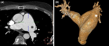
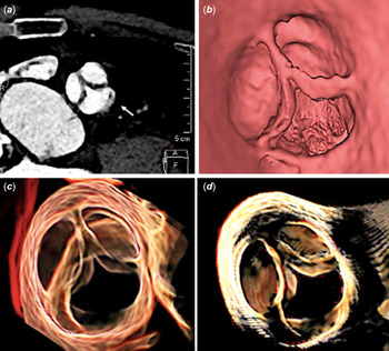
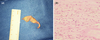
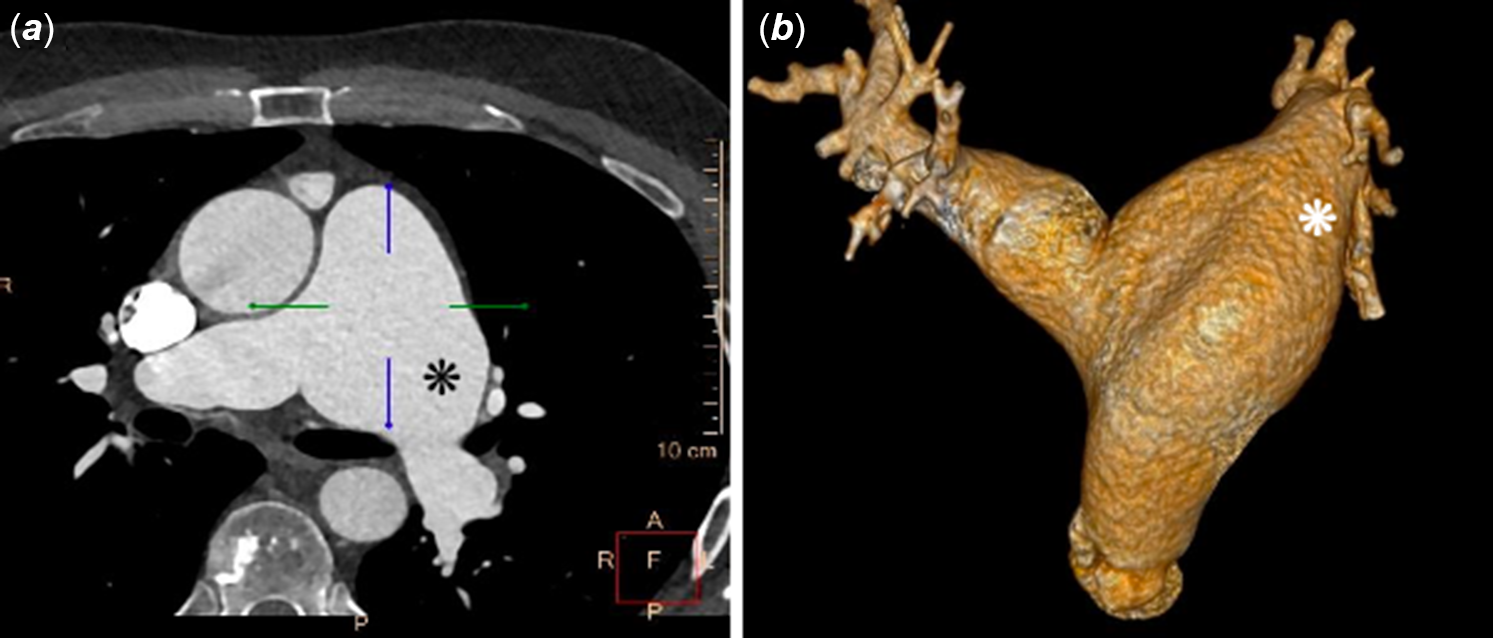
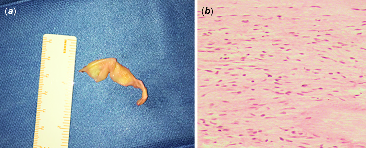
A 46-year-old male patient, complaining of moderate dyspnoea and fatigue, was referred to our centre for pulmonary valve regurgitation and left pulmonary artery aneurysm. CT confirmed a 50 × 65 mm left pulmonary artery aneurysm, located at the junction between the pulmonary trunk and the left pulmonary artery. The pulmonary valve annulus was normal (25 mm). The pulmonary trunk was normal only in the proximal tract (35 mm) and dilated at bifurcation (Fig 1a and b). Multiplanar reformation (Fig 2a, arrow), virtual angioscopy (Fig 2b), and volume rendering reconstruction (Fig 2c and d) showed the complete absence of the left-posterior pulmonary valve cusp. The other two cusps appeared normal. This pulmonary valve anomaly was not detected at previous echocardiograms. Right ventricle presented a significant dilatation (basal right ventricle diameter: 20.8 mm, mid right ventricle diameter: 46.3 mm, base-to-apex length: 73.7 mm). The patient underwent surgical operation by complete sternotomy and cardiopulmonary bypass. At surgical inspection, radiological findings were confirmed. Insufficient pulmonary valve was excised (Fig 3a) and replaced with a 23 mm Inspiris Resilia bioprosthesis (Edwards Lifesciences, CA, United States of America). Pulmonary reductive elliptical left arterioplasty was also performed. Microscopic examination showed only a mild focal myxoid degeneration of two pulmonary cusps, otherwise normal (Fig 3b). Postoperative course was uneventful. Congenital absence of one pulmonary valve cusp with completely normal size and morphology of both the other two cusps is a very rare finding. Indeed, no signs of atrophy or dysplasia were observed in the remaining two cusps. In the only two similar cases previously reported in 1997 and 2000,Reference Westaby and Katsumata1,Reference Sayger, Lewis, Arcilla and Ilbawi2 patients became symptomatic and were operated at a younger age (21 and 16 years old) than our patient. Furthermore, differently from our case, both previous patients presented also an aneurysm of the first tract of the pulmonary trunk as a concomitant cause of pulmonary regurgitation. We present the first images of three-dimensional pulmonary valve reconstruction in this very unusual scenario. In the current era, CT three-dimensional reconstructions could be determinant for diagnosis and may represent a very useful tool for an optimal preoperative evaluation of pulmonary valve morphology.
Two-dimensional ( a ) and three-dimensional ( b ) reconstruction showing the aneurysm of the left pulmonary artery (asterisk) with normal pulmonary trunk.
Multiplanar reformation ( a ), virtual angioscopy ( b ), and volume rendering reconstruction ( c and d ) showing the congenital absence of the left-posterior pulmonary valve cusp (white arrow in Fig 2A) with normal morphology of the other two cusps.

Surgical specimen of the pulmonary valve removed ( a ) and microscopic examination ( b ).
Financial support
This work received no specific grant from any funding agency.
Conflicts of interest
None.
Ethical standards
The authors declare that they comply with appropriate Istitutional and international guidelines for ethical guidelines.