Introduction
Thisbemys brevicrista has a highly crenulated enamel pattern, which makes this little-known species distinct from other medium-sized ischyromyids found in the middle Eocene Bridger Formation, Green River Basin, Wyoming (McGrew et al., Reference Mcgrew, Berman, Hecht, Hummel, Simpson and Wood1959; Wood, Reference Wood1962; McGrew and Sullivan, Reference Mcgrew and Sullivan1970; West and Atkins, Reference West and Atkins1970; Gazin, Reference Gazin1976; West and Hutchison, Reference West and Hutchison1981; Gunnell and Bartels, Reference Gunnell and Bartels1994; Gunnell, Reference Gunnell1998). Previously, only a single specimen of T. brevicrista was known, a partial maxilla with the upper dentition. This holotype (KUVP 14233), designated by Ostrander (Reference Ostrander1986) when he originally named the species, was recovered from the Lonetree White Layer, Upper Bridger Formation (biochron Br3; Gunnell et al., Reference Gunnell, Murphey, Stucky, Townsend, Robinson, Zonneveld and Bartels2009), Uinta County, Wyoming. Features that set T. brevicrista apart from other species of Thisbemys include an additional loph on the anterior two upper molars, attachment of the protoloph to the center of the protocone on M1-M2, and various features of the mesostyle on P4-M3.
Ostrander (Reference Ostrander1986) suggested that T. brevicrista was derived from T. plicatus. His conclusion was based on a combination of morphological and stratigraphic evidence as well as a phylogeny presented by Wood (Reference Wood1962, p. 243) showing the upper Bridger species T. corrugatus evolving from the lower Bridger species T. plicatus. The morphological evidence that Ostrander (Reference Ostrander1986) presented included similarities in the condition of the mesostyle. Isolation of the M1 mesostyle is a character shared with T. plicatus, while having the M2 mesostyle connected to the paracone is a character shared with T. corrugatus. This puts T. brevicrista between T. plicatus and T. corrugatus. Since the holotype of T. brevicrista was found at approximately the same stratigraphic level as specimens of T. corrugatus (Wood, Reference Wood1962; Ostrander, Reference Ostrander1986), derivation from T. plicatus is a logical conclusion. Further analysis of these evolutionary relationships has been hindered by the lack of additional specimens, in particular, the absence of any mandibles and lower dentition of T. brevicrista.
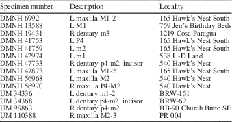
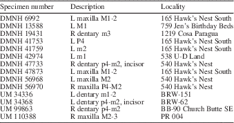
Here, I document the discovery of 14 new specimens of T. brevicrista, including examples of the mandibular cheek teeth, recovered from the Bridger Formation, Green River Basin (Table 1, Fig. 1). In addition to the mandibles with teeth, there are maxillae with teeth and isolated upper and lower cheek teeth. The majority of specimens, including the neotype (DMNH 56970), were recovered from the Blacks Fork Member (Fig. 2). However, a single mandible was collected from the Twin Buttes Member, the same rock unit from which the original type specimen was recovered. The Bridger Formation specimens do not include any associated upper and lower cheek teeth of T. brevicrista. Thus, species identification is based on a correlation between the morphology of the lower premolars and molars, which correspond closely in size and morphology for proper occlusion with the known upper molars. Examples of this type of species-specific correlation have been demonstrated in previous studies of fossil rodents (Lazzari et al., Reference Lazzari, Charles, Tafforeau, Vianey-Liaud and Aguilar2008, Reference Lazzari, Aguilar and Michaux2010). Likewise, functional studies suggest that the occlusion between the upper and lower teeth constrains dental evolution such that variations in the shape of the upper and lower dentition is apparently integrated (Butler, Reference Butler1985). This knowledge, plus the fact that these lower teeth were recovered from the same localities as some of the uppers, makes it possible to identify lower cheek teeth of T. brevicrista even though associated upper and lower teeth have yet to be recovered.
Fossil specimens used in this study were found in the Bridger Basin in the southeastern part of the Greater Green River Basin, southern Wyoming, USA (modified with permission from Murphey and Evanoff, Reference Murphey and Evanoff2007, and Gunnell et al., Reference Gunnell, Murphey, Stucky, Townsend, Robinson, Zonneveld and Bartels2009).

Temporal distribution and relative stratigraphic positions of Bridgerian species of Thisbemys. Relative thickness of lithostratigraphic units is roughly estimated. Note T. nini also has been found from the San Jose and Huerfano Formations.
Neotype DMNH 56970 and referred specimens of Thisbemys brevicrista from Sweetwater County, Wyoming, Bridger Formation. All specimens were recovered from the Black Forks Member (Br2–Br1b) except UM 34336, which is from the Twin Buttes Member (Br3). Localities and Bridgerian biochrons follow Gunnell et al., (Reference Gunnell, Murphey, Stucky, Townsend, Robinson, Zonneveld and Bartels2009). (More detailed locality information is available to qualified researchers by contacting either the Denver Museum of Nature and Science or the Museum of Paleontology at the University of Michigan.)
The newly discovered specimens of T. brevicrista are of particular importance for several reasons. First, the original type specimen of T. brevicrista Ostrander, Reference Ostrander1986, appears to be lost (Desui, personal commun., 2012) and research-quality casts of it were never produced. From the new material, a neotype is designated to clarify the alpha taxonomy and to provide criteria for recognizing the species in the future. Second, this sample includes the first known mandibles and lower dentition of T. brevicrista, prompting a species re-diagnosis. Third, the new specimens include a maxillary fragment with a partial zygomatic arch that preserves the relationship of the arch to the fourth premolar and first upper molar, an aspect of the species previously unknown. Finally, discovery of T. brevicrista from a middle Bridgerian locality (biochron Br2) documents the co-existence of T. brevicrista with T. corrugatus, T. plicatus (Wood, Reference Wood1962), T. perditus, and T. nini.
Materials and methods
The sample from the Bridger Formation, of the Green River Basin, Wyoming, includes 14 specimens of medium-sized ischyromyids referred to Thisbemys brevicrista (Table 1). Specimens come from a variety of Bridger Formation localities, one Twin Buttes Member (biochron Br3) locality (BRW-151), and several Blacks Fork Member (biochron Br2) localities (540 Hawks Nest, 165 Hawks Nest South, 1219 Cosa Paragua, 538 U-D Land, 2308 Turtle Heaven, PR 004, and BB090). Comparisons were made to the holotypes of Thisbemys corrugatus, T. plicatus, T. perditus, and T. nini. Since the holotype of T. brevicrista is lost, stereophotos in the original paper by Ostrander (Reference Ostrander1986) were used to identify specimens with maxillary teeth.
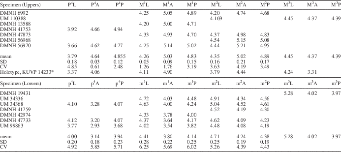
Dental terminology used in this paper (Fig. 3) is modified after Rose and Koenigswald (Reference Rose and Koenigswald2007), Wood and Wilson (Reference Wood and Wilson1936), and Wood (Reference Wood1962). Teeth were measured to the nearest 0.01 mm using an optical micrometer fitted to a Bausch and Lomb dissection microscope. Measurements of lower molars were taken as described by Wood (Reference Wood1962). Upper molar length (AP) measurements were taken at the center of the crown. Width was measured along the protoloph (buccal margin of the paracone to base of protocone) and metaloph (buccal margin of metacone to base of protocone). In the case of the second breadth measurement, the protocone was chosen in lieu of the hypocone because of the greater repeatability of measurements. Measurements and basic statistics of the sample are provided in Table 2.
Diagram of cheek teeth of Thisbemys showing occlusal morphology of the (1) left upper molar, and (2) left lower molar and related nomenclature (modified after Wood and Wilson, Reference Wood and Wilson1936).
Measurements (in mm) of the dentition of Thisbemys brevicrista. Abbreviations: L, length; A, anterior breadth; P, posterior breadth; CV, coefficient of variation; SD, standard deviation; *measurements from Ostrander (Reference Ostrander1986).
Systematic paleontology
Institutional acronyms are as follows: AMNH, American Museum of Natural History, New York; CNHM, The Field Museum, Chicago; DMNH, Denver Museum of Nature and Science, Denver; KUVP, University of Kansas Museum of Natural History, Lawrence; UM, University of Michigan Museum of Paleontology, Ann Arbor; NMNH, National Museum of Natural History, Smithsonian Institution, Washington, D.C.
Class Mammalia Linnaeus, Reference Linnaeus1758
Order Rodentia Bowditch, Reference Bowditch1821
Family Ischyromyidae Miller And Gidley, Reference Miller and Gidley1918
Genus Thisbemys Wood, 1952
Thisbemys brevicrista (Ostrander, Reference Ostrander1986)
Figures 4, 5.1–5.8, 5.15, 5.16, 6.1–6.3, 7.1–7.7, 8.1
Right (reversed) P4-M2 and partial zygomatic arch of neotype of Thisbemys brevicrista, DMNH 56970 (teeth coated with ammonium chloride).
Lower teeth of Thisbemys brevicrista (1–8, 15, 16), T. plicatus (9, 13), T. corrugatus (10, 14), and T. nini (11) from the Bridger Formation and T. perditus (12) from the Wind River Formation. (1) UM 34368, left p4-m2; (2) UM 34336, left m1-2; (3) UM 99863, right p4-m2; (4) DMNH 47733, right p4-m2; (5) UM 34368, left m2; (6) DMNH 41759, left m2; (7) DMNH 42974, left m1; (8) DMNH 19431, right m3; (9) AMNH cast 11565, left p4-m3; (10) UM 34323, right p4-m3; (11) AMNH 17454, left p4-m3; (12) CNHM P-26897, right m2-m3; (13) NMNH 363554, left incisor; (14) UM 34311, left incisor; (15) DMNH 47733, right incisor; (16) UM 34368, left incisor. Pulp cavity indicated by dotted line. Anterior at top; specimens 1, 2, 5–7, 9, and 11 are reversed; all specimens are coated with ammonium chloride except 5, 13, and 14. Common scale bar for 1–5, 6–8, 9–12, 13–16.
Buccal view of three lower mandibles of Thisbemys brevicrista from the Bridger Formation. (1) UM 34368 left dentary; (2) UM 99863 right dentary; (3) DMNH 47733 right dentary. Position of angle between the oblique line and alveolar ridge (A) illustrated on the right mandible of T. brevicrista, (4) DMNH 47733, buccal view; common scale with (3). Specimens coated with ammonium chloride.
Maxillary teeth of Thisbemys brevicrista (1–7), T. plicatus (9,10), and T. corrugatus (8, 11–13) from the Bridger Formation. (1) DMNH 56970, right P4-M2; (2) UM 110388, right M2-3; (3) DMNH 47873, left M1-2; (4) DMNH 6992, left M1-2; (5) DMNH 56968, left M2; (6) DMNH 13588, left M1; (7) DMNH 41753, left P4; (8) UM 98782, left P4; (9) DMNH 47695, right P4-M2; (10) DMNH 110597, left M2-3; (11) DMNH 10312, right M1; (12) DMNH 40123, left M2; (13) UM 34311, left M2-3. Anterior on left; specimens 1, 2, 10, and 11 are reversed; 2 mm scale bar in 1–5 and 8–13; 1 mm scale bar in 6, 7. Specimens are coated with ammonium chloride.
Zygomatic arches of (1) T. brevicrista, DMNH 56970, right arch; (2) T. corrugatus, NMNH 17165, right arch; and (3) T. plicatus, AMNH 11560 (cast), right arch. Scale=3 mm.
Neotype
DMNH 56970 (Fig. 4), partial maxilla with right P3 alveolus, P4-M2 and part of the anterior root of the zygomatic arch.
Neotype locality and horizon
DMNH locality 540 Hawk’s Nest, Sweetwater County, Wyoming, Blacks Fork Member, Bridger Formation, earliest middle Eocene, Bridgerian Biochronological Zone Br2.
Other material examined
KUVP number 14233 (holotype); UM numbers 110388, 34336, 34368, 99863; DMNH numbers 6992, 13588, 19431, 41753, 41759, 42974, 47733, 47873, 56968.
Occurrence
Middle Bridgerian, Bridgerian biochron Br2–latest Bridgerian, Bridgerian biochron Br3.
Diagnosis
Differs from all other species of Thisbemys in having an accessory loph anterior to the protoloph on M1-M2, complete metalophid on m1-3, protoloph attached to the center of the protocone on P4-2, and metaloph and protoloph of P4-M3 corrugated nearly the entire width of the tooth by a series of fine anteroposteriorly directed ridges; differs from T. nini in absence of a hypocone on P4, having the posterior aspect of the anterior root of the zygomatic arch in line with posterior cingulum of P4 and a lower incisor that is broad and nearly triangular in cross section; differs from T. perditus in having deeply incised corrugations, a less distinct hypocone on M1, and m2 with a rhomboid outline; differs from T. plicatus in having shallower lingual grooves separating hypocone from protocone and absence of a parastyle in M1-2, a deep lingual gorge continued as a valley part way across talonid basin, and second protoconid arm connects protoconid to mesoconid; differs from T. corrugatus in having a buccolingually elongate mesostyle on P4, the mesostyle of M2 connected to paracone but separate from metacone, transversally oriented metaloph on M3, and significantly longer than wide m3 with an AP elongate and open external valley.
Description of neotype DMNH 56970
Cheek teeth highly rugose, with distinct anteroposteriorly oriented ridges defining the protoloph and metaloph, filling the central basin of P4-M2. Teeth increase in size from P4-M2. P3 alveolus indicates that this tooth was small and conical. A relatively short anterior cingulum ends parallel to the well-defined paracone. The P4 protoloph is shorter than the metaloph. The convex metaloph connects to the posterior aspect of the protocone via a weakly developed ridge. The P4 mesostyle is small and connected to both paracone and metacone. The mesostyle of M1 is isolated on both sides by buccal valleys. A distinct, rounded metaconule occurs on P4-M2. The anterior accessory loph is longer on M2 than M1. Two pillars of enamel extend lingually from the AP elongate mesostyle of M1-2, the posterior one nearly twice as long as the anterior. The posterior aspect of the anterior region of the zygomatic arch is level with the posterior cingulum of P4.
Remarks
The initial and only account of Thisbemys brevicrista (Ostrander, Reference Ostrander1986) was based on a single specimen collected from the Lone Tree White Layer, Twin Buttes Member of the Bridger Formation, Uinta County, Wyoming. Unfortunately, Ostrander passed away shortly after the 1986 publication and the exact location of the holotype is now unknown. Until this paper, no new specimens of T. brevicrista have been reported. Given that the holotype cannot be located, specimen DMNH 56970, which preserves the right P3 alveolus, P4-M2 and part of the anterior root of the zygomatic arch, is designated as the neotype.
Ostrander’s (Reference Ostrander1986) description is sufficient to distinguish T. brevicrista from any other species of Thisbemys. His description and stereophotos of the original holotype were used to classify new individuals recovered from the Blacks Fork Member of the Bridger Formation. Previously, it was thought that species of Thisbemys formed an evolutionary lineage demarcated by stratigraphic levels with little to no morphological differences (Wood, Reference Wood1962). Specimens from Twin Buttes Member were classified as T. corrugatus and those from Blacks Fork Member were classified as T. plicatus (Eaton, Reference Eaton1982). Formal recognition of the new specimens as a distinct species, and clarification of the alpha taxonomy, necessitate the redescription of T. brevicrista and the designation of a neotype. In accordance with the International Code of Zoological Nomenclature (ICZN) Article 75, the specimen selected as the neotype originated from the lower Bridger Formation, as nearly as practicable to the type locality.
Description of mandible and mandibular dentition
Mandible
The mandible is best preserved in three specimens: UM 34368, 99863, DMNH 47733 (Fig. 6). The horizontal ramus is comparable in size to that of T. corrugatus with an increase in thickness posteriorly. The masseteric scar is well defined and extends anteriorly to a distinct, rounded knob below the center of m2. The prominent superior ridge (= oblique line) of the masseter scar forms approximately a 65-degree angle to the alveolar ridge (Fig. 6.4, angle A). Masseter fossa depth ranges from deep in DMNH 47733 to moderate in UM 34368 to shallow in UM 99863. There is a sharp crest along the dorsal surface of the diastema. On the lateral surface of the horizontal ramus there are two mental foraminae. A large oval-shaped mental foramen is located anterior and inferior to p4, and a smaller and rounded mental foramen is present below the anterior root of p4, consistent with Thisbemys’ generic description (Wood, Reference Wood1962). Due to breakage, it is not possible to determine if there was a chin eminence along the anteroventral edge of the mandible.
The lower incisor is broad and approximately triangular in outline. The incisor pulp cavity shows two morphs: one is broad with the same shape as the tooth as a whole (e.g., DMNH 47733; Fig. 5.1) the other is T-shaped (e.g., UM 34368; Fig. 5.2). A thin layer of enamel extends across the anterior face of the lower incisor just past the widest point on the lingual face and about 1/3 of the way onto the buccal face.
Mandibular dentition
Lower teeth are represented in seven specimens: UM 34336, 34368, 99863; DMNH 19431, 41759, 42974, 47733 (Fig. 5).
The p4 is present in three specimens, and in each case, significantly smaller than m1 (e.g., UM 34368, UM 99863, DMNH 47733; Fig. 5). The anterior width is distinctly narrower than the posterior width making the tooth rhomboid in outline. The metaconid is the tallest cusp, its buccal face expanded into a sloping, rounded ridge that extends into the trigonid basin, leaving only a narrow slit-like opening anterior to the metaconid. The trigonid basin extends posteriorly via a narrow short vale, but is isolated from the talonid basin by a weak, low metalophid. A well-developed anterolophid joins the metaconid and protoconid and defines the anterior aspect of p4.
A mesoconid is present, but relatively indistinct, as a swelling at the anterior aspect of the essentially straight ectolophid. Originating from the ectolophid, just inferior to the mesoconid, there are two anteroposterior elongate enamel extensions that reach the center of the trigonid basin. The external valley, located buccal to the ectolophid, is shaped like a lambda, with the lingual arm extending further posteriorly than the buccal arm. The lingual gorge (Fig. 3), a deep valley between the metaconid and entoconid, is well defined. It is intermediate to the deep lingual gorge of T. corrugatus (e.g., UM 34323; Fig. 5.10) and shallower gorge of T. plicatus (e.g., AMNH 11565; Fig. 5.9). Several well-defined ridges characterize the posterolophid near the entoconid. A hypoconulid is absent. In unworn specimens (e.g., UM 99863; Fig. 5.3), the wrinkles in the enamel of the talonid basin are deep.
The m1 is known from five specimens that encompass considerable variation in wear. The tooth is elongate and rhomboid in outline, with nearly equal anterior and posterior widths.
The metaconid is the tallest cusp, as on p4. The buccal, rounded ridge-like extension of the metaconid meets up with a shorter lingual extension of the protoconid to form a strong, complete metalophid (e.g., UM 34336, UM 99863; Fig. 5). A very narrow opening may develop between the metalophid and protoconid arm, thereby connecting the trigonid and talonid basins, in well-worn specimens (e.g., DMNH 47733; Fig. 5.4). The trigonid basin is transversally elongated and anteroposteriorly constricted due to the steep angle of the metalophid and aspects of the anterior cingulum. Buccally, the anterolophid is narrow; lingually it is inflated. The mesoconid is a distinct swelling at the midway point of an essentially straight ectolophid, which is continuous between the protoconid and hypoconid.
Along the buccal wall of the mesoconid, an enamel crest extends from the surface of the mesoconid to the root of the tooth, creating a lambda-shaped external valley. The posterior arm of the lambda is about twice as long as the anterior arm and forked where it meets the hypoconid. A deep lingual gorge extends one-third of the distance across the talonid basin. The anterior protoconid arm forms the metalophid, in part, while the other protoconid arm connects to the mesoconid (e.g., UM 34368, UM 99863; Fig. 5). The somewhat flattened posterolophid has a distinct hypoconulid and several well-defined ridges of enamel extending into the talonid basin from its lingual aspect.
The m2 is preserved in five specimens and is morphologically similar to m1 except for the following differences. The metaconid arm is broad and elongate, forming a stronger metalophid than that of m1 (e.g., UM 34336; Fig. 5.2). The external valley is deeper on each side of the mesoconid crest and the indentations of the distal aspect of the posterior arm of the lambda are deeper as well.
The m3 is preserved only in DMNH 19431 (Fig. 5.8), a mandibular fragment with m3 in situ. The strong, complete metalophid crest, deep lingual gorge, and lambda-shaped external valley makes it possible to positively identify the specimen as T. brevicrista. The tooth is roughly rectangular in outline, the anterior width somewhat greater than the posterior width. The metaconid is the highest cusp, the entoconid is the second-tallest cusp, elevated well above the talonid basin. Medially, a ridge-like extension of the metaconid meets up with the anterior protoconid arm to form a strong, complete metalophid isolating the trigonid basin from the talonid basin. The narrow trigonid basin extends from the anterobuccal aspect of the metaconid to the anterolingual side of the protoconid. The mesoconid, present but indistinct, is located at the anterior aspect of the essentially straight ectolophid; it is nearly isolated from the second protoconid arm.
Buccal to the mesoconid, the external valley is open, without the enamel crest seen in m1-2 of T. brevicrista. Two indentations define the distal aspect of this valley, anterior to the hypoconid (Fig. 5.8), a feature common to m1-2. The lingual gorge between the metaconid and entoconid is nearly as deep as that of m1-2, and extends partially into the talonid basin. A distinct hypoconulid, at the lingual aspect of the posterolophid, is isolated from the entoconid by a very shallow groove. There is a rounded heel with two distinct wrinkles near the distal-most aspect of m3.
Comparisons
The thick mandible of Thisbemys brevicrista has a well-defined masseteric scar. This is similar to that of T. corrugatus (Wood, Reference Wood1962; fig. 36A, C) and T. nini, whose mandibles are thicker than the slender mandibles of T. plicatus, Uintaparamys parvus, Paramys delicatus, and P. delicatior. It differs from the mandible of T. perditus, which is thick, but has a shallow masseteric scar. The angle of the oblique line is steeper than that of Uintaparamys parvus at approximately 50° and somewhat less steep than that of P. delicatus and P. delicatior at approximately 70°.
The mandibular incisor of T. brevicrista is intermediate in shape to T. corrugatus, which has the outline of a broad, nearly equilateral triangle (e.g., UM 34311; Fig. 5.14) and T. plicatus, whose outline is most similar to an isosceles triangle with a rounded anterolateral corner (e.g., NMNH 363554; Fig. 5.13). The triangular-shaped mandibular incisor of T. brevicrista differs from that of T. perditus, which is compressed transversally and elongated anteroposteriorly (Wood, Reference Wood1962; fig. 37H), and the roughly oval-shaped lower incisor of T. nini (Wood, Reference Wood1962; fig. 37O). All of these species have a thin layer of enamel extending across the anterior face of the incisor, distributed as described above for T. brevicrista.
Crenulations of the cheek teeth of Thisbemys brevicrista are deeply incised and remain detectable after moderate wear (e.g., DMNH 47733; Fig. 5.4), similar to those of T. corrugatus (e.g., UM 34323; Fig. 5.10). One specimen of T. brevicrista shows that these crenulations are limited to the relatively thick enamel and do not include dentine cores (UM 34368, Fig. 5.5). In lieu of developing a sharp cutting edge at the enamel-dentin interface, as seen in modern rodents, Thisbemys has a variable crenulation pattern and depth of grooves. The reason that the corrugations wear away more quickly in other species of Thisbemys, such as T. plicatus, T. nini, and T. perditus is because they are shallow initially.
The crenulation pattern of T. brevicrista is most similar to that of T. corrugatus. In addition to having the deepest crenulations and a deep lingual gorge, both species have a p4 with a lambda-shaped external valley. The complete metalophids of T. brevicrista set the two apart. Crenulations of T. plicatus are less well defined, especially near the center of the talonid basin where they resemble rounded bumps instead of ridges (e.g., AMNH 11565; Fig. 5.9). The lingual gorge, while present, is less well defined for T. plicatus, T. nini, and T. perditus. Relatively smooth enamel is found in both T. nini (e.g., AMNH 17454; Fig. 5.11) and T. perditus (e.g., CNHM P-26897; Fig. 5.12), with some evidence of crenulations in the posterobuccal aspect of the talonid basins. Both species are significantly smaller than T. brevicrista.
Features of p4 common to T. brevicrista and T. corrugatus include the nearly straight ectolophid; two enamel extensions from the ectolophid, one from the posterolophid, and several from the entoconid, all directed towards the central basin. Absence of an enamel crest along the buccal aspect of the mesoconid in T. brevicrista (e.g., UM 99863, Fig. 5.3) makes the two species distinct.
The m1 is rectangular in outline, distinct from the nearly square m1 of T. corrugatus (e.g., UM 34323; Fig. 5.10) and T. nini (e.g., Fig. 5.11); more elongate than that of T. plicatus (e.g., AMNH 11565; Fig. 5.9), and T. perditus (e.g., Fig. 5.12). The m1 trigonid is narrow and constricted, similar to that of T. plicatus and T. nini, and distinct from the more open trigonid of T. corrugatus and T. perditus. The anterolophid is most similar to that of T. plicatus and T. perditus. The complete metalophid of T. brevicrista persists even in moderately worn specimens (e.g., UM 34368; Fig. 5.1). The ectolophid of T. brevicrista is complete, whereas in T. plicatus, corrugatus, and T. nini, there is a break between the mesoconid and protoconid. The presence of indentations anterior to the hypoconid set T. brevicrista, T. corrguatus, and T. plicatus apart from other Bridgerian species of Thisbemys. Possession of a deep lingual gorge, a distinct hypoconulid, and long-lasting crenulations of the talonid basin are features shared with T. corrugatus.
In m2, the strong, complete metalophid is highlighted due to the greater anterior width (e.g., UM 34368, UM 99863; Fig. 5). The m2 rhomboid outline makes T. brevicrista distinct from that of T. corrugatus and T. plicatus, in which the m2 outline is relatively square (e.g., UM 34323, AMNH 11565; Fig. 5). The well-defined lingual gorge of m2 sets T. brevicrista apart from T. nini and T. perditus.
The rhomboid outline of m3 (AP/TR=1.43; DMNH 19431, Fig. 5.8) is similar to that of T. plicatus (AP/TR=1.43; AMNH 11565, Fig. 5.9), and distinct from the less elongate, nearly square m3 of T. corrugatus (AP/TR=1.22; UM 34323, Fig. 5.10). The highly elevated entoconid, nearly as high as the metaconid, is a feature shared with T. corrugatus, but is not found in other Bridgerian species of Thisbemys. Extension of the trigonid basin from the metaconid to the protoconid is a feature common to T. corrugatus, T. plicatus, and T. perditus, distinct from T. nini. The m3 mesoconid is not as well defined as that of other species of Thisbemys. Separation from the protoconid by a small valley is a common feature among Thisbemys sp.
Possession of a broad, open m3 external valley in T. brevicrista is unique among the Bridgerian species of Thisbemys, all of which share a small enamel extension (anterior to the hypoconid) that subdivides the external valley, creating the lambda shape characteristic of the p4-m3 of T. corrugatus (e.g., UM 34323; Fig. 5.10). This enamel ridge is reduced in length in T. brevicrista relative to that of T. corrugatus and T. plicatus. It is similar in length to that of T. perditus (e.g., CNHM P-26897; Fig. 5.12), and more well defined than the relatively small bump of enamel found in T. nini (e.g., AMNH 17454; Fig. 5.11). The lingual gorge of T. brevicrista is most similar to that of T. plicatus (e.g., AMNH 11565; Fig. 5.9) filled, in part, by a series of enamel ridges; distinct from the lingual gorge of T. corrugatus, which extends one-third of the way into the talonid basin. Presence of a hypoconulid and a rounded heel is common to T. corrugatus and T. perditus.
Wrinkles of enamel fill the talonid basin of m3 forming a pattern most similar to that of T. plicatus and T. corrugatus. Here is what they have in common: there are several enamel ridges arranged diagonally from the protoconid to the lingual gorge. Equally prominent, but fewer and shorter, are ridges extending from the metaconid towards the center of the talonid basin. Three enamel ridges, two diverging from the entoconid and one from the inner lingual aspect of the posterolophid, all terminate at the posterolingual aspect of the talonid basin center. Altogether, what appears at first glance to be random, does have a pattern of symmetry.
Description and comparison of zygomatic arch and maxillary teeth
Zygomatic Arch
The shape and position of the zygomatic arch of Thisbemys brevicrista is distinct from that found in other species of Thisbemys. The posterior aspect of the anterior part of the zygomatic arch of T. brevicrista resembles the tapered end of an ellipse, most similar to that of T. nini (Wood, Reference Wood1962; fig. 37K), distinct from the relatively broad arch in T. corrugatus and T. plicatus (Fig. 8). In T. corrugatus and T. plicatus, the posterior aspect of the anterior part of the zygomatic arch lines up with the metaloph of P4. In contrast, for T. brevicrista and T. nini, this part of the zygomatic arch is level with the posterior cingulum of P4.
Maxillary teeth
Maxillary teeth are represented in seven specimens: DMNH 6992, 13588, 41753, 47873, 56968, 56970; UM 110388 (Fig. 7). In the first and second molars of these specimens, there is an extra loph present anterior to the protoloph, as in the holotype, KUVP 14233. In his diagnosis, Ostrander (Reference Ostrander1986) described this extra loph as the key feature that set Thisbemys brevicrista apart from other members of the genus. Ostrander’s (Reference Ostrander1986) diagnosis included several other distinctive characters of the species. Ostrander (Reference Ostrander1986) observed that the protoloph met up with the center of the protocone on M1-M2 and was strongly developed on M3. For P4, he noted that it had a buccolingually elongated mesostyle, which sets T. brevicrista apart from T. corrugatus. Ostrander (Reference Ostrander1986) also observed that P4, M1, M3 mesostyles were isolated by buccally opening valleys, while that of M2 was isolated from the metacone and connected to the paracone.
The P4 is preserved in two specimens (DMNH 56970, Fig. 7.1; UM 98782, Fig. 7.8). P4 is significantly smaller than M1-2 in the holotype and neotype. As noted by Ostrander (Reference Ostrander1986), the mesostyle is buccolingually elongated and isolated by narrow valleys on each side, but the valleys do not open buccally, as described by Ostrander (Reference Ostrander1986). There is a connection between the mesostyle and metacone. Absence of a hypocone, a relatively short anterior cingulum, and curvature of the metaloph gives the premolar a rounded outline. The protoloph, which connects to the protocone uninterrupted by a protoconule, is approximately equal in length to the metaloph (e.g., DMNH 56970; Fig. 7.1). The anterior cingulum reaches the anterolingual aspect of the paracone, separated from the protoloph by a narrow valley. The slender posterior cingulum of T. brevicrista curves forward slightly to meet up with the posterior aspect of the metacone,
The M1 is preserved in four specimens. The M1 has the diagnostic feature of T. brevicrista, an additional loph anterior to the protoloph (e.g., DMNH 56970, DMNH 47873; Fig. 7), which shifts the attachment of the protoloph to the center of the protocone. The mesostyle is buccolingually elongate, centrally located, and isolated by more definite valleys than those of P4, as noted by Ostrander (Reference Ostrander1986). A buccolingual extension of enamel joins the mesostyle to the metacone. The metaconule is well developed. The anterior and posterior cingula are essentially parallel, giving the tooth a somewhat rounded rather than rhomboid outline. The posterior cingulum of T. brevicrista reaches past the metacone to its buccal aspect.
The M2 is preserved in five specimens. The M2 is similar to M1, with the anterior accessory loph (shifted protoconule) somewhat larger and more isolated from the protoloph (e.g., DMNH 47873; Fig. 7.3). The mesostyle is less isolated than in M1, and there is a ridge of enamel extending from the paracone, but a direct connection is not always apparent. A deep valley cuts across the center of the metaloph, making the metaconule more distinctive than in M1, and isolates the cuspule with wear (e.g., DMNH 6992; Fig. 7.4).
The M3 is known from one specimen, UM 110388 (Fig. 7.2). As noted by Ostrander (Reference Ostrander1986), the protoloph is well developed, the mesostyle appears isolated from the paracone, and the metaloph is directed toward the protocone. The lingual aspect of M3 is broad and rounded with no hypocone. Anterior and posterior cingula are essentially parallel, similar to the molars, and both appear to reach the buccal aspect of the tooth (this is difficult to discern for the posterior cingulum due to wear, but was true for the original type specimen). The valley anterior to the protoloph is filled with enamel extensions from the anterior cingulum and the protoloph. The protoconule is not well developed or isolated, similar to the P4.
Comparisons
The P4 of Thisbemys brevicrista most closely resembles that of T. corrugatus, except for the condition of the mesostyle, which is anteroposteriorly elongated in T. corrugatus (e.g., UM 98782; Fig. 7.8). Connection of the mesostyle to the metacone is a feature shared with T. plicatus and T. nini. Distinct from T. nini is the absence of a hypocone, a feature common to T. brevicrista and all other Bridgerian species of Thisbemys. In T. plicatus, a valley located 2/3 across the surface of the tooth, lingual to the paracone, crosses the protoloph before it meets up with the protocone. Unlike T. perditus, there is no connection between the anterior cingulum and the protoconule. More numerous enamel extensions of the protoloph and metaloph, which nearly fill the central valley (e.g., DMNH 56970; Fig. 7.1), make T. brevicrista distinct from all other species of Thisbemys. The metaconule, although present, is not as well defined as that of T. corrugatus, T. plicatus, and T. nini. The anterior and posterior cingula are similar to those of T. plicatus and T. corrugatus, respectively.
The M1-2 morphology is most similar to that of T. corrugatus (e.g., DMNH 10312, DMNH 40123; Fig. 7). The ‘extra’ loph anterior to the protoloph distinguishes T. brevicrista from any other species of Thisbemys (e.g., DMNH 56970; Fig. 7.1). Possession of well-defined valleys on either side of the mesostyle is a feature shared with T. corrugatus and T. plicatus. The M1-2 metaconules are well developed, but not as large as those of T. corrugatus (e.g., DMNH 10312; Fig. 7.11) and T. plicatus (e.g, DMNH 47695; Fig. 7.9). The nearly parallel cingula most closely resemble T. plicatus and T. nini, while the M1 anterior cingulum of T. corrugatus is directed anteriorly at its buccal aspect, producing a rhomboid outline in M1. A relatively shorter posterior cingulum accentuates this distinction. The M2 of T. brevicrista, T. corrugatus and T. plicatus are very similar morphologically.
The M3 morphology is intermediate to that of T. plicatus and T. corrugatus. The outline is boxlike, similar to T. plicatus (e.g., DMNH 110597; Fig. 7.10), vs. rounded like the M3 of T. corrugatus (e.g., UM 34311; Fig. 7.13) and T. perditus (e.g., Wood, Reference Wood1962; fig. 37G). And distinct from T. nini (Wood, Reference Wood1962; fig. 37I), there is no anteroposterior elongation of M3. The metaloph appears well developed on the holotype specimen figured by Ostrander (Reference Ostrander1986). However, it is difficult to discern on the worn specimen included in this report.
Remarks
The original diagnosis of Thisbemys brevicrista was based on the maxillary dentition and a partial maxilla known from the Lone Tree White Layer, upper Bridger Formation (biochron Br3; Ostrander, Reference Ostrander1986). Seven of the referred specimens are lower teeth (Fig. 5.1–5.7) and cannot be directly compared with the holotype. Their assignment here is justified because the enamel pattern includes deeply incised, long-lasting crenulations similar to those found in the maxillary teeth. These well-defined crenulations and complete metaloph complement the accessory loph and the comb-like metaloph and protoloph ridges, which distinguish the maxillary teeth from those found in other species of Thisbemys. In addition, the lower molars come from the same general area and stratigraphic interval as the neotype, they are similar in size, and they are clearly distinct from all other known members of Thisbemys, and thus must belong to T. brevicrista or to a new, otherwise unknown species.
Ostrander (Reference Ostrander1986) suggested that M1-2 protoloph’s posterior shift in its attachment to the protocone is due to the development of an accessory loph anterior to the protoloph. However, a more likely explanation is that the protoloph retained its connection to the protocone’s anterior face, as in T. corrugatus, and a new accessory loph developed as a central extension of the protocone along its buccal face, thereby anteriorly displacing the now isolated protoconule. Such an anterior shift and isolation of the protoconule is foreshadowed in some specimens of T. plicatus (e.g., DMNH 47695, DMNH 110597; Fig. 7) and T. corrugatus (e.g., DMNH, 10312, 40123; Fig. 7.).
Discussion
Discovery and description of the lower dentition, mandibles, and the anterior root of the zygomatic arch of T. brevicrista not only clarifies the alpha taxonomy of this species, but also increases its utility for studies of phylogenetic relationships and for documenting Eocene mammalian diversity patterns. Prior to this, the lower cheek tooth morphology of T. brevicrista was unknown, and the only known specimen of the species, a maxillary fragment with P3-M3 but no zygomatic arch fragment, was from Br3. This lack of knowledge about lower dental morphology and the absence of T. brevicrista specimens from Br2 led to incorrect classifications and species lumping. For example, T. brevicrista specimens from Br1 and Br2 were erroneously classified as T. plicatus, and T. corrugatus, respectively, based primarily on their stratigraphic position (Eaton, Reference Eaton1982; Gunnell, Reference Gunnell1998). The new characters and measurements identified in this study (namely the complete metalophid on m1-3 and protoconid arms connected to deeply incised crenulations on m1-2, narrow posterior aspect of the anterior root of the zygomatic arch at the M1 anterior cingulum) can be used to distinguish T. brevicrista from the two most closely related, contemporaneous, species, T. corrugatus and T. plicatus.
These new specimens of T. brevicrista, also allow for re-evaluation of its phylogenetic affinities. Ostrander (Reference Ostrander1986) proposed that T. brevicrista was derived from T. plicatus, a decision he based on stratigraphic position and morphological similarity of the mesostyle. Ostrander was following Wood (Reference Wood1962) and Eaton (Reference Eaton1982), delimiting species of Thisbemys using stratigraphy. New specimens of T. brevicrista from Br2 provide evidence that several species of Thisbemys were contemporaneous and not limited to a particular stratigraphic level. The fact that T. brevicrista was found throughout the middle to late Bridgerian (Br2–3), and displays qualities intermediate to those of T. corrugatus and T. plicatus, suggests that all three species likely evolved from a common ancestor prior to Br2 (Fig. 2).
Thisbemys brevicrista makes its first appearance at the beginning of the middle Bridgerian when the early Bridgerian expansion in mammalian diversity was just ending (Woodburne et al., Reference Woodburne, Gunnell and Stucky2009a, Reference Woodburne, Gunnell and Stuckyb). This origination coincides with the beginning of a dramatic decline in generic diversity, which spans the interval from Br1b–Br3. Given that there were few immigrants during this time period, the decrease in diversity is attributed to a deteriorating climate of cooler temperatures and increased aridity (Woodburne et al., Reference Woodburne, Gunnell and Stucky2009a). The accessory lophs, prominent and persistent crenulations, and larger size relative to other species of Thisbemys may have contributed to the success of T. brevicrista at a time when Eocene mammalian diversity was rapidly declining.
The distinctive pattern of crenulations of T. brevicrista, which include a complete metalophid and extra lophs, may reflect adaptations to a novel diet. A highly crenulated enamel pattern increases the surface area, a feature presumably related to dietary preferences in other mammals (Hunter and Jernvall, Reference Hunter and Jernvall1995; Boyer, Reference Boyer2008; Boyer et al., Reference Boyer, Evans and Jernvall2010). For example, fossil primates with bunodont molars and a similar wrinkling of the enamel reportedly used their molars for crushing fruit pulp and seeds (Rosenberger and Kinzey, Reference Rosenberger and Kinzey1976; Norconk et al., Reference Norconk, Grafton and Conklin-Brittain1998). Extensive crenulations of the molar surface is a progressive character among ischyromyids (Wood, Reference Wood1962). Only one other genus of ischyromyid rodent, Lophiparamys, known from the Early Eocene of the Bighorn Basin, Wyoming (Wood, Reference Wood1962), has crenulations as distinct as those of Thisbemys. Considering the significant separation in time, geographic location, and differences in the particular pattern of the crenulations, this similarity is attributed to parallelism, a well-known phenomenon among ischyromyids (Anderson, Reference Anderson2008).
In addition to dental morphology, differences among contemporaneous species of Thisbemys in the anatomy of the anterior root of the zygomatic arch may reflect functional differences related to dietary preferences. Previous studies have established that the zygomatic arch is the most highly stressed region of the rodent skull (Cox et al., Reference Cox, Rayfield, Fagan, Herrel, Pataky and Jeffery2012). In addition, the shape, position, and size of jaw muscles is affected by the anatomy of the zygomatic arch, which translates into differences in bite-force efficiency and in feeding habit. A correlation between craniodental morphology and ecological variation was discovered in sympatric species of squirrels: the species that ate hard seeds (vs. fruits and insects) had a more robust zygomatic arch designed to resist against stresses generated by the masseter during chewing (Koyabu et al., Reference Koyabu, Oshida, Dang, Can, Kimura, Sasaki, Motokawa, Son, Hayashida, Shintaku and Endo2009). Morphological differences in the anterior root of the zygomatic arch may reflect reduced overlap of resources among the various species of Thisbemys, which may have facilitated sympatry in the face of ecological deterioration during the “Bridgerian Crash” of Br2 (Woodburne et al., Reference Woodburne, Gunnell and Stucky2009a).
A response to Eocene climatic changes may explain the co-occurrence of several species of Thisbemys and the increase in inter- and intraspecific variation among specimens recovered from Br2. A variety of studies of fossil mammals have described the influence of climate on patterns of molar evolution (Reumer, Reference Reumer1995; Renaud et al., Reference Renaud, Benammi and Jaeger1999, Reference Renaud, Michaux, Schmidt, Aguilar, Mein and Auffray2005). Environmental changes have been shown to indirectly favor morphological specialization in one species while another species, subject to the same changes, remains stable (Renaud et al., Reference Renaud, Michaux, Schmidt, Aguilar, Mein and Auffray2005). Potential selection factors for maintaining the new morphology include decreased susceptibility to extinction (Kolbe et al., Reference Kolbe, Lockwood and Hunt2011; Pfenning and Pfenning, Reference Pfennig and Pfennig2012) during a time of climatic deterioration, like that of Br2. Development of cooler temperatures and an increase in aridity may explain the apparent increase in body size (inferred from teeth; Gould and Garwood, Reference Gould and Garwood1969; Gould, Reference Gould1975; Gingerich, Reference Gingerich1979) of T. brevicrista from Br1–Br3. Although the sample size is small, the specimen from low in the Blacks Fork Member (Br2; UM 99863) is significantly smaller than Blacks Fork Member specimens recovered from a stratigraphically higher position, which, in turn, are smaller than the new specimen from the Twin Buttes Member (Br3; UM 34336). A similar pattern has been found in other species of Thisbemys (T. corrugatus, T. plicatus, and T. nini; unpublished data).
In summary, discovery of new specimens of T. brevicrista makes it possible to clarify the alpha taxonomy for this species and reevaluate its phylogenetic position. During the middle Bridgerian (Br2), Thisbemys brevicrista was contemporaneous with several other species of Thisbemys. Among these, only T. brevicrista and T. corrugatus continued into the latest Bridgerian (Br3). Relatively high species diversity may have contributed to the resistance to extinction of the genus Thisbemys at a time of decline for other taxa. Possession of persistent crenulations and a relatively large size may have been an advantage during the changing climatic conditions of the early to middle Eocene. However, large size was not enough to carry the genus past the Uintan NALMA, when the last two species of Thisbemys became extinct. These patterns of dental evolution in Thisbemys are part of a larger story that highlights the interplay between climate change and Eocene mammals of North America.
Acknowledgments
I thank G. F. Gunnell for providing mentorship and guidance throughout the project. Thanks are due to R. Stucky for support and encouragement. I also thank the following people for access to specimens in their care: M. Brett-Surman, G. F. Gunnell, J. Meng, C. Norris, W. Stanley, and R. Stucky. Special thanks are due to S. Coursin for the illustrations in the manuscript and help with Photoshop. Grateful thanks are due to G. F. Gunnell, K. E. B. Townsend, M. Silcox, A. Chew, J. Damuth, and C. Kissman, who reviewed earlier versions of this manuscript and contributed immensely to its improvement.