Introduction
The mesoporous silicon dioxide has been concerned for its high adsorption capacity, high chemical stability, modification property, and controllable aperture morphology. Many effective means have been discovered to synthesize the mesoporous silicon dioxides [Reference Li and Zhao1, Reference Maraumoto, Misran and Tsutsumi2, Reference Zhao, Liu, Chen, Gao and Xu3]. Since the MCM-41 synthesis [Reference Araujo and Jaroniec4], many morphology mesoporous materials have been synthesized. Yu et al. synthesized particular symmetrical mesoporous silicon dioxide sphere [Reference Yu, Xia and Zhao5]. The mesoporous materials have large surface area, regular arrangement, and adjustable pores [Reference Lee, Deka, Cheng, Lu, Saikia, Yang and Kao6, Reference Zhao, Lu and Millar7]. These properties make mesoporous silicon dioxide an ideal hosts for the adsorption sites of active molecules, and it has been devised in some fields, such as applications in battery and photocatalysis. In addition, a large number of electrons are bound to the inside and outside surfaces of the pores of mesoporous SiO2, which effectively inhibit fleetly recombination of photogenerated electron–hole pairs. This porous structure facilitates the production of active sites, and improves the efficiency of catalytic. Similarly, this specific surface area expanding idea was also applied in the field of absorbing waves [Reference Yang, Xia, Zhang, Shi, Huang, Zhong, Zhang, Wang, Zhang and Wen8, Reference Xia, Zhang, Yang, Zhang, Zhong, Zhang and Wang9]. The porous silicon dioxides are crystalline or amorphous materials with uniform mesoporous.
Since Fujishima and Honda’s report [Reference Fujishima and Honda10], the researches on semiconductor-based photocatalysis has been motivated, and the metal oxide nanomaterials have been used to clean up organic pollutants through photodegradation. Semiconductor-based photocatalysis has attracted increasing attention as a potential environmental technology for removing pollutants [Reference Chen, Xu, Xie and Shen11, Reference Zhen, Sheng, Zhang, Li and Xu12, Reference Ribeirinha, Mateos-Pedrero, Boaventura, Sousa and Mendes13, Reference Kamat14, Reference Liu, Yu, Hu, Guo, Zhang and Lou15, Reference Ferreira, Garcia, Gurgel, Nascimento, Godinho, Bomio, Motta and Rodrigues16, Reference Felipe, Laitinen, Pirilä, Keiski and Ojala17, Reference Li, Yu, Jaroniec and Chen18, Reference Li, Yu and Jaroniec19, Reference Li, Xie, Jiang, Yu and Zhang20, Reference Shen, Jiang, Xiang, Xie and Li21]. The wide band gap metal oxide semiconductor materials, such as TiO2 [Reference Challagulla and Roy22, Reference Gao, Zhu, Ong, Wang and Ho23], ZnO [Reference Liu, Hu, Zhai, Yang, Liu and Jia24, Reference Rashid, Barakat, Salah and Habib25], SnO2 [Reference You, Liu, Liang, Yang, Wang, Lu and Ding26, Reference Wu, Zhang, Ren, Xiao, Zhou and Jiang27], Fe2O3 [Reference Wang, Zhang, Su and Guo28], and MoS2 [Reference Zhang, Wan, Li, Dong and Zhou29], have been proved to be potential photocatalytic materials. Although great progress and efforts have been made, there are still several challenging issues, such as photocatalytic effect is executed only under ultraviolet (UV) light. It is well known that the UV region occupies only about 4% of all the solar spectrum, while the visible light region accounts for 45% of all the solar spectrum.
Graphitic carbon nitride (g-C3N4) has been used as a promising green metal-free photocatalyst for applications in photo-splitting water, photodegradation of organic contaminants, and photosynthesis under visible light irradiation [Reference Mahzoon, Nowee and Haghighi30, Reference Feng, Zeng, Chen, Ma, Zhao, Jin and Lu31, Reference Fu, He, Wu, Wu and Wu32, Reference Wang, Fang, Tang, Zhang, Huang, Liu and Wu33, Reference Wen, Xie, Chen and Li34, Reference Yu, Liu and Yang35]. As an n-type polymer semiconductor, g-C3N4 has stimulated the research interest in photocatalysis field since 2009. [Reference Wang, Maeda, Thomas, Takanabe, Xin, Carlsson, Domen and Antonietti36, Reference Wang, Li, Wang, Zhang, Li and Gong37, Reference Han, Ge, Chen, Li, Xiao, Zhang and Guo38, Reference Yin, Han, Zhou and Xu39, Reference Akple, Low, Wageh and Yu40]. As a kind of organic semiconductor, g-C3N4 has a 2.7 eV band gap, which has been an ideal photocatalyst due to its promising electronic band structure, high thermal and chemical stability, and perfect acid–alkali resistance [Reference Liu and Chen41, Reference Naseri, Samadi, Pourjavadi, Moshfegh and Ramakrishna42]. g-C3N4 could be synthesized easily by melamine, urea, and dicyandiamide [Reference Cui, Yang and Zhao43, Reference Ye, Yang, Wang and Feng44]. Kong et al. have prepared low-cost Ni/g-C3N4 photocatalyst, and the H2 production rate of 4318 μ mol/(g h) was obtained when Ni loaded content was to 7.40 wt% [Reference Kong, Dong, Jiang, Wang, Zhang and Zhao45]. However, g-C3N4 has particular deficiencies, which include the poor dispersion, poor adsorption, and small specific surface areas. The overcoming of these disadvantages has become one of the research focuses.
When the exciting light energy is greater or equal to forbidden bandwidth of the semiconductor, the electrons (e−) on the valence bands are excited into corresponding conduction bands and leave positively charged holes (h+) in the valence band. These photogenerated electrons and holes compose an oxidation–reduction system, which has ability to degrade the organic contaminants on the surface of the semiconductor. But, the photogenerated electrons and holes recombine on the surface or interior of semiconductor under space charge layer effect, which leads to deactivation, and the energy will be released in the form of radiation or heat. Electrons and holes will be more transferred to the surface of the semiconductor and difficult to recombine if the system has a suitable trapping agent, surface defects, or other effects. Organic matters are adsorbed on the surface of the semiconductor, which will be more conducive to the transfer of electrons. However, the process will be influenced by recombination of electrons and holes. The former and the latter form competitive relationship. It is an effective method to improve the photocatalytic effect that reduces the recombination probability of electrons and holes.
G-C3N4 is uniformly loaded into mesoporous silicon dioxide with the stable structure, which can solve the shortcomings of g-C3N4. The better absorption performance of mesoporous SiO2 with high specific surface area and pores structure promotes the charge transfer speed between the excited dye molecules and g-C3N4 molecules accelerant. Wang et al. made relevant reports that g-C3N4 was used as a matrix and synthesized SiO2/g-C3N4 catalyst by milling and heating method. The prepared photocatalyst exhibited the much higher photocatalytic activity to the photodegradation of rhodamine B (RhB) [Reference Wang, Wang, Hu, Cai, Zhang, Dong, Zhao and He46].
In our work, the porous SiO2 aerogel was used as a matrix, g-C3N4 guest material was doped into the pores of the mesoporous SiO2 aerogel nanostructures by sol–gel method, and g-C3N4/SiO2 photocatalyst was prepared. By this method, the guest material (g-C3N4) could be completely introduced into the pores of the host material (porous SiO2), which vastly enhanced the stability and catalytic activity of the catalyst. The prepared g-C3N4/SiO2 was characterized through powder X-ray diffraction (XRD), scanning electron microscopy (SEM), transmission electron microscope (TEM), ultraviolet-visible spectrum (UV-vis), and N2 adsorption–desorption measurements. The g-C3N4/SiO2 photocatalyst exhibited the much higher photocatalytic activity in the photodegradation of RhB under visible light. The photocatalytic activity of the heterogeneous structure g-C3N4/SiO2 was much higher than that of pure g-C3N4, and g-C3N4/SiO2 had much better stability, which opened a new avenue for potential industrial applications. The prepared composite could be applied to degrade the organic contaminants in wastewater.
Results and discussion
Structural characterizations
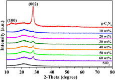
The XRD patterns of samples are shown in Fig. 1. Figure 1 shows that pure g-C3N4 exhibited two characteristic diffraction peaks at 13.7° and 27.6°, which were assignable to (100) and (002) planes of the graphite-like carbon nitride [Reference Wen, Xie, Chen and Li34]. The structure was obtained from the accumulation of aromatic layers [Reference Shiraishi, Kanazawa and Sugano47, Reference Li, Zhang and Liu48]. The diffraction peak of g-C3N4/SiO2 sample appeared in 2θ = 27.6°, which could be attributed to the (002) crystal plane diffraction peak of g-C3N4. The above results indicated that the g-C3N4 existed in the prepared materials. In this study, the guest material (g-C3N4) was completely introduced into the pores of the host material (porous SiO2), and the host material was amorphous, which led to weakening of the characteristic diffraction peaks of g-C3N4.
XRD patterns of g-C3N4 and g-C3N4/SiO2 composites with 10 wt%, 20 wt%, 30 wt%, 40 wt% 50 wt%, and 60 wt% g-C3N4 samples.

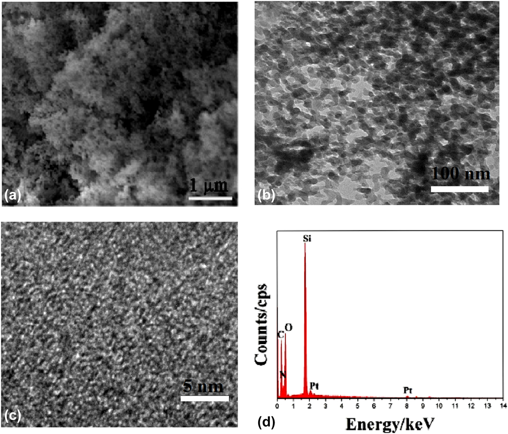
The SEM image [Fig. 2(a)] of g-C3N4/SiO2 (50 wt%) sample showed the fluffy, sponge-like porous structure. The TEM image [Fig. 2(b)] showed that the disordered porous structures were formed in the g-C3N4/SiO2 (50 wt%) sample. Figure 2(c) showed high-resolution TEM image of g-C3N4/SiO2 (50 wt%) sample, and no obvious lattice fringe was observed; this indicates that the sample was amorphous, which was consistent with the XRD measurement. All elements in the sample were checked by energy-dispersive spectrometer (EDS); the EDS spectrum [Fig. 2(d)] shows that the sample contained C, N, Si, and O elements.
(a) SEM image, (b) TEM image, (c) high-resolution TEM image, and (d) EDS spectrum of the prepared g-C3N4/SiO2 (50 wt%) samples.
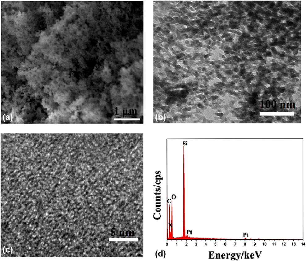
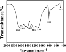
Figure 3 shows the fourier transform infrared (FT-IR) spectrum of the g-C3N4/SiO2 (50 wt%) sample. The absorption peaks at 1641 cm−1, 1569 cm−1, 1325 cm−1, and 1243 cm−1 belonged to the stretching vibration characteristic absorption peaks of the six membered ring of C–N bond. The characteristic absorption peaks at 483 cm−1, 808 cm−1, and 1130 cm−1 pertained to the bending vibration, symmetric stretching vibration, and antisymmetric stretching vibration of [SiO4], respectively. The above conclusions showed that g-C3N4/SiO2 composites had been prepared.
FT-IR spectra of the g-C3N4/SiO2 (50 wt%) sample.

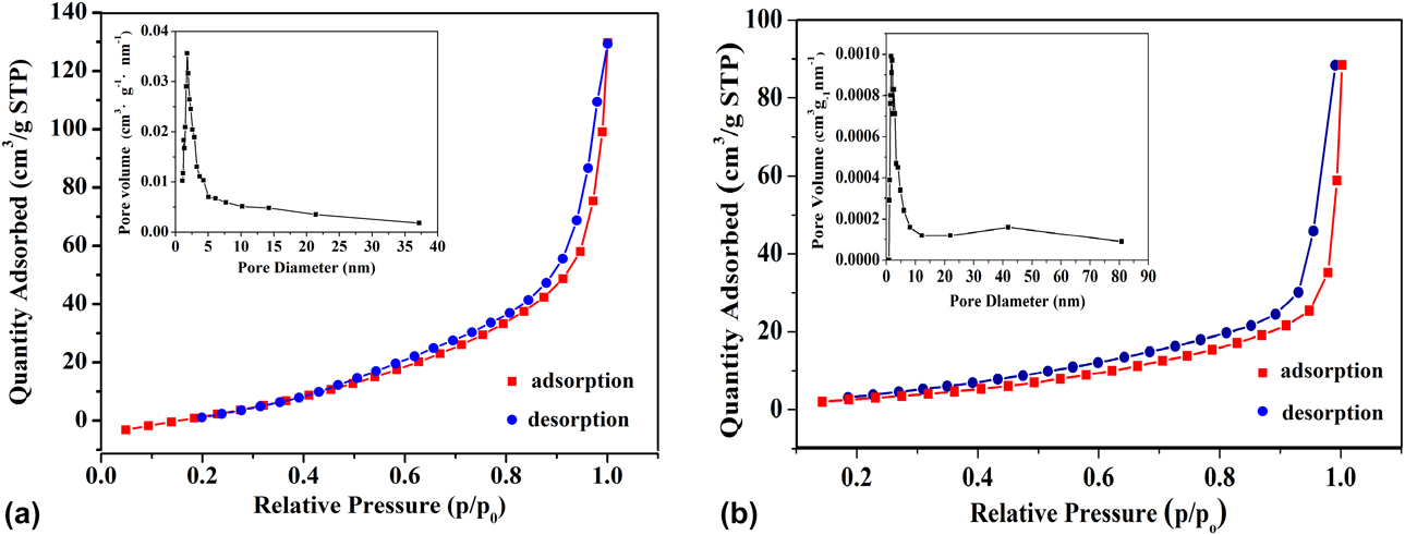
The N2 adsorption–desorption isotherm and pore size distributions of pure SiO2 aerogel and g-C3N4/SiO2 (50%) samples are shown in Fig. 4. The isotherm was of the typical IV type isotherm with distinct H1 hysteresis loop, which indicated the presence of cylindrical mesoporous in the g-C3N4/SiO2 sample. The Brunner-Emmet-Teller (BET) surface area of pure SiO2 aerogel and g-C3N4/SiO2 (50%) samples was 513.78 m2/g and 106.02 m2/g, respectively. The pore size parameter was determined by the Barrett–Joyner–Halenda (BJH) method. The inset in Fig. 4 shows the pore size distribution of pure SiO2 aerogel and g-C3N4/SiO2 (50%) samples, and the average pore diameter was about 3.88 nm and 2.98 nm, respectively. The specific surface area and pore size were significantly reduced, which could be attributed that g-C3N4 entered the pores of SiO2 aerogel.
Nitrogen adsorption–desorption isotherms and pore size distribution curves (inset) of the (a) pure SiO2 aerogel and (b) g-C3N4/SiO2 (50 wt%) samples.

Photocatalytic experiment
The photocatalytic activity of the g-C3N4/SiO2 samples with different g-C3N4 content was evaluated by the degradation rate of RhB under visible light. The concentration of RhB was evaluated by calculating the characteristic absorption of RhB at 550 nm. The degradation rate of RhB was calculated from the following equation [Eq. (1)]:
 $${\rm{Degradation}}\;{\rm{rate\ }} = {\rm{\ }}{{\left( {{A_0} - {A_{\rm{t}}}} \right)} / {{A_0}}} \times 100\% \quad ,$$
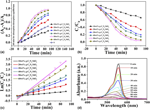
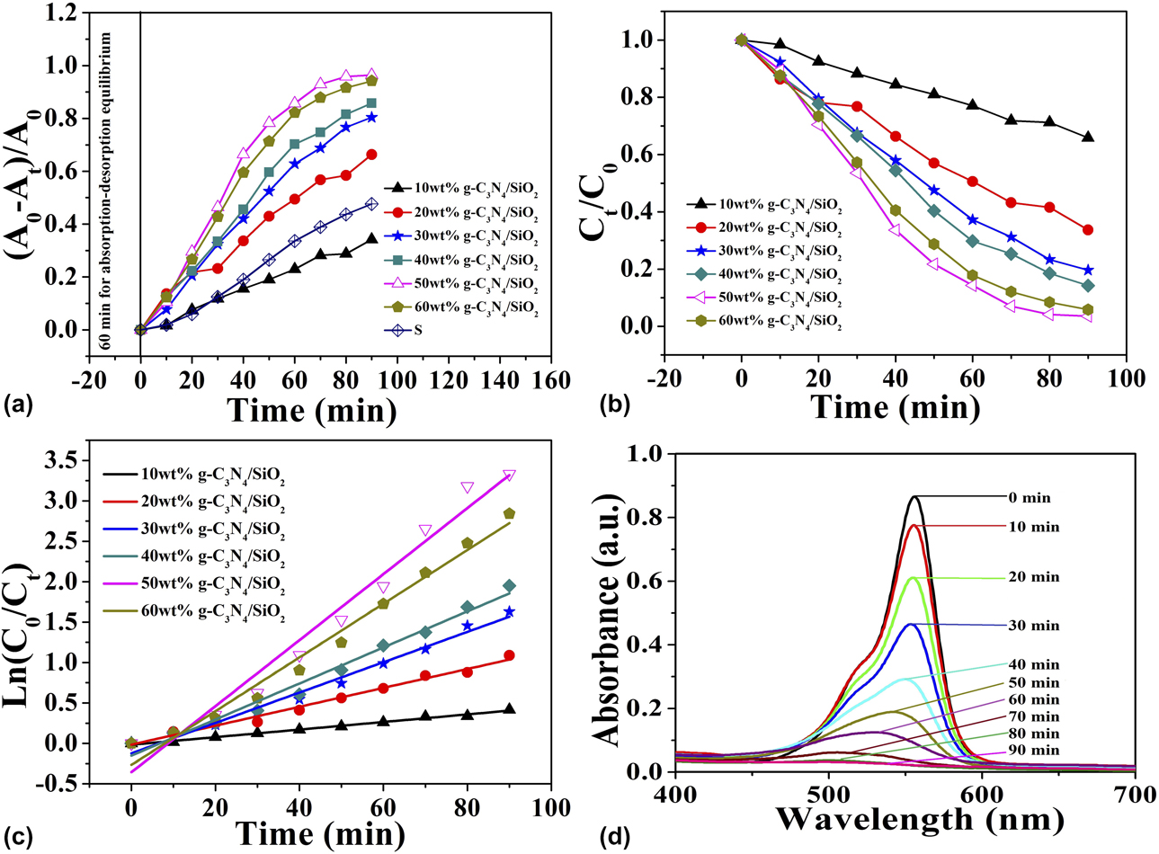
where A 0 is the initial absorption of RhB solutions before light irradiation and A t is the absorption of RhB solutions at selected time interval under visible light irradiation. The degradation time was fixed at 90 min. The amount of SiO2 aerogel was fixed, and the different proportion of g-C3N4 (0 wt%, 10 wt%, 20 wt%, 30 w%, 40 wt%, 50 wt%, and 60 wt%) was doped into SiO2 aerogel matrix. Figure 5(a) shows that the doped amount of g-C3N4 had a great influence on degradation of RhB in the visible light range. When g-C3N4 content was 0 wt%, the RhB had almost no degradation under visible light irradiation. When the content of g-C3N4 was gradually increased from 10 wt% to 50 wt%, the photocatalytic degradation efficiencies of g-C3N4/SiO2 composites were accordingly increased, which were 34.16%, 66.32%, 80.38%, 85.77%, and 96.42%, respectively. When the content of g-C3N4 was 60 wt%, the degradation rate was slightly decreased. Therefore, the optimal doped content of g-C3N4 was 50 wt% in the g-C3N4/SiO2 composite. For comparison, S was used to represent a mechanical mixture of g-C3N4 and SiO2 matrix according to the proportion of g-C3N4/SiO2 (50 wt%), a comparison experiment was studied. It was found that the photocatalytic efficiency of S sample was lower than those of all samples, except 60 wt% g-C3N4/SiO2. The normalized temporal concentration changes (C t/C 0) of RhB during the photocatalytic process are proportional to the normalized maximum absorbance changes (A t/A 0), which can be derived from the change in the RhB absorption profile at a given time interval. The RhB concentration variations with time of the different g-C3N4 concentration samples are shown in Fig. 5(b), which is consistent with Fig. 5(a). To study the reaction kinetics of the photocatalytic degradation, the test data were fitted by the pseudo-first kinetics as Eq. (2):
 $$\left( {{{{C_0}} / {{C_{\rm{t}}}}}} \right) = kt\quad ,$$
where k is the reaction rate constants, C 0 is the initial concentration of RhB, and C t is the actual concentration of RhB at time t. The linear relationships between Ln (C 0/C t) and the irradiation time are shown in Fig. 5(c), which was used to study the photocatalytic activity as Eq. (3).
 $${\rm{Ln}}\left( {{{{C_0}} / {{C_{\rm{t}}}}}} \right) = kt\quad ,$$
where k is the degradation rate constant. The k of g-C3N4/SiO2 (10 wt%, 20 wt%, 30 wt%. 40 wt%, 50 wt%, and 60 wt%) samples were 0.005, 0.012, 0.019, 0.022, 0.041, and 0.033 min−1, respectively. When the g-C3N4 amount was 60 wt%, the photocatalytic effect was lower than 50% sample. It is because the reunite occurred among the particles of the sample when the g-C3N4 amount was too large, and the separation efficiency of electrons and holes was weakened, which led to the weak photocatalytic effect. Figure 5(d) shows the RhB degradation curves by the prepared g-C3N4/SiO2 (50%) sample in different visible light radiation time. With the visible light radiation time lengthening, the RhB content decreased, which was almost completely degraded when degradation time was 90 min and the degradation rate reached 96.42%. The blue shift of the maximum absorption value showed that the degradation process was a chemical reaction process.
(a) Photocatalytic activity degradation curve of g-C3N4/SiO2 (10 wt%, 20 wt%, 30 wt%. 40 wt%, 50 wt%, and 60 wt%) and mechanical mixture(s). (b) Time dependency of photocatalytic degradation of RhB over composites under visible light. (c) Plots of Ln (C 0/C t) versus irradiation time t for the samples. (d) UV-vis absorption spectrum of RhB under catalysis of g-C3N4/SiO2 (50 wt%) at different visible light radiation time.
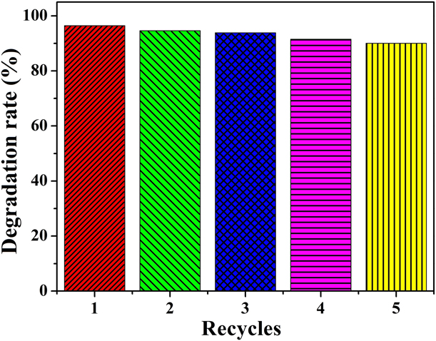
To investigate the stability of the prepared catalyst, a cycle stability experiment was needed. The degradation rate of g-C3N4/SiO2 (50%) was still above 90% after five consecutive cycles under visible light, and the results are presented in Fig. 6. During the recycling experiment, the catalyst was collected by centrifugation, washed with deionized water, dried at 60 °C, and reused in the next cycles. It indicated that the prepared material had better stability.
Cycling stability test result of g-C3N4/SiO2 (50%) sample.

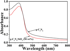
The UV-vis absorption spectra of the prepared g-C3N4/SiO2 (50 wt%) sample are shown in Fig. 7. The prepared sample had strong absorption in the visible region. For a crystalline semiconductor, the band edge can be calculated by the following formulas:
 $$\alpha h\nu = A{\left( {h\nu - {E_{\rm{g}}}} \right)^{{n / 2}}}\quad ,$$
 $${E_{\rm{g}}} = {{1240} / {{\lambda _{\rm{g}}}}}\quad ,$$
where α, ν, E g, and A are absorption coefficient, light frequency, band gap, and constant, respectively. It should be noted that the n is determined by the type of optical transition of a semiconductor. Equation (5) was applied to estimate the E g of the g-C3N4/SiO2 (50 wt%) sample, in which λg was the wavelength of the absorption edge. The absorption wavelength range of the prepared g-C3N4/SiO2 (50 wt%) composites could reach about 450–550 nm, and the band gap was E g = 2.66 eV. The absorption wavelength range of the prepared g-C3N4/SiO2 (50 wt%) sample was wider than that of the prepared g-C3N4. Therefore, the prepared composite had the better optical absorption in the UV and visible range. At the same time, g-C3N4 was highly dispersed in the pores of the mesoporous material, which resulted in a high dispersion degree of the electron–hole pair, improved dispersion of electron–hole pairs, reduced electron-mutual contact and impact of the probability, prevented the recombination of the photogenerated electron–hole pairs, and improved the photocatalytic efficiency of the material.
UV-vis absorption spectra of the g-C3N4/SiO2 (50 wt%) sample.

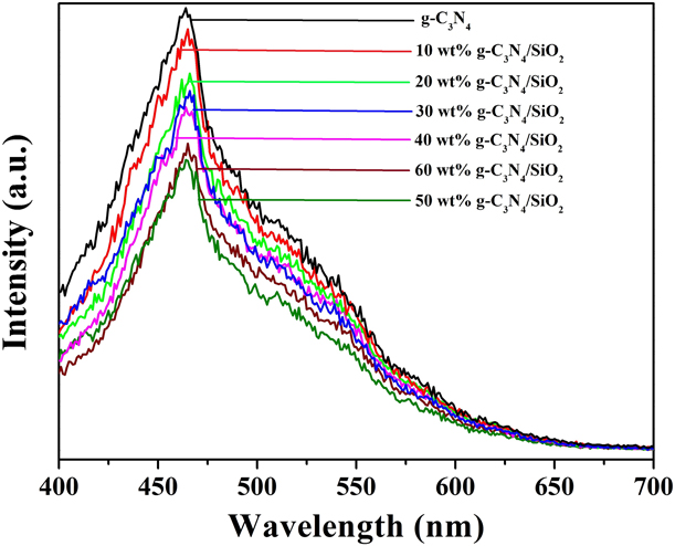
It is well known that the faster photogenerated electron–hole pair transfer rates and the slower photogenerated electron and hole recombination rates are beneficial to improve the photocatalytic properties of the samples. To evaluate the transfer efficiency of photogenerated electrons and holes, the photoluminescence (PL) spectrum is an ideal test method. The better photocatalytic performance will be obtained for the sample with the lower PL intensity. Figure 8 shows the PL spectra of g-C3N4 and g-C3N4/SiO2 (10 wt%, 20 wt%, 30 wt%. 40 wt%, 50 wt%, and 60 wt%) samples. It could be found that the pure g-C3N4 had the strongest PL intensity, and the PL intensity was gradually decreased with the increase in the amount of g-C3N4 doped into the g-C3N4/SiO2 samples. When the g-C3N4 doped amount was 50 wt%, the PL intensity was the lowest. When the g-C3N4 doped amount was 60 wt%, the PL intensity was slightly enhanced. When the g-C3N4 content was increased, the superfluous g-C3N4 would to lead to the reunite of the sample particles, which made that the heterojunction interface was reduced, the electron–hole recombination rate was increased, and the photocatalytic characters were reduced. The PL intensity of the g-C3N4/SiO2 (50 wt%) sample was the weakest, whose charge separation rate was the best and its photocatalytic characters were the highest.
Room temperature PL spectra of g-C3N4 and g-C3N4/SiO2 composites (10 wt%, 20 wt%, 30 wt%. 40 wt%, 50 wt%, and 60 wt%).

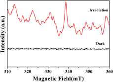
In the photocatalytic degradation process, some active oxidation species were produced. To further prove the existence of these active oxidation species, electron spin resonance (ESR) is an ideal test method. As shown in Fig. 9, the signal of DMPO–•O2− could be detected under light irradiation. On the contrary, no signals were detected in dark, which indicated that the •O2− plays a leading role in photocatalytic process.
ESR spectra of •O2− trapped by DMPO for g-C3N4/SiO2 (50 wt%) sample.

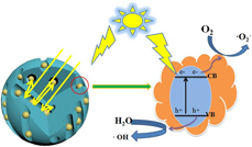
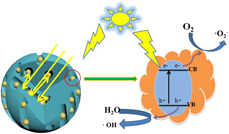
The photocatalytic degradation of organic pollutants needs to go through four processes: (i) light irradiation, (ii) charges were generated by photoexcitation, (iii) charges and holes were separated and transferred to the surface of catalyst, and (iv) oxidation–reduction reaction of electrons, holes, and pollutants adsorbed on the catalyst surface. A lot of pores were produced in the preparation process of material in this study. When the light enters the channels, the emission and refraction of light are increased due to the special porous structure, which stores more energy of the light and generates more photoelectrons and holes. Meanwhile, the disordered porous structure increases the specific surface area of the composite, which provides more active sites for catalytic degradation reactions. The valence band electron absorption energy is excited to conduction band when g-C3N4 is subjected to the radiation of light energy. The energy is greater than forbidden bandwidth of g-C3N4 and generates photogenerated electrons (e−) in the conduction band, leaves the positively charged holes (h+) in the valence band. The electrons and holes can successfully reach to the surface of particles, which occurs oxidation–reduction reaction with the adsorbed organic pollutant on the surface of the particles. In this process, first, oxygen as electron acceptor turns into superoxide anions (•O2−) after reduction reaction with photogenerated electrons. Subsequently, superoxide anions oxidize the more organic pollutants and can react with hydrogen ions to produce hydrogen peroxide (H2O2). Holes will also react with water and hydroxyl (OH−) group to produce high activity hydroxyl radical (•OH). The organic matter (D), hydrogen peroxide, and hydroxyl can be completely mineralized into carbon dioxide molecules and water molecules on the surface of the catalyst [Reference Wang, Feng, Chen, Wang, Su, Zhang, Zeng, Xie, Liu, Liu, Lv and Liu49]. Specific reaction process is given as Eqs. (6)–(10). The mechanism is shown in Fig. 10.
 $${{\rm{C}}_3}{{\rm{N}}_4} + h\nu \left( {{\rm{ > 2}}{\rm{.7}}\;{\rm{eV}}} \right) \to {{\rm{h}}^ + } + {{\rm{e}}^ - }\quad ,$$
 $${{\rm{e}}^ - } + {{\rm{O}}_2} \to {\;^ \bullet }{{\rm{O}}_2}^ - \quad ,$$
 $$^ \bullet {{\rm{O}}_2}^ - + {{\rm{H}}^ + } \to {{\rm{H}}_2}{{\rm{O}}_2}\quad ,$$
 $${{\rm{H}}_{\rm{2}}}{{\rm{O}}_2} + {{\rm{e}}^ - } \to {\rm{O}}{{\rm{H}}^ - } + {\;^ \bullet }{\rm{OH}}\quad ,$$
 $${\rm{D}} + {\;^ \bullet }{\rm{OH}} \to \cdots \to {\rm{C}}{{\rm{O}}_2} + {{\rm{H}}_2}{\rm{O}} + {{\rm{N}}_2} + \cdots \quad .$$
Schematic illustration of photocatalytic reaction and charge transfer of the g-C3N4/SiO2 sample under visible light irradiation.

Conclusions
In summary, the g-C3N4/SiO2 nanostructures have been successfully fabricated by sol–gel method, and the loose and porous structures were formed. The composite material exhibited much better photocatalytic activity in photodegradation of RhB, and degradation rate reached 96.42% in 90 min under the irradiation of visible light. The prepared material has a promising application in the wastewater treatment.
Experimental section
All chemicals were of analytical grade (AR) and used without further purification. Tetraethyl orthosilicate (TEOS) and melamine were purchased from Tianjin Guangfu Fine Chemical Research Institute (Tianjin, China). Hydrofluoric acid was purchased from Beijing Chemical Works (Beijing, China). Deionized water and absolute alcohol were used throughout dissolution and filtration process.
Preparation of porous g-C3N4/SiO2 composites
G-C3N4 was prepared according to the procedure of literature reported [Reference Sun, Qi, Jia, Jin and Fan50]. Melamine was calcined at 550 °C for 2 h at a heat rate of 10 °C/min, and g-C3N4 was obtained.
The g-C3N4/SiO2 composite was synthesized by a sol–gel method. The detailed synthesis process is as follows: a certain percentage of g-C3N4 (0 wt%, 10 wt%, 20 wt%, 30 wt%, 40 wt%, 50 wt%, and 60 wt%) was uniformly dispersed into the mixed solution of 6.2 mL ethanol and 3.8 mL deionized water with stirring. Then, 5.7 mL of TEOS was added into the solution to form the sol, and the sol was continuously stirred for 60 min. The hydrofluoric acid was dropwise added into the sol under stirring, and the gel was obtained. The gel was aged for 24 h in room temperature. The aged gel was soaked in deionized water to continue age for 2–4 days at 70 °C. The deionized water was dumped, a lot of n-hexane was added, the gel was aged in hexane solution for 2 days at 50 °C, and the n-hexane solution was replaced once every 12 h. The gel was dried at 60 °C, and the target product was obtained.
Characterizations
Powder XRD patterns of the samples were collected on a Siemens D5005 diffractometer (Siemens, Munich, Germany) using Cu Kα radiation (λ = 5 1.5418 Å and operating at 30 kV and 20 mA). TEM images were recorded on a JEOL 2010 TEM instrument (JEOL, Tokyo, Japan). The microstructures of the g-C3N4/SiO2 were investigated by a JEOL JSM-5600L SEM instrument (JEOL, Tokyo, Japan). The elementary compositions of samples were inspected by Oxford ISIS-300 energy dispersive spectrometer (EDS) (JEOL, Tokyo, Japan) attached to the SEM. The specific surface area and pore size of the prepared g-C3N4/SiO2 samples were measured by nitrogen adsorption-desorption isotherm in a Micromeritics ASAP 2010 nitrogen adsorption-desorption analyzer (Micromeritics, Norcross, Georgia), and the samples were degassed at 500 K for 12 h. The surface area was determined from the nitrogen adsorption-desorption isotherm by BET method, while average pore sizes of the samples were determined from desorption isotherm by the BJH method. The UV-vis absorption spectra for the evaluation of photophysical properties were recorded in the diffuse reflection mode, and the absorption spectra were measured through a Shimadzu UV-3600 spectrophotometer (Shimadzu, Kyoto, Japan). The ultraviolet-visible-near infrared spectrum of sample was determined on a UV-1240 ultraviolet-visible spectrophotometer (Shimadzu, Kyoto, Japan). All the measurements were carried out at room temperature.
Photodegradation character measurement
The photocatalytic activities of the prepared g-C3N4/SiO2 composite were evaluated by the photodegradation efficiency of rhodamine B (RhB) solution response to visible light by using a 500 W Xe lamp with a filters to remove wavelengths less than 380 nm at room temperature. First, 0.01 g/L RhB solution was prepared. Five milligrams of the prepared g-C3N4/SiO2 composite was soaked in 95 mL deionized water, and 5 mL RhB solution (0.01 g/L) was added into the above solution. The mixture was continuously stirred at room temperature in the dark for 1 h to ensure adsorption–desorption equilibrium of RhB on the sample surfaces. Then, the mixed solution was radiated by a 500 W xenon lamp with continuous stirring. The distance between the beaker and the lamp was 15 cm, and the beaker was placed in the circulating water to ensure a constant temperature during the test. Every 10 min, 4 mL of suspension liquid was measured out and centrifuged to remove the particles of catalyst. The concentration of RhB solution was determined by measuring the maximum absorbance at 550 nm using a UV-3600 spectrophotometer.
Acknowledgments
We gratefully acknowledge the support of the work by the National Natural Science Foundation of China (Grant No. 51572034).