History
Right ventricular to pulmonary artery (RV-PA) conduit dysfunction is a common long-term complication in patients with complex CHD, often necessitating surgical or transcatheter interventions. Reference Mery, Guzman-Pruneda and De Leon1–Reference Cheatham, Hellenbrand and Zahn3 We present the case of a 14-year-old girl with an extensive cardiac history, including a diagnosis of posterior malalignment ventricular septal defect and interrupted aortic arch. She previously underwent three sternotomies, beginning with a neonatal Yasui procedure utilising a 13 mm RV-PA conduit, followed by subsequent conduit replacement with a 20 mm Gore-Tex conduit (Gore Medical, USA). Her medical history was further complicated by partial DiGeorge syndrome with hypocalcaemia and lymphopenia, feeding intolerance requiring Nissen fundoplication and gastrostomy tube placement, and a history of clubfoot repair.
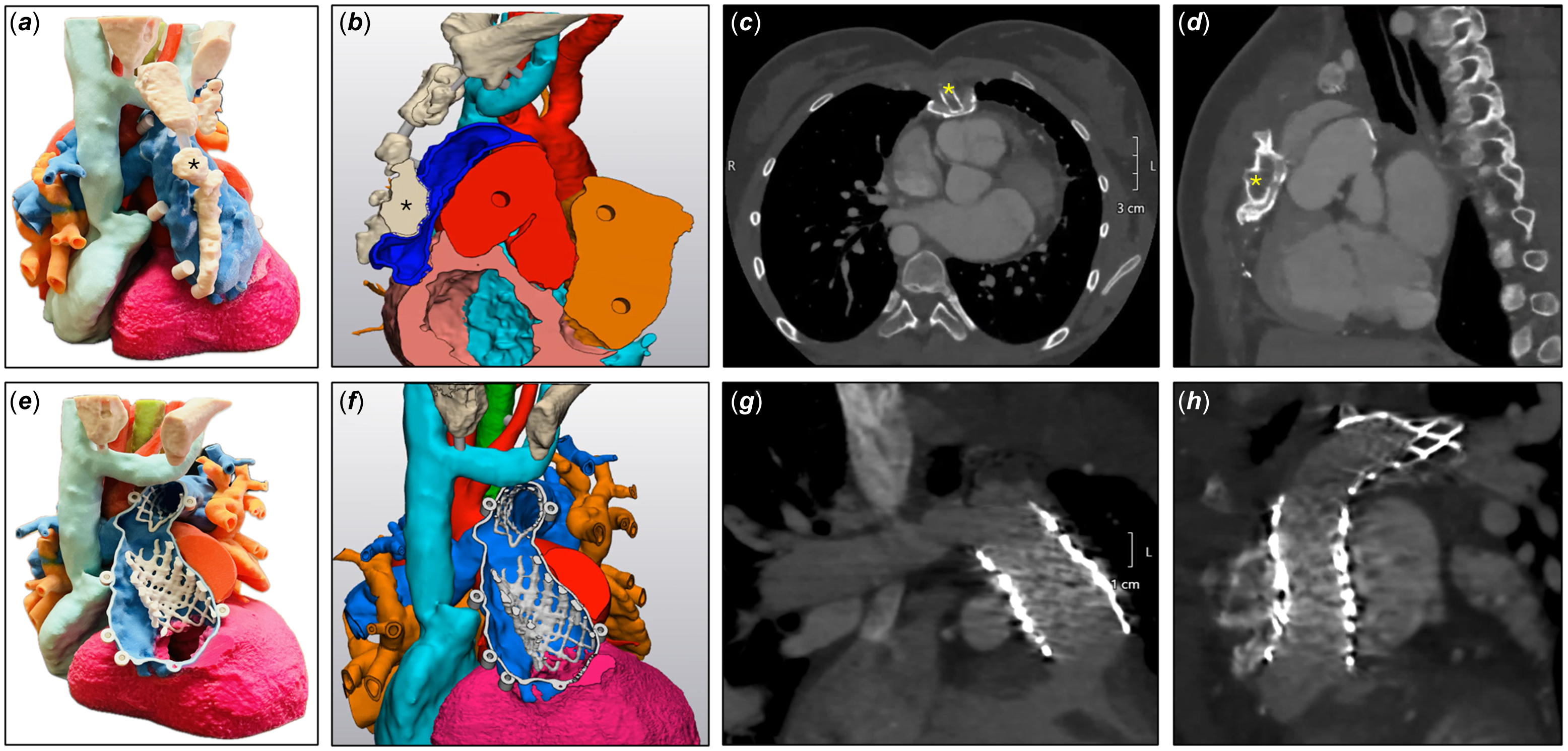
She was referred to the catheterisation team for progressive RV-PA conduit dysfunction, which had demonstrated suboptimal response to balloon angioplasty performed five years prior. A CT scan, reviewed in a multidisciplinary surgical conference, revealed that the midportion of the conduit was significantly compressed along nearly its entire length by a posteriorly protruding sternum (Figure 1a–d, Video 1). Given the anatomical complexity, the surgical team determined that a redo sternotomy posed a significant risk of complications, with a possible need for sternectomy. After thorough discussion of these risks during shared decision-making with the patient’s family, a percutaneous approach was chosen for transcatheter pulmonary valve replacement (TPVR).
Pre- and post-procedural imaging. ( a ),( b ) Pre-procedure AP and sagittal projections of 3D model print; ( c ),( d ) Pre-procedure axial and sagittal CT slices demonstrating sternal protrusion into conduit; ( e ),( f ) Post-procedure 3D model print demonstrating stents within the leftward aspect of the conduit and left pulmonary artery; ( g ),( h ) Post-procedure near coronal and sagittal CT slices (* = sternum).

Case
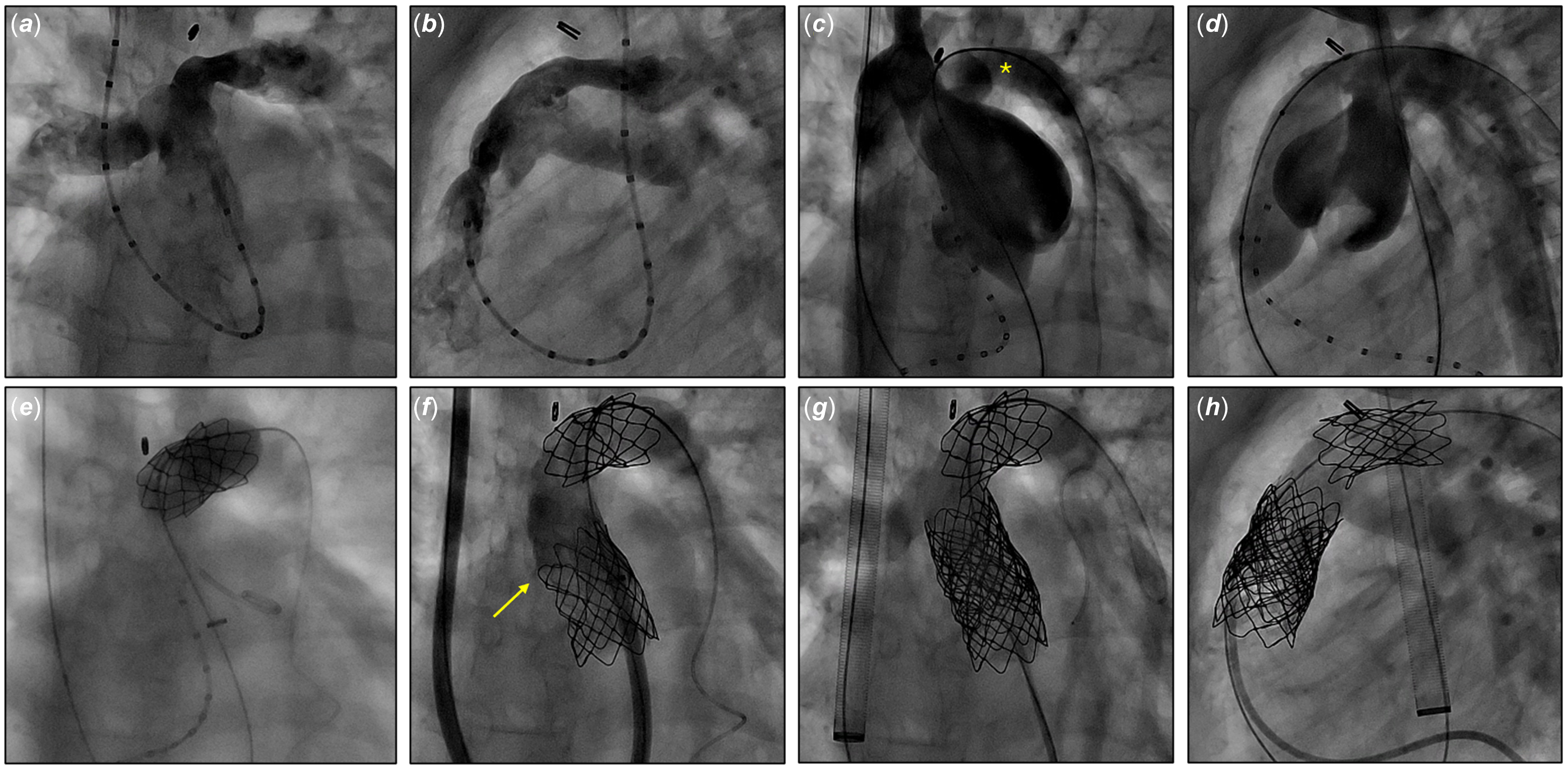
Vascular access was obtained via the right internal jugular vein (RIJ), right femoral vein (RFV), and right femoral artery. Angiography revealed that the RFV was too small to accommodate the large sheath required for valve delivery, necessitating that most interventions be performed via the RIJ. Hemodynamic assessment demonstrated suprasystemic right ventricular pressures with normal bilateral filling pressures and a gradient across the conduit of 101 mmHg. Angiography confirmed severe RV-PA conduit stenosis, along with proximal left pulmonary artery (LPA) stenosis and coronary arteries positioned remote from the conduit (Figure 2a,b).
Intraprocedural cineangiography. ( a ),( b ) AP and lateral projections of cineangiography in the RV-PA conduit; ( c ),( d ) AP and lateral projections of simultaneous aortic root angiography and balloon inflation in the RV-PA conduit demonstrating aortopulmonary fistula to the LPA; ( e ) Covered stent deployment in the proximal LPA; ( f ) Angiography demonstrating para-stent leak around sternum to the rightward aspect of conduit; ( g ),( h ) Angiography following valve deployment demonstrating no residual insufficiency or paravalvular leak (* = LPA; arrow = para-stent leak).

Following balloon angioplasty of the proximal LPA, serial high-pressure, non-compliant balloon angioplasty of the RV-PA conduit was performed using 16 mm, 18 mm, and 20 mm balloons. Notably, each inflation failed to produce the expected drop in systemic blood pressure, with cardiac output maintained throughout. Aortic root angiography performed with simultaneous 20 mm balloon inflation in the conduit revealed the development of a new aortopulmonary fistulous connection between the aorta and LPA (Figure 2c,d). The patient remained hemodynamically stable, and consultation with the surgical team reaffirmed that a redo sternotomy remained a high-risk option. Given this, transcatheter intervention on the LPA and RV-PA conduit was continued.
To seal the aortopulmonary fistula, an 18 mm x 3 cm covered Cheatham Platinum (CP) stent (B. Braun, USA) was deployed in the proximal LPA (Figure 2e), achieving successful closure. Attention then shifted to TPVR. A pre-mounted 20 mm x 4 cm covered CP stent was deployed within the conduit, followed by deployment of a 4010 Palmaz XL stent (Cordis, USA), both of which were dilated with a 20 mm high-pressure balloon. However, angiography revealed persistent leakage around the rightward aspect of the stents, indicating that only the leftward portion of the conduit had been successfully expanded, while the sternum remained adherent to the conduit and prevented full radial expansion. To address this, a second 20 mm x 4 cm covered CP stent was placed distally in the conduit, successfully sealing the leak.
Following this, a 22 mm Melody valve (Medtronic, USA) was crimped onto an Ensemble delivery system along with a 4110 Palmaz XL stent and deployed within the stented RV-PA conduit, which was then post-dilated with the 20 mm high-pressure balloon. Angiography confirmed a well-functioning valve with no paravalvular leak (Figure 2g,h). The final gradient across the valve was 6 mmHg.
Follow-up
The patient was admitted overnight for observation. Imaging the following day demonstrated no residual pulmonary valve stenosis or insufficiency, and she was discharged home in stable condition. A follow-up CT scan performed two months later confirmed the absence of residual aortopulmonary fistula and showed that the pulmonary valve remained positioned in the portion of the conduit leftward of the sternum (Figure 1e–h, Video 2). In addition, no leakage into the rightward portion of the conduit was observed. At six-month follow-up, the patient remained asymptomatic with no evidence of pulmonary valve stenosis or insufficiency on echocardiography.
Conclusion
This case highlights several important findings: (1) the first reported instance of sternal compression of an RV-PA conduit successfully managed via transcatheter intervention; (2) Gore-Tex conduits can be significantly expanded beyond their nominal diameter using current stent and balloon technologies, as only a portion of this conduit was able to expand to the original nominal diameter of the conduit; (3) Iatrogenic aortopulmonary fistula can be effectively treated with covered stents via transcatheter approach; and 4) Advanced imaging, including CT and 3D printing, is essential for diagnosis, procedural planning, and post-procedural evaluation. Despite complex challenges—including severe conduit stenosis, limited ability to displace the conduit from the sternum, development of a fistula, and para-stent leakage—multimodal imaging enabled precise planning and successful management.
Acknowledgements
The authors thank Eric Braden for his contributions to pre- and post-procedural 3D modelling, as well as the Arkansas Children’s Foundation for financially supporting our 3D modelling programme.
Financial support
This research received no specific grant from any funding agency, commercial, or not-for-profit sectors.
Competing interests
The authors have no conflicts of interest to disclose.