Introduction
Osteoporosis is a skeletal disorder characterized by low bone mass and micro-architectural deterioration of bone tissue with a consequent increase in bone fragility and susceptibility to fracture. 1 It is a widespread condition, often unrecognized in clinical practice, which may have devastating health consequences through its association with fragility fractures. The risk of osteoporotic fracture ultimately depends on two factors: the mechanical strength of bone and the forces applied to it. Bone mass (a composite measure including contributions from bone size and from its volumetric mineral density) is an established determinant of bone strength, and the bone mass of an individual in later life depends upon the peak attained during skeletal growth and the subsequent rate of bone loss. The foetus accretes 80% of the required 30 g calcium during the last trimester in human pregnancies,Reference Kovacs 2 and then bone mass increases largely as a result of increase in bone size throughout childhood. During the adolescent growth spurt, around 25% of the final peak bone mass is achieved.Reference Gilsanz and Nelson 3 Peak bone mass is reached in the twenties, but exact timing appears to vary with skeletal site and with sex.Reference Hui, Slemenda and Johnston 4 After the achievement of peak bone mass, bone density starts to decline and this is accelerated after the menopause in women.Reference Dennison, Cole and Cooper 5
The skeletal growth trajectory becomes set early in life, such that a child’s size, relative to its peers, remains constant until adult size is reached. This is known as ‘tracking’, and gives rise to childhood centile growth charts. It is known from animal work that altering dietary intake after this trajectory has been set may temporarily alter growth, but once normal diet is restored, the original trajectory is regained. Before the point of setting, the pattern of growth can be altered.Reference Barker 6 Clearly, early life influences are most likely to influence bone mass in old age by an effect on peak bone mass, and evidence is accruing for the importance of peak bone mass as a determinant of osteoporotic fracture risk. Hui et al.Reference Hui, Slemenda and Johnston 4 explored the relative contributions of peak bone mass and bone loss, and found that at 70 years of age, they each explained 50% of the variance in bone mineral density (BMD). A more recent modelling study from Hernandez et al.Reference Hernandez, Beaupre and Carter 7 showed that peak bone mass was six times more powerful a predictor of the age of onset of osteoporosis than the age of menopause or rate of bone loss.
Evidence has been accruing that for diseases such as osteoporosis, which increase dramatically in prevalence with age, and for which heredity can only partly explain, there is an interaction between the genome and the environment in the expression of the disease. Thus, given a particular genetic predisposition, the environmental factors to which a subject is exposed during early life are a critical determinant of later health and disease. This phenomenon is known as ‘programming’. Programming is defined as ‘persisting changes in structure and function caused by environmental stimuli acting at critical periods during early development.’Reference Barker 8 There is no doubt that the skeleton can be permanently changed by an adverse early environment – rickets is a very visible example. This study will review the evidence for programming of osteoporosis, highlighting epidemiological studies of BMD and fracture in cohorts where birth details are known, physiological studies and studies of potential underlying mechanisms.
Normal skeletal growth – bone growth in utero
The foetal skeleton develops in two distinct components, intramembranous (the skull and facial bones) and endochondral (the remainder of the skeleton) ossification. Endochondral ossification is responsible for the formation of the bones that are the main sites of fragility fracture in later life. This form of ossification depends on a pre-existing cartilaginous model that undergoes invasion by osteoblasts and is only subsequently mineralized. The development of this cartilage model can be seen by 5 weeks of gestation with the migration and condensation of mesenchymal cells in areas destined to form the bone.Reference DeLise, Fischer and Tuan 9 These pre-cartilaginous analgen reflect the shape, size, position and number of skeletal elements that will be present in the mature skeleton. There is then an ordered differentiation of mesenchymal stem cells into chondrocyte precursors, proliferative chondrocytes, prehypertrophic chondrocytes and hypertrophic chondrocytes. During these stages of differentiation, there is expansion of the bony template and production of an extracellular matrix rich in cytokines that facilitate vascular invasion and mineralization. The major regulator of the proliferation of chondrocytes is parathyroid hormone-related protein,Reference Karaplis, Luz and Glowacki 10 which is secreted by the perichondral cells; other proliferative stimuli include cytokines of the growth hormone/insulin-like growth factor-1 (GH/IGF-1).Reference Bhaumick and Bala 11 The 1,25 (OH)2 vitamin D3 Reference Sylvia, Del Toro and Hardin 12 and tri-iodothyronineReference Quarto, Campanile, Cancedda and Dozin 13 are stimuli for the differentiation of the chondrocytes through different stages. Once the cartilage model has been formed, vascular growth factors embedded in the matrix are released by chondrocyte metalloproteinases. This stimulates angiogenesis and, under the influence of core-binding factor alpha-1,Reference Ducy 14 osteoblasts from the perichondrium invade and lay down matrix which is then mineralized.
To supply foetal calcium demand, there is a requirement for (1) an adequate maternal supply of calcium to the placenta and (2) increased placental calcium transfer to maintain a higher foetal serum calcium concentration than the mother.Reference Schauberger and Pitkin 15 This materno-foetal gradient emerges as early as 20 weeks of gestation.Reference Forester, Daffos, Rainaut, Bruneau and Trivin 16 A rise in maternal serum PTHrP and 1,25 (OH)2 vitamin D3 Reference Ardawi, Nasrat and BA’Aqueel 17 is thought to drive the maternal supply of calcium to the foetus. Active calcium transfer across the placenta takes place in the cytotrophoblasts and involves storage of calcium by calcium-binding proteins in the cytoplasm and in the endoplasmic reticulum.Reference Hosking 18 While in the mother, 1,25 (OH)2 vitamin D3 is the principal stimulus for calcium absorption, the mid-portion of PTHrP is essential at the placenta for the maintenance of the maternofetal gradient.Reference Kovacs, Lanske and Hunzelman 19 Secretion of PTHrP by the foetal parathyroid glands also enhances foetal renal calcium reabsorption. The rate of materno-foetal calcium transfer increases dramatically after 24 weeks, such that around two-thirds of total body calcium, phosphorous and magnesium are accumulated in a healthy-term human foetus during this period. Factors which increase placental calcium transport capacity as gestation proceeds are only partly genetically controlled, and are achieved through regulatory hormones including 1,25 (OH)2 vitamin D3, parathyroid hormone, PTH-related peptide and calcitonin.
As the majority of foetal bone is gained during the last trimester, one of the major variables affecting bone mass at birth is gestational age. Other factors known to influence neonatal bone mineral content (BMC) include environmental variables such as season of birth and maternal lifestyle; newborn total BMC has been shown to be lower among winter births than among infants born during the summer.Reference Namgung, Tsang and Li 20 This may be a consequence of lower cord serum 25 (OH) vitamin D concentrations observed during winter months due to maternal vitamin D deficiency. Other maternal factors that may contribute to impaired bone mineral acquisition during intrauterine life include maternal smoking, alcohol consumption, caffeine intake and diabetes mellitus.Reference Specker, Namgung and Tsang 21
Intrauterine programming of osteoporosis and fracture
The evidence that the risk of osteoporosis might be modified by environmental influences during early life stems from four groups of studies: (a) bone mineral measurements undertaken in cohorts of adults whose detailed birth and/or childhood records have been preserved; (b) detailed physiological studies exploring the relationship between candidate endocrine systems which might be programmed (GH/IGF-1; hypothalamic-pituitary adrenal and gonadal steroid) and age-related bone loss; (c) studies characterizing the nutrition, body build and lifestyle of pregnant women and relating these to the bone mass of their newborn offspring; and (d) studies relating childhood growth rates to the later risk of hip fracture.
Epidemiological studies
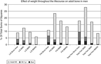
The first epidemiological evidence that osteoporosis risk might be programmed came from a study of 153 women born in Bath during 1968–1969 who were traced and studied at 21 years of age.Reference Cooper, Cawley and Bhalla 22 Data on childhood growth were obtained from linked birth and school health records. There were statistically significant (P < 0.05) associations between weight at 1 year and BMC, at the lumbar spine and femoral neck; these relationships were independent of adult weight and body mass index. The data suggested a discordance between the processes that govern skeletal growth, and those which influence mineralization. They also provide direct evidence that the trajectory of bone growth might be programmed. The association between weight in infancy and adult bone mass was replicated in a second cohort study of 238 men and 201 women aged 60–75 years, who were born and still lived in Hertfordshire.Reference Cooper, Fall and Egger 23 In this study, there were highly significant relationships between weight at 1 year and adult bone area at the spine and hip (P < 0.005); the relationships with BMC at these two sites were weaker, but remained statistically significant (P < 0.02). They also remained after adjustment for lifestyle characteristics in adulthood that might have influenced bone mass (physical activity, dietary calcium intake, cigarette smoking and alcohol consumption). Similar relationships have now been replicated in a younger Hertfordshire cohort;Reference Dennison, Syddall, Aihie Sayer, Gilbody and Cooper 24 in a conditional analysis of this group, 18% of adult bone mass was explained by a model that included birthweight, weight at 1 year and adult weight, with relative contributions attributable to each being 2.8%, 6.8% and 8.2% respectively (Fig. 1). The contributions of early growth were greatest for bone area, and clearly apparent for BMC, but not as evident for areal BMD (aBMD) or derived volumetric BMD. The findings of studies performed by other groups also suggest the importance of the early environment in the determination of later bone mass.Reference Yarbrough, Barrett-Connor and Morton 25 , Reference Jones, Riley and Dwyer 26
Effect of weight through the lifecourse on bone mass among 468 adult men from the Hertfordshire cohort study. Data derived from Dennison et al.Reference Dennison, Syddall, Aihie Sayer, Gilbody and Cooper 24

Javaid et al.Reference Javaid, Lekamwasam and Clark 27 undertook analysis of the dual-energy X-ray absorptiometry (DXA) scan images acquired in 300 Hertfordshire Cohort Study participants, using automated hip structure analysis to derive estimates of femoral neck length, width, cross-sectional moment of inertia (CSMI) and buckling ratio. There were significant (P < 0.002) relationships between weight at 1 year and measures of femoral width, as well as intertrochanteric CSMI, but not with femoral neck length. The associations with measures of femoral width remained after adjusting for adult body weight and were independent of proximal femoral BMC. These findings suggest that poor growth in utero and during the first year of postnatal life is associated with disproportion of the proximal femur in later life (narrower neck but preserved axis length), with a corresponding reduction in mechanical strength of the region, over and above that attributable to reduced BMC per se.
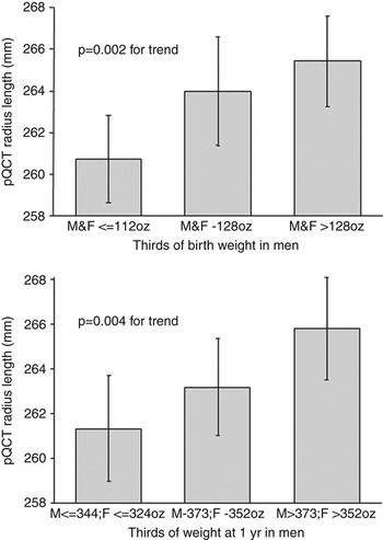
In the third detailed analysis of bone density, geometry and strength among participants from the Hertfordshire Cohort Study, peripheral computed tomography was utilized to evaluate cortical and trabecular bone density, strength–strain index (SSI) and fracture load in the distal radius and tibia (Stratec XCT2000 instrument).Reference Oliver, Jameson, Aihie Sayer, Cooper and Dennison 28 There were strong (P < 0.01) associations between birthweight, conditionally adjusted weight at 1 year and each of bone width, length, area, fracture load and SSI, at the tibia among both men and women, with less marked associations in a similar direction for the proximal radius (Fig. 2). The relationships were again robust to adjustment for age, body mass index, social class, cigarette smoking, alcohol consumption, physical activity, dietary calcium intake and hormone replacement therapy use among women. Women who had sustained fractures since 45 years of age had lower radial total (P = 0.0001), cortical (P < 0.005) and trabecular (P = 0.0002) bone density, poorer forearm bone strength (polar SSI, P = 0.006), fracture load (P = 0.02) and lower tibial total (P < 0.001), cortical (P = 0.008) and trabecular (P < 0.0001) bone density. The data add to those from hip structure analysis; thus, poor growth in utero and during the first year of postnatal life is associated with alterations in bone architecture, cortical size and geometry, in addition to a deficit in densitometrically measured bone mineral content, resulting in compromized bone strength and increased risk of later fracture.
Bone strength according to the birthweight and weight at 1 year among men from the Hertfordshire cohort study. Data derived from Oliver et al.Reference Oliver, Jameson, Aihie Sayer, Cooper and Dennison 28

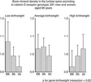
Detailed analyses of the interaction between polymorphism in the gene for the vitamin D receptor (VDR), birthweight and BMD in the Hertfordshire cohort showed a relationship between lumbar spine BMD and VDR genotype according to birthweight (Fig. 3). Among individuals in the lowest third of birthweight, spine BMD was higher (P = 0.01) among individuals of genotype ‘BB’ after adjustment for age, sex and weight at baseline.Reference Dennison, Arden and Keen 29 In contrast, spine BMD was reduced (P = 0.04) in individuals of the same genotype who were in the highest third of the birthweight distribution. A statistically significant (P = 0.02) interaction was also found between VDR genotype and birthweight as determinants of BMD. A similar study evaluated the association between single nucleotide polymorphisms in the human growth hormone gene (GH1) and weight in infancy, adult bone mass and bone loss rates.Reference Dennison, Syddall and Rodriguez 30 Homozygotes at loci GH1 A5157G and T6331A displayed lower BMD and accelerated bone loss; there was again a statistically significant (P = 0.04) interaction between GH1 genotype, weight at 1 year and bone loss. Furthermore, when 24-h circulating profiles of GH in samples of the men and women studied were analysed, polymorphism at the A5157G locus within the GH1 promoter showed a graded association with basal GH concentration, suggesting that the polymorphism was functional.
Relation between VDR genotype (BB, Bb and bb), birthweight and lumbar spine BMD among 165 men and 126 women resident in Hertfordshire. Data derived from Dennison et al.Reference Dennison, Arden and Keen 29

Physiological studies
To explore further the potential role of hypothalamic–pituitary function and its relevance to the pathogenesis of osteoporosis, profiles of circulating GH and cortisol were compared with bone density among groups of men and women whose birth records had been preserved. These studies revealed that birthweight and weight in infancy were predictors of basal levels of GH and cortisol during late adult life.Reference Dennison, Syddall and Rodriguez 30 – Reference Dennison, Hindmarsh and Fall 32 The levels of these two skeletally active hormones were also found to be determinants of prospectively determined bone loss rate. The data are compatible with the hypothesis that environmental stressors during intrauterine or early postnatal life alter the sensitivity of the growth plate to GH and cortisol. The consequence of such endocrine programming would be to reduce peak skeletal size, perhaps also to reduce mineralization, and to predispose to an accelerated rate of bone loss during later life.
Maternal nutrition, lifestyle and neonatal bone mineral
The third piece of epidemiological evidence that osteoporosis might be programmed stems from investigation of a series of mothers through pregnancy; anthropometric and lifestyle maternal characteristics were related to the bone mineral of their newborn offspring.Reference Dennison, Godfrey and Wheeler 33 – Reference Godfrey, Walker-Bone and Robinson 34 After adjusting for sex and gestational age, neonatal bone mass was strongly, positively associated with birthweight, birth length and placental weight. Other determinants included maternal and paternal birthweight, and maternal triceps skinfold thickness at 28 weeks. Maternal smoking and energy intake at 18-week gestation were negatively associated with neonatal BMC at both the spine and whole body. More recently, paternal skeletal size and areal density were shown to be positively associated with intrauterine bone mineral accrual, but more strongly in female infants.Reference Harvey, Javaid and Poole 35 The influence of the father on skeletal development in the offspring is necessarily genetic (e.g. through the imprinting of growth-promoting genes such as IGF-2), in contrast to the combined genetic and environmental influences in the mother. Work has also shown the importance of a well-balanced diet during pregnancy.Reference Cole, Gale and Javaid 36 Hence, maternal dietary pattern (assessed by principal component analysis) was associated with bone size, BMC and BMD in the offspring aged 9 years.
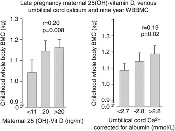
Javaid et al. Reference Javaid, Crozier and Harvey 37 utilized a population-based study of maternal nutrition and foetal growth to show that reduced maternal height, lower preconception maternal weight, reduced maternal fat stores during late pregnancy, a history of maternal smoking during pregnancy and lower maternal social class were all associated with reduced whole body BMC of the child at 9 years of age (Fig. 4). Lower ionized calcium concentration in umbilical venous serum also predicted reduced childhood bone mass (r = 0.19, P = 0.02); this association appeared to mediate the effects of maternal fat stores, smoking and socioeconomic status on the bone mass of the children at 9 years of age. Around 31% of the mothers had insufficient and 18% had deficient circulating concentrations of 25(OH)-vitamin D during late pregnancy (11–20 and <11 nmol/l, respectively). Reduced concentration of 25(OH)-vitamin D in mothers during late pregnancy was associated with reduced whole body (r = 0.21, P = 0.009) and lumbar spine (r = 0.17, P = 0.03) BMC in children at 9 years of age (Fig. 4). Maternal vitamin D status was also statistically significantly associated (P < 0.01) with childhood bone area and aBMD. The associations of maternal vitamin D status with childhood bone mass remained robust after adjusting for childhood calcium intake and physical activity, as well as for maternal smoking and body build. In bivariate regression models, part of the maternal vitamin D effect on childhood bone mass could be explained by umbilical venous calcium concentration. Adjunctive evidence supporting a role for maternal vitamin D status was obtained in the Southampton Women’s Survey, where maternal vitamin D concentrations again correlated with neonatal bone mass,Reference Harvey, Javaid and Poole 35 and where women who were pregnant over winter months had greater apparent decline in both calcaneal speed of sound and broadband ultrasound attenuation during their pregnancy.Reference Javaid, Crozier and Harvey 38 The findings suggest that vitamin D supplementation of pregnant women, especially during winter months, could lead to long-lasting reductions in the risk of osteoporotic fracture in the offspring.
Nine year whole body BMC by maternal 25(OH) vitamin D during late pregnancy and corrected cord-blood calcium. Data derived from Javaid et al.Reference Javaid, Crozier and Harvey 37

Mechanistic work using placental samples from the Southampton Women’s Survey has shown that mRNA expression of the key active plasma membrane calcium transporter (PMCA3) is significantly (P < 0.05) related to bone mass of the newborn offspringReference Martin, Harvey and Crozier 39 consistent with the hypothesis that maternal vitamin D insufficiency might alter placental calcium transport.
Childhood growth and hip fracture
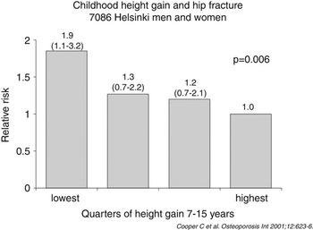
The clinically important consequence of reduced bone mass is fracture. Data are now available that directly link growth rates in childhood with the risk of later hip fracture.Reference Cooper, Eriksson and Forsén 40 Studies of a unique Finnish cohort in whom birth and childhood growth data were linked to later hospital discharge records for hip fracture have permitted follow-up of around 7,000 men and women who were born in Helsinki during 1924–1933. Body size at birth was recorded and an average of 10 measurements were obtained of height and weight throughout childhood. Hip fracture incidence was assessed in this cohort using the Finnish hospital discharge registration system. After adjustment for age and sex, there were two major determinants of hip fracture risk: tall maternal height (P < 0.001) and low rate of childhood growth (height, P = 0.006; weight, P = 0.01; Fig. 5), which was statistically independent of each other. More important, hip fracture risk was also elevated (P = 0.05) among babies born short. These data are compatible with endocrine programming influencing the risk of hip fracture. In addition, the observation that fracture subjects were shorter at birth, but of average height by 7 years of age, suggests that hip fracture risk might be particularly elevated among children in whom growth of the skeletal envelope is forced ahead of the capacity to mineralize, a phenomenon which is accelerated during pubertal growth.
Rate of childhood height gain between 7 and 15 years of age, and later risk of hip fracture among 3639 men and 3447 women born in the Helsinki University Central Hospital between 1924 and 1933.

Animal models
There is good evidence from animal models that undernutrition during pregnancy leads to an adverse outcome for the offspring. Recent work in rats has led to a deeper understanding of the possible underlying mechanisms, in addition to providing experimental evidence of programming. Ammann et al.Reference Ammann, Bourrin, Bonjour, Meyer and Rizzoli 41 investigated the effects of four isocaloric diets with varying levels of protein content on aBMD, bone ultimate strength, histomorphometry, biochemical markers of bone remodelling, plasma IGF-I and sex hormone status in adult female rats. After 16 weeks on a low protein diet, BMD was significantly decreased at skeletal sites containing trabecular or cortical bone. Mehta et al. Reference Mehta, Roach and Langley-Evans 42 also explored the effect of maternal undernutrition using a rat maternal protein deficiency model. They found that offspring of protein-restricted mothers had a reduction in bone area, but not BMD, compared to the offspring of mothers fed a normal diet during pregnancy. It was also found that the offspring of the protein-restricted mothers had abnormally widened the growth plates compared with the offspring of controls (P < 0.001). Thus, maternal undernourishment appears to modulate skeletal growth in the offspring by delaying maturation of the growth plate, with subsequent ‘catch-up’ growth.
Mechanisms of programming
There are three cellular mechanisms for the induction of programming. First, the nutrient environment may permanently alter gene expression; one example of this is permanent change in the activity of metabolic enzymes such as HMG-CoA reductase.Reference Brown, Rogers, Dunn, Gotto and Patsch 43 Second, early nutrition may permanently reduce cell numbers. The small, but normally proportioned, rat produced by undernutrition before weaning has been shown to have fewer cells in its organs and tissues.Reference McLeod, Goldrick and Whyte 44 Growth-retarded human babies have reduced numbers of cells in their pancreas, which may limit insulin secretion.Reference Snoeck, Remacle, Reusens and Hoet 45 Third, certain clones of cells may be altered by environmental adversity during development; for example, an altered balance of the TH-1 and TH-2 lymphocyte subtypes might predispose to atopic disease in later life.Reference Godfrey, Barker and Osmond 46 Evidence from the human studies outlined above suggests that skeletal development might be programmed as a consequence of the first two of these mechanisms.
Prematurity and the importance of body size
Follow-up studies in former premature infants have shown height growth below expected and low bone mass for age, although proportional to height or lean mass.Reference Weiler, Yuen and Seshia 47 , Reference Fewtrell, Prentice, Jones, Bishop and Stirling 48 By contrast, a recent study by Wang et al.Reference Wang, Vandermeulen and Atkinson 49 reported that many very low-birthweight infants do not attain optimal growth and body composition by 6–9 years, and speculated that the use of dexamethasone may be important in this group. It is important to note that in general the epidemiological observations made in Hertfordshire and many other studies relate to individuals born at, or close to, term. It is apparent that the study of pre-term infants will be important as the number surviving to adulthood increases; in this group, appropriate adjustment for skeletal size will be particularly important as BMC determined by DXA is a two-dimensional measurement reflecting bone area and not bone volume; this method may artificially overestimate relative BMC in individuals with larger bones and underestimate BMC in individuals with smaller bones – a correction for height may be necessary in a correct interpretation of the results. Studies of Hertfordshire individuals (born at term and whose birthweight fell within the normal range) have included volumetric assessment of bone density using peripheral quantitative computed tomography in an attempt to correct for body size.Reference Oliver, Jameson, Aihie Sayer, Cooper and Dennison 28
Conclusions
Over the last 20 years it has become increasingly apparent that for diseases such as osteoporosis, there are significant environmental factors, in addition to genetic inheritance, which lead to the expression of the disease. Although initial work in BathReference Cooper, Cawley and Bhalla 22 and Hertfordshire, UK,Reference Cooper, Fall and Egger 23 showed relationships between weight at 1 year and BMC, more recent studiesReference Dennison, Syddall, Aihie Sayer, Gilbody and Cooper 24 – Reference Jones, Riley and Dwyer 26 , Reference Gale, Martyn, Kellingray, Eastell and Cooper 50 have also found evidence of an association between birthweight and adult bone mass; these relationships have been shown by a variety of researchers in different populations. Evidence is accruing that the genome does not work in isolation, and that there is a critical interaction, early in life, between the genome and the environment. Undernutrition and other adverse influences, arising in foetal life or immediately after birth, may have an important effect on later risk of osteoporotic fracture; body weight in infancy is a determinant of adult bone mineral content, as well as of the basal levels of activity of the GH/IGF-1 and HPA axes, and recent work has suggested a central role for vitamin D. Epidemiological studies have shown that maternal smoking and nutrition during pregnancy influence intrauterine skeletal mineralization. Childhood growth rates have been directly linked to the risk of hip fracture many decades later, and now, evidence is emerging from experimental animal studies that support the observational data. Further study is needed to use this approach to develop novel therapeutic and preventative strategies to reduce the burden of osteoporotic fractures in the population.
Acknowledgements
We are grateful to the Medical Research Council; the Wellcome Trust, the Arthritis Research Campaign and the National Osteoporosis Society for support of our research programme into the foetal origins of osteoporotic fracture. Dr Zoe Cole is a receipt of an ARC Clinical Research Fellowship.
Statement of interest
None.