Introduction
An anterior skull base defect creates an opening between the nasal cavity and the subarachnoid space, which often results in cerebrospinal fluid (CSF) rhinorrhoea. This CSF rhinorrhoea may be primary (i.e. congenital or spontaneous) or secondary (i.e. iatrogenic or due to cranial injury).Reference Lescanne, Bakhos, Aesch, Celebi, Maheut-Lourmière and Cottier1 Rarely, CSF rhinorrhoea can lead to the diagnosis of an ethmoido-sphenoidal foreign body. An endoscopic approach is commonly used to remove these foreign bodies, but in some cases an external approach is needed.
We present a case of unilateral rhinorrhoea caused by a foreign body which had been lodged in the ethmoido-sphenoidal sinus for 38 years, and we discuss the radiological and surgical characteristics of this unusual case.
Case report
A 40-year-old woman presented with right-sided, clear rhinorrhoea, occipital headache and periorbital pain, which she had experienced for three weeks. She was referred to our ENT department.
According to her past medical history, at two years of age she had fallen, causing the impaction of a pen through the right medial canthus. An ophthalmologist had performed the extraction. She had subsequently suffered no specific symptoms, apart from occipital headache, until the current presentation.
On clinical examination, she had no fever. She had spontaneous, unilateral, clear rhinorrhoea without meningeal signs.
Nasal endoscopy failed to localise the origin of the CSF leakage.
However, a β2-transferrin detection test performed on a sample of the rhinorrhoea fluid was positive.
A pneumococcal vaccination was administered.
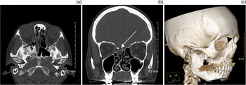
A computed tomography (CT) scan of the anterior skull base showed defects of the right lamina papyracea and the right posterior ethmoidal sinus roof, together with a foreign body in the right ethmoido-sphenoidal sinus (Figure 1).
(a) Axial computed tomography (CT) scan of the ethmoid region showing a foreign body in the right ethmoido-sphenoidal sinus. (b) Coronal CT scan of the ethmoid region showing a defect of the roof of the right ethmoidal sinus. (c) Three-dimensional CT scan reconstruction showing the impact of the original foreign body in the right medial canthus. R = right; A = anterior; F = feet

Magnetic resonance imaging (MRI) confirmed the defects and the presence of a foreign body in the right ethmoidal sinus, projecting into the subarachnoid space (Figure 2).
Coronal magnetic resonance imaging scan demonstrating the foreign body in the right ethmoidal sinus, without a meningo-encephalocoele.

Endoscopic surgery using an endonasal approach was performed to extract the foreign body and to reconstruct the anterior skull base defect. After resection of a meningocoele, a 5 × 5 mm osteo-meningeal defect was identified, and the foreign body was visualised. However, the foreign body could not be extracted during this endoscopic procedure, because the object had become calcified and incorporated into the ethmoido-sphenoidal sinus. Therefore, the defect was closed with a graft of inferior turbinate mucosa placed in underlay. The patient was discharged on post-operative day 4.
Fourteen days post-operatively, the patient re-presented to our department with recurrent CSF rhinorrhoea.
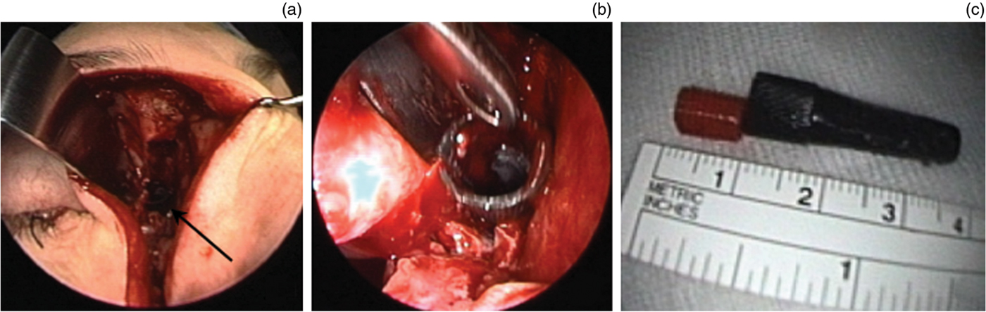
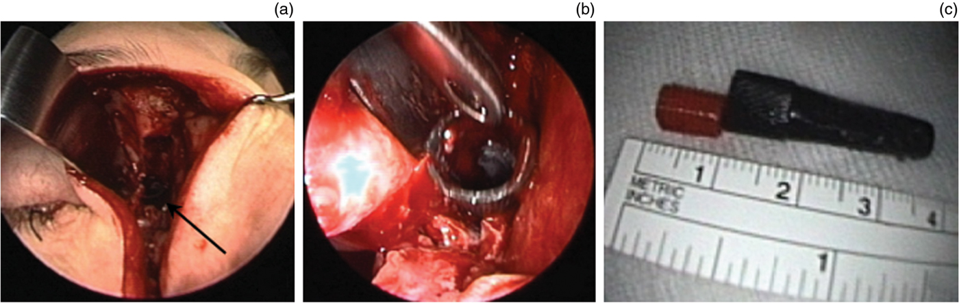
Surgery was performed two months later by both ENT surgeons and neurosurgeons, using a combined external and endoscopic approach. A paralateronasal incision enabled the completion of the ethmoidectomy and the location and extraction of the foreign body (Figures 3a and 3b), which was identified as a pen cap (Figure 3c). An overlay graft (3 × 1 cm) harvested from rectus muscle fascia was affixed with abdominal fat and Surgicel (Ethicon, Somerville, New Jersey, USA) to rebuild the roof of the ethmoid. An external lumbar drain was placed, and remained in situ for 5 days.
(a) Surgical photograph showing the paralateronasal incision, which permitted visualisation of the foreign body. (b) Endoscopic image showing extraction of the foreign body located in the ethmoido-sphenoidal sinus. (c) The extracted pen cap, approximately 4 cm in length.
The post-operative course was uneventful, allowing discharge on day 9 post-operatively. Silastic (Dow Corning, Midland, Michigan, USA) wicks were withdrawn on day 20 post-operatively. After two years of follow up, the patient had suffered no recurrence of CSF rhinorrhoea.
Discussion
Foreign bodies of the ethmoid and sphenoid sinuses are rarely reported. Various objects have been described, including firearm bullets,Reference Akhaddar, Abouchadi, Jidal, Elmostarchid, Naana and Rzin2 woodReference Kitajiri, Tabuchi and Hiraumi3 and pens.Reference Fajardo, Chavolla, Loza, Ysunza and Solorio4 Several authorsReference Fajardo, Chavolla, Loza, Ysunza and Solorio4–Reference Bhattacharayya and Wenokkur7 have reported violent trauma as the cause of a penetrating foreign body in the nasal cavity at the level of the orbit or the medial canthus, as in our case.
In cases of persistent intra-sphenoidal foreign body, recurrent sinusitis can appear several years after the causative trauma.Reference Fajardo, Chavolla, Loza, Ysunza and Solorio4, Reference Bhattacharayya and Wenokkur7 Foreign body impaction within the paranasal sinuses can lead to intracranial complicationsReference Kitajiri, Tabuchi and Hiraumi3 such as: cerebral, subdural or extradural haemorrhage; pneumocephalus; CSF rhinorrhoea; meningitis; and brain abscess.Reference Akhaddar, Abouchadi, Jidal, Elmostarchid, Naana and Rzin2 When such complications are present,Reference Akhaddar, Abouchadi, Jidal, Elmostarchid, Naana and Rzin2, Reference Kitajiri, Tabuchi and Hiraumi3, Reference Tsao, Kao, Wang, Chin and Moe8 the underlying condition must be managed immediately in order to prevent further developments.
In our case, the patient reported no specific symptoms for 38 years, except for occipital headache. The osteo-meningeal defect appeared to have been formed gradually by a process of chronic inflammation, with bone remodelling of the ethmoid roof adjacent to the foreign body. Furthermore, the foreign body did not extend into the nasal cavity because it was completely impacted within the ethmoido-sphenoidal sinus.
• This patient had cerebrospinal fluid rhinorrhoea due to a traumatic ethmoid sinus foreign body impaction 38 years prior
• Endoscopic, endonasal surgery could not remove the object
• Thus, a combined external–endonasal approach, by ENT and neurosurgery, was used
A CT scan with coronal and sagittal reconstructions is essential in order to diagnose osteo-meningeal defects and to identify foreign bodies and associated complications. A MRI scan enables identification of isodense objectsReference Akhaddar, Abouchadi, Jidal, Elmostarchid, Naana and Rzin2 and facilitates surgical management in the presence of a meningocoele or meningo-encephalocoele.
Extracting a foreign body embedded in a paranasal sinus presents a considerable challenge because of the proximity of vital structures (e.g. brain, orbit, ethmoidal arteries and internal carotid arteries).Reference Fajardo, Chavolla, Loza, Ysunza and Solorio4, Reference Bhattacharayya and Wenokkur7 Transnasal endoscopy is an essential step in identifying and confirming the location of the foreign body as well as the osteo-meningeal defect.Reference Bhattacharayya and Wenokkur7 However, this approach does not always enable visualisation of the object. In our case, the foreign body was completely impacted within the ethmoido-sphenoidal sinus. Therefore, a combined external and endoscopic approach, performed by ENT surgeons and neurosurgeons, was necessary to control the distal end of the pen cap.
Post-operative follow up with nasal endoscopy, CT and MRI is important to exclude recurrent CSF rhinorrhoea.
Conclusion
The occurrence of clear, unilateral rhinorrhoea should always prompt an examination to exclude an osteo-meningeal defect. The presented patient had suffered a traumatic external penetrating foreign body as a two-year-old child, which was brought to medical attention and diagnosed 38 years later as a result of an ethmoid skull base CSF leak. Computed tomography and MRI scanning can aid the diagnosis, localisation and extraction of such a foreign body. Despite its limitations, the endonasal approach remains the treatment of first choice for osteo-meningeal defects, because of its minimal invasiveness and high success rate. However, when this approach fails, a combined approach in collaboration with the neurosurgeons should be considered.