INTRODUCTION
Histomonas meleagridis is a flagellated protozoan parasite causing histomonosis in poultry, also known as blackhead disease (McDougald, 2003). Beside Histomonas meleagridis other flagellated and non-flagellated protozoa are reported to infect poultry, for example Tetratrichomonas gallinarum and Blastocystis spp.. Whereas infections with Histomonas meleagridis result in high losses, especially in turkey flocks, little is known about the prevalence and relevance of the other protozoa. Up to now it is not clear whether Tetratrichomonas gallinarum should be regarded as primary pathogen in poultry, even though it was already described nearly hundred years ago (Martin and Robertson, 1911). Some of this controversy can be attributed to the frequent occurrence of mixed infections with different protozoa, as already described by Allen (1936). Additionally, the presence of cryptic species within the Tetratrichomonas gallinarum contributes to these different observations (Cepicka et al. 2005). In comparison to the flagellated protozan parasites, Blastocystis spp. are thought to be non-pathogenic for birds with zoonotic potential due to their low host specificity (Stenzel and Boreham, 1996). Consequently, an animal model is required in order to investigate the clinical relevance of infections with Blastocystis in humans and animals (Tan, 2004).
Since the introduction of highly efficient pharmaceuticals in the poultry industry in the 1960s, research about flagellated protozoan parasites from poultry is very limited. In the EU the situation has changed recently with the ban of all chemicals used as feed additives in food-producing animals (EEC Regulation/1756/2002). As a consequence, complete loss of turkey flocks is reported (Hess et al. 2004). The lack of licensed drugs against flagellates is an increasing problem in all countries where poultry are kept. The actual situation underlines the need for basic research in this area and data about these microbes needs to be generated. Optimization of cultures is a general aim for parasitic protists reviewed by Clark and Diamond (2002). Therefore, the establishment of more defined cultures was our main target.
Clonal cultures would be the ideal tool to address general questions about the biology, e.g. pathogenicity, epidemiology and evolution of these parasites. The current investigation was performed in order to develop a protocol for the establishment of clonal cultures of Histomonas meleagridis, Tetratrichomonas gallinarum and a Blastocystis sp. obtained from turkeys. This investigation includes the separation technique followed by the proliferation in vitro to establish clonal cultures containing high numbers of protists.
MATERIALS AND METHODS
Cultures
Two different progenitor cultures (A and B) were used as a source to establish the clonal cultures. The first culture (A) originated from a 10-week-old dead turkey taken from an outbreak of histomonosis in a meat turkey flock consisting of 5000 birds of which more than 40% died. Approximately 1 g of caecal content together with some material scraped from the caecal wall were placed in 9 ml of Medium 199 supplied with Earle's salts, L-glutamine, 25 mM HEPES and L-amino acids (Gibco™, Invitrogen). In addition, 11 mg of rice starch (Sigma Aldrich), 15% FCS (Gibco™, Invitrogen), antibiotics (200 international units penicillin and 200 μg streptomycin per ml medium) and an antimycoticum (2·5 μg amphotericin B/ml medium) were added. This medium was kept as a standard in all further investigations. Passages were performed every second or third day by transferring 1 ml into a new sterile 50 ml tube (Sarstedt) containing 9 ml of the standard medium. At passage number (P) 117 the culture was taken for micromanipulation.
Progenitor culture B was established in the same way as culture A, from a bronze turkey kept in a backyard flock. Of the 6 turkeys, 2 died during the first 3 months of rearing and another died at about 20 weeks of age. From this bird faeces and caecal material was taken and processed as described for culture A. This culture was taken for micromanipulation at P7.
Feeder medium for propagation of cloned protozoa
Prior to micromanipulation the bacterial flora of each culture was determined in order to establish a suitable ‘feeder’ substrate for propagating the extracted protozoa. Some of the material of the non-axenic cultures was put on commercial Columbia (+5% blood), McConkey, Salmonella Detection and Identification Media (SMID) and Sabouraud agar plates (all: bioMerieux, Marcy l'Etoile, France). Identification of bacteria was done using the API-20 E Microsystem (bio Merieux, Marcy l'Etoile, France).
For cloning of parasites 300 μl of the culture medium (described above) were transferred into an Eppendorf tube, together with 1 mg of rice starch, and 1 loop of the bacterial culture taken from the Columbia agar plate.
Micromanipulation
Glass micropipettes were prepared using a borosilicate capillary (1·0 mm outer diameter, Hilgenberg, Germany) pulled on a pipette puller (P97, Sutter, USA) and cut to an outer diameter of 25 μm (microforge deFonbrune, Bachofer, Germany). The pipette was statically fixed at the microscope and suction was achieved by connecting the glass pipette through a flexible tube with an empty syringe. For separation, progenitor cultures were diluted 1[ratio ]100 to achieve a clear single cell suspension of protozoa. Individual parasites were selected randomly out of 100 μl culture drops on a cover-glass. Parasites were released from the pipette each into a single drop of culture medium on the cover-glass and transferred with the medium into an Eppendorf tube. The whole procedure was carried out using Narishige micromanipulators (Narishige, Japan) and an inverted microscope (Diaphot 300, Nikon, Austria) under 400-fold magnification. Additional monitoring was conducted with a CCD camera and a monitor (both Sony, Japan) connected to the microscope to ensure that only single cells were transferred. After the isolation procedure the protozoa were incubated at 40 °C up to 4 days in individual Eppendorf tubes. Growth of protozoa was monitored by light microscopy on days 2, 3 and 4 using light microscopy. Any positive clone was transferred into 9·7 ml of the standard medium in a 50 ml plastic tube (Sarstedt) and closed tightly whereas all negative tubes were destroyed.
Propagation of the cloned parasites
Following the separation procedure all parasites were maintained and subcultivated in the same way. At 3–4 days after incubation 1 ml of the respective clone material was transferred into 9 ml of fresh culture medium, kept once again in 50 ml plastic tubes and passaged as described for the progenitor cultures. At different passage levels cultures were incubated for up to 4 days in order to monitor the growth behaviour. Numbers of parasites were calculated using a Neubauer cell counting chamber. Live and dead cells were differentiated by vital colouration with Trypan blue (0·4%) recording only the live cells.
PCR and nucleotide sequencing
The presence of different parasites in the progenitor cultures and the clonality of cloned parasites were demonstrated by PCR and nucleic acid sequencing. The whole process of DNA extraction, primer development and cycle conditions followed the procedures described earlier (Grabensteiner and Hess, 2005). Briefly, DNA was isolated from 1 ml of the progenitor cultures or an early passage of each cloned parasite. To demonstrate clonality the cultures were retested again at later passages. Using DNA extracted from the same number of parasites (10000) of Histomonas meleagridis (P55), Tetratrichomonas gallinarum (P75) and Blastocystis sp. (P71) cross-over reactions were performed by species-specific PCRs.
For construction of non-specific primers nucleotide sequences of the small subunit ribosomal RNA genes of several parabasalids were aligned using a standard software program (DNASTAR, Inc. WI, USA). Based on this alignment a forward (5′-aggaagcacactatggtcatag-3′) and a reverse (5′- cgt-taccttgttacgacttctcctt-3′) primer were developed suitable to hybridize to the relevant gene of H. meleagridis and T. gallinarum. The obtained fragments were cloned with the TOPO TA Cloning® Kit for Sequencing and one Shot® TOP10 chemically competent Escherichia coli (Invitrogen) cells, according to the manufacturer's instructions. For the Blastocystis clone a separate specific primer pair was used (Grabensteiner and Hess, 2005). Nucleotide sequences were determined using an ABI 373 automatic sequencing apparatus as described by the manufacturer. The determined nucleotide sequences were submitted to the EMBL database and the Accession numbers for the respective sequence of H. meleadgridis (AJ920323), T. gallinarum (AJ920324) and Blastocystis sp. (AJ920322) were assigned.
The obtained nucleotide sequences of the small subunit rRNA gene sequences were aligned with the respective nucleotide sequences available in the data bank, namely AF293056 (H. meleagridis) and AF124608 (T. gallinarum) (Delgado-Viscogliosi et al. 2000; Gerbod et al. 2001). Nucleotide sequences of the following Blastocystis spp., all of them isolated from chickens or turkeys, were used for homology studies: (AB070993/94; AB091240-42; AY135409/10; AY135411) (Arisue et al. 2003; Noel et al. 2003). Homology studies were performed using the software program mentioned above.
RESULTS
Characterization of progenitor cultures and micromanipulation
Various protozoan parasites were identified in progenitor cultures A and B (data not shown). The bacteriological investigation of culture A revealed a multi-resistant Escherichia coli strain with susceptibility against colistin and trimethoprim/sulfamethoxazole. This antibiotic was not supplied in the culture medium. In addition, some fungi were isolated on the Sabouraud agar plates. An Escherichia coli isolate was also obtained from culture B showing resistance against colistin and neomycin.
Following micromanipulation multiplication of T. gallinarum and the Blastocystis sp. was observed after 3 days. For positive growth of H. meleagridis the initial culture had to be incubated for 4 days after separation.
Morphological characterization of cloned protozoa
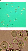
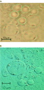
In a first step, cultures were first characterized by light microscopy to identify the relevant microorganisms. In the progenitor culture 3 different organisms, namely H. meleagridis, T. gallinarum and Blastocysts were identified. In the cloned cultures most of the H. meleagridis parasites had rice starch incorporated and size variations of the parasites could be noticed (Fig. 1). For Trichomonads different forms were noticed in the culture with the majority of parasites resembling the trophozoite form with the anterior flagella, the typical undulating membrane and a clear forward movement (Fig. 2A). However, some of the cells displayed a round form similar to what was described as pseudocysts. After adding a drop of Lugol's solution the development of such cysts was induced for a single parasite and documented in a continuing series of pictures (Fig. 2B–D). Those pseudocysts were rotating around themselves in a slow forward movement. Various forms were observed for the Blastocystis sp., characterized by high transparency of the protozoa and dissemination of the nuclei at the rim of the cytoplasm (Fig. 3A,B).

Fig. 1. Light micrographs of the cloned Histomonas meleagrids culture using lower (A) and higher (B) magnification. Rice starch in the medium, incorporated by the majority of parasites, is clearly visible.

Fig. 2. Trophozoite of Tetratrichomonas gallinarum displayed in brightfield (A). Pseudocyst formation of an individual parasite after adding a drop of Lugol's solution to the microscope slide (B–D). The rounding and alteration of the cell in the series of pictures (B–D) is clearly visible.

Fig. 3. Microscopic picture of Blastocystis sp. using normal light (A) and phase-contrast (B) microscopy. Several nuclei are present at the surface coats of the cells.
Growth behaviour of cloned parasites
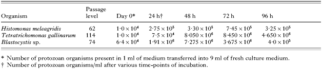
From each species of parasite a single culture was chosen for investigation of the growth behaviour. Viability and propagation of the cloned protozoa was assessed by counting the number of cells present over 4 continuous days and the result is given in Table 1. Peak levels for H. meleagridis and T. gallinarum were reached at 72h, with a decline at the 4th day of incubation. The highest numbers of T. gallinarum were 8·450×106/ml viable cells, whereas 7·45×103/ml viable Histomonads were counted at day 3 of incubation. For Blastocystis sp. the highest number of parasites was noticed already 48h after incubation with 7·275×106/ml viable cells.

PCR and nucleotide sequencing
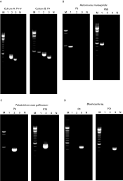
The clonality of the cultures was demonstrated by species-specific PCRs as shown in Fig. 4. Both of the progenitor cultures contained several protozoa (Fig. 4A). Whereas T. gallinarum together with Blastocystis sp. were identified in culture A, culture B contained also H. meleagridis. All of the clonal cultures contained only a single protozoon species independent of the passage number (Fig. 4B–D).

Fig. 4. PCR experiments to demonstrate the presence of protozoon species in progenitor (A) and clonal cultures (B–D). Specific primers for Histomonas meleagridis (lane 1), Tetratrichomonas gallinarum (lane 2) and Blastocystis spp. (lane 3) were used to demonstrate the presence of only a single parasite. M: molecular size marker (100-bp ladder); N: negative PCR control without DNA template.
Using non-specific oligonucleotides parts of the small subunit ribosomal RNA genes were amplified from all clones. No differences were noticed between the nucleotide sequences of the 3 Tetratrichomonas clones, isolated from culture A. Sequence identities between 97 and 97·9% could be determined with the respective nucleotide sequences of the small subunit RNA genes of T. gallinarum (AF124608), Blastocystis sp. (AB1079731) and H. meleagridis (AJ920323), available in the data bank.
DISCUSSION
So far, only limited data are available about some of the most important protozoan parasites in poultry, namely Histomonas meleagridis, Tetratrichomonas gallinarum and Blastocystis spp. To gain more basic knowledge optimization of the culture systems and in vitro propagation is elementary, in order (i) to investigate the pathogenicity in various hosts; (ii) to establish and evaluate more defined diagnostic tools and (iii) to investigate general principles of morphology, genetic variation and evolution of a specific parasite. To fulfil all the aforementioned needs a clonal culture is desirable.
Different approaches are described to develop clonal cultures of protozoan parasites, with the limited dilution method as the least precise one. Entamoeba species were cloned using their ability to form different colonies in soft agar (Gillin and Diamond, 1978). The same approach was reported to clone Blastocystis hominis (Tan et al. 1996). However, this method depends mainly on the growth behaviour of the respective micro-organism on agar plates and there is only limited information available about the growth behaviour of H. meleagridis on this kind of substrate (Bayon and Bishop, 1937). In addition, this method may neglect those strains within a species which lack the ability to form colonies on soft agar. This disadvantage can be prevented by using the micromanipulation approach, which was used to establish clonal cultures of Entamoeba histolytica (Farri, 1978). Oduola et al. (1988) used this method to investigate the heterogeneity of Plasmodium falciparum isolates. Later on, Bushek et al. (2000) described the cloning of the oyster pathogen Perkinsus marinus through micromanipulation. In poultry science the approach of using a deFonbrune micropipette was already described some time ago to obtain single sporozoites of Eimeria tenella (Shirley and Millard, 1976). Micromanipulation offers the advantage that individual cells can be selected, as the whole procedure is controlled visually.
Several difficulties had to be overcome in the current experiments. H. meleagridis itself has a very low resistance outside the host and cysts as protective forms are not reported (Tyzzer, 1919). As a consequence, survival outside the host is very limited and micromanipulation has to occur in a short time-frame to sustain viability and to prevent the organism from drying out. After selecting individual cells a suitable ‘feeder’ substrate has to be used in order to create optimal growth conditions for the micro-organism. In the present investigation only mixed cultures of various protozoan parasites were available as demonstrated for the progenitor cultures, a situation reflecting the variety of protists to be found in the chicken intestine. The presence of different flagellates isolated in vitro from outbreaks of Histomonosis was already described in the early report presented by Tyzzer (1920), in which the aetiology of the disease was described. This emphasizes the necessity to establish clonal cultures for principle biological investigations.
In the present study the priority was mainly set towards the establishment of clonal cultures of H. meleagridis. So far two different liquid media were reported facilitating the growth of H. meleagridis (Dwyer, 1970; Stepkowski and Klimont, 1979). In principal, the protocol developed by Stepkowski and Klimont (1979) was followed, introducing some minor modifications. Firstly, antibiotics and an antimycoticum were added to suppress the bacterial and fungal growth in the cultures. Secondly, the chicken embryo extract described by Stepkowski and Klimont (1979) was found to be dispensable. Anyhow, further optimization of the culture conditions is needed, as T. gallinarum and Blastocystis spp. should be able to multiply under axenic culture conditions (Zierdt and Williams, 1974; Clark and Diamond, 2002). This scenario is probably rather unrealistic for H. meleagridis, keeping in mind that the phylogenetic closely related protozoa Dientamoeba fragilis can only be maintained in xenic cultures (Gerbod et al. 2001; Clark and Diamond, 2002).
The addition of antibiotics and an antimycoticum in the culture medium had an adverse effect on the Blastocystis sp.. Amphotericin B, beside other factors, is known to induce the production of the granular form of Blastocystis hominis (Stenzel and Boreham, 1996). This form was predominantly noticed in the present cultures prior to and after micromanipulation. Trichomonads are protists described as rapidly moving flagellates with 4 anterior flagella and a recurrent flagellum (Honigberg and Brugerolle, 1990). However, in the present investigation flagellated round forms were noticed as well. Such forms are known as pseudocysts and they are described in various Trichomonads, including Tetratrichomonas gallinarum (Friedhoff et al. 1991). In the present investigation the formation of pseudocysts in T. gallinarum under visual control was demonstrated for the first time. Granger et al. (2000) described that cooling down of cultures may provoke the reversible process of pseudocyst formation in Tritrichomonas foetus, a situation obviously present during the micromanipulation approach. In the present investigation a drop of Lugol's solution was used as a fixative and, accidentally, pseudocysts were induced. The close resemblance of these pseudocysts to Blastocysts and Histomonads may complicate the microscopic diagnosis of protozoan parasites in faeces of diseased animals.
Nucleotide sequences revealed for the cloned parasites showed a close phylogentic relationship with the relevant sequences in the data bank. The nucleotide sequence of the small subunit RNA gene from the protozoan T. gallinarum which was isolated from turkey faeces showed very good homology with the same gene obtained from a duck isolate (Delgado-Viscogliosi et al. 2000). A similar observation was made comparing the genes of H. meleagridis cloned from turkeys in this investigation and the only published sequence determined from a chicken isolate (Gerbod et al. 2001). Final characterization was done by species-specific PCRs that confirmed the clonality of the established cultures over a series of passages.
In conclusion, isolation of H. meleagridis is a laborious process and no protocol has been reported so far to establish clonal cultures that can be traced back to a single organism, as reported in the present investigation. In addition, clonal cultures of T. gallinarum and a Blastocystis sp. were established successfully, providing some of the most important protists known to infect poultry. By having clonal cultures available, different questions with regard to genetics, morphology and pathogenicity of those poultry protozoan parasites can be addressed much more accurately.
The authors wish to thank the Austrian Ministry for Agriculture, Forestry, Environment and Water Management and the Austrian Ministry for Health and Women's Issues for financial support to perform the present investigations.