INTRODUCTION
Although anomuran crustaceans (Chirostylidae: Galatheidae: Lithodidae) are well represented in hydrothermal vent communities (Martin & Haney, Reference Martin and Haney2005), records of hermit crabs (Paguroidea) are very scarce (Chevaldonné & Olu, Reference Chevaldonné and Olu1996; Lemaitre, Reference Lemaitre2004). Only the parapagurid, Paragiopagurus ventilatus Lemaitre, Reference Lemaitre2004, is known from hydrothermal environments (off north-eastern Taiwan), although it remains to be determined whether the species is an obligate associate of hydrothermal vent ecosystems.
During the NT09-05 cruise by RV ‘Natsushima’ of the Japan Agency for Marine–Earth Science and Technology (JAMSTEC) (a cruise report is available at http://docsrv.godac.jp/MSV2_DATA/23/NT09-05_leg2_all.pdf), samples were collected by the remote operating vehicle (ROV) ‘Hyper-dolphin’ at hydrothermal vent fields on the Nikko Seamount, Mariana Trough in the north-western Pacific. Among the collected materials are some specimens of a parapagurid hermit crab utilizing empty tubes of the siboglinid tubeworm Lamellibrachia satsuma Miura, Tsukahara & Hashimoto, 1997 for housing, which represent the first example of siboglinid tubes as carcinoecia by hermit crab. Comparison of the specimens with those from Taiwan based on morphology and partial sequence of COI gene revealed that specimens from the Nikko Seamount belonged to Paragiopagurus ventilatus, which significantly extended the geographical range to the east. Our observations support Lemaitre's (2004) conclusion that P. ventilatus is associated with hydrothermal environments.
MATERIALS AND METHODS
Specimens of Paragiopagurus ventilatus were collected on a bush of Lamellibrachia satsuma using a slurp gun or manipulator equipped on ROV ‘Hyper-dolphin’. Seven specimens of the hermit crab were kept alive in an aquarium (0.251 m3, water temperature 13–15°C) at the Enoshima Aquarium. The substratum of the aquarium was fine sand, on which several empty tubes of L. satsuma were laid. One specimen was dead after two days of arrival at this aquarium. Four specimens were preserved 30 to 150 days after the arrival, three of which were sent to the first author for identification.
The specimens examined are deposited in the Natural History Museum and Institute, Chiba (CBM). The measurement provided is shield length (SL), measured in millimetres from the tip of the rostral lobe to the midpoint of the posterior margin of the shield. The terminology follows McLaughlin (Reference McLaughlin2003) for the most part, with exception of numbered thoracic sternites and dactylus (dactyli) for dactyl (dactyls). An abbreviated description is given to show diagnostically important features and variation not mentioned by Lemaitre (Reference Lemaitre2004).
SYSTEMATICS
Paragiopagurus ventilatus Lemaitre, Reference Lemaitre2004: 326, figures 1–5; Lemaitre, Reference Lemaitre, Desbruyères, Segonzac and Bright2006: 435, 2 unnumbered figures; McLaughlin et al., Reference McLaughlin, Rahayu, Komai and Chan2007: 320, 321, unnumbered figures.
Paragiopagurus ventilatus Lemaitre, Reference Lemaitre2004, male (SL 3.3 mm), Nikko Seamount, CBM-ZC 9743. (A) Shield and cephalic appendages, dorsal view; (B) ocular acicles, dorsal view; (C) sixth thoracic sternite, ventral view; (D) posterior part of sixth pleonal tergite, telson and uropods, dorsal view; (E) telson, dorsal view. Scale bars: 1 mm for A, D & E; 0.5 mm for B & C.

Paragiopagurus ventilatus Lemaitre, Reference Lemaitre2004, male (SL 3.3 mm), Nikko Seamount, CBM-ZC 9743. (A) Chela and carpus of right cheliped, dorsal view (setae omitted); (B) right cheliped, mesial view (bacteriophore setae simplified); (C) chela of right cheliped, lateral view (setae omitted); (D) chela and carpus of left cheliped, dorsal view (setae omitted); (E) left cheliped, mesial view (bacteriophore setae simplified). Scale bar: 1 mm.

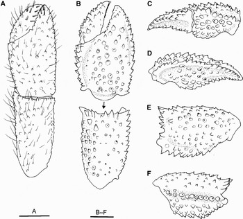
Paragiopagurus ventilatus Lemaitre, Reference Lemaitre2004. (A) Female (SL 2.1 mm), Nikko Seamount, CBM-ZC 9743; (B–F) badly damaged specimen of unknown sex (could not be measured); (A & B) chela and carpus of right cheliped, dorsal view; (C) chela of right cheliped, mesial view; (D) same, lateral view; (E) carpus of right cheliped, lateral view; (F) same, mesial view. Scale bars: 1 mm.

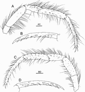
Paragiopagurus ventilatus Lemaitre, Reference Lemaitre2004, male (SL 3.3 mm), Nikko Seamount, CBM-ZC 9743. (A) Right second pereopod, lateral view (bacteriophore setae simplified); (B) same, dactylus, mesial view (only mesial setae shown); (C) left third pereopod, lateral view (bacteriophore setae simplified); (D) same, dactylus, mesial view. Scale bars: 1 mm.

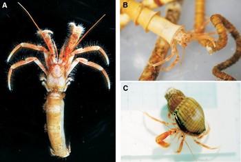
Paragiopagurus ventilatus Lemaitre, Reference Lemaitre2004. (A) Male (SL 3.3 mm), CBM-ZC 9743, entire animal in dorsal view; (B) specimen not preserved, just after capture, in dwelling tube of Lamellibrachia satsuma; (C) male (SL 3.3 mm), CBM-ZC 9743, kept in aquarium after changing housing to a gastropod shell.

MATERIAL EXAMINED
RV ‘Natsushima’, NT09-05 cruise, ROV ‘Hyper-dolphin’ dive No. 968, Nikko Seamount, Mariana Trough, 23°04.890′N 142°19.497′E, 406–472 m, hydrothermal vent field, bush of Lamellibrachia satsuma, 15 April 2009, 2 males (SL 3.3, 3.3 mm), 1 female (SL 2.1 mm), fragments from more than one specimen, CBM-ZC 9743.
Gueishandao, Yilan County, Taiwan, Station KS 13, 24°51.231′N 121°59.204′E to 24°49.928′N 122°00.671′E, 252–275 m, 4 September 2008, 3 males (SL 5.3–6.2 mm), 3 ovigerous females (SL 4.0–4.6 mm), CBM-ZC 9744.
DESCRIPTION OF SPECIMENS FROM NIKKO SEAMOUNT
Eleven pairs of biserial gills. Shield (Figure 1A) almost as long as broad; rostrum broadly rounded; anterolateral margin between rostrum and lateral projections slightly concave; lateral projections obtusely triangular, with or without terminal spine.
Ocular peduncles (Figure 1A) 0.7–0.8 times as long as shield; corneas very slightly dilated. Ocular acicles (Figure 1B) triangular, terminating in a strong, simple spine with transverse striae on the dorsal surface. Antennular peduncles (Figure 1A) overreaching the distal corneal margins by nearly entire lengths of ultimate segments. Antennal peduncles (Figure 1A) reaching nearly to distal corneal margins; third segment with strong ventromesial distal spine; second segment with dorsolateral distal angle terminating in strong, simple spine; antennal acicle nearly straight, slightly falling short of distal corneal margin, mesial margin with row of 3 or 4 spines.
Epistome has a very small median spine.
Third maxilliped having numerous bacteriophore setae (see Lemaitre, Reference Lemaitre2004) on the ventral margins of ischium and merus in male specimens, but such setae absent in small female.
Right chela of male specimens (Figure 2A–C) 2.0–2.5 times longer than wide, weakly flattened dorsoventrally; dorsal and ventral surfaces with scattered numerous, small blunt tubercles and tufts of stiff setae; fingers slightly curved ventrally. Dactylus shorter than palm, set at a weakly oblique angle to longitudinal axis of palm; palm longer than broad; dorsal surface slightly convex, dorsolateral margin weakly delimited, dorsomesial margin not delimited. Carpus 2.0–2.2 times longer than wide, with scattered small, blunt or subacute tubercles on surfaces, setation similar to that of palm; ventrolateral margin not delimited; ventromesial distal margin with row of small tubercles. Merus also having scattered small tubercles and setae; ventromesial margin with row of small spines; mesial and ventral surfaces with dense bacteriophore setae. Ischium having dense bacteriophore setae on mesial surface.
Right chela of female specimen (Figure 3A) about 1.9 times longer than wide; dorsal and ventral surfaces with scattered numerous blunt or subacute tubercles (size of these tubercles relatively larger than in male specimen) and short stiff setae. Dactylus shorter than palm, dorsomesial margin delimited by row of small, corneous-tipped spines. Palm longer than broad; dorsal surface slightly convex, dorsolateral margin delimited by row of small corneous-tipped spines, dorsomesial margin not delimited. Carpus about 2.2 times longer than wide, with scattered blunt or subacute tubercles on surfaces, setation similar to that of palm; dorsodistal margin with row of moderately small spines or tubercles; ventrolateral margin not delimited; ventromesial distal margin with row of small tubercles. Merus also has scattered small tubercles and setae; ventromesial margin with row of small spines; mesial and ventral surfaces without bacteriophore setae. Ischium without setae on mesial surface.
Right cheliped of unknown occupant clearly different from those of other two specimens (Figure 3B–F). Chela strongly flattened dorsoventrally, 1.7 times longer than broad, with sparse setae. Dactylus slightly longer than palm; dorsal surface with irregular longitudinal row of acute, corneous-tipped spines; mesial margin delimited by double row of sharp spines, ventromesial face concave; ventral surface also with row of sharp spines on midline. Palm with scattered small to moderately large, corneous-tipped spines, dorsolateral and dorsomesial margins slightly upturned, both delimited by row of sharp spines; lateral, mesial and ventral faces with covering of spines or blunt tubercles. Carpus 1.6 times longer than broad; surfaces covered with small spines or spinulose tubercles, dorsodistal margin with row of large, obliquely erect spines, lateral margin delimited by row of prominent spines; ventromesial margin also delimited by row of prominent spines. Merus missing.
Left cheliped (Figure 2D, E) slender. Dactylus longer than palm, slightly curved ventrally; surface unarmed, but with tufts of setae. Palm with some small, rounded tubercles on dorsal surface; dorsolateral and dorsomesial margins not delimited; surfaces with tufts of setae. Carpus 2.9 times longer than broad; dorsal surface with irregular rows of small tubercles, dorsolateral face oblique; lateral, mesial and ventral surfaces with tufts of setae. Merus with numerous setae on dorsal surface; ventrolateral and ventromesial margins without conspicuous spines. Bacteriophore setae present on merus and ischium in two male specimens, while absent in female specimen.
Ambulatory legs (Figure 4A, C) generally similar. Dactyli about 1.4 times as long as propodi, weakly curved ventrally, terminating in sharp, strong corneous claws; dorsal margins with tufts of stiff setae; ventromesial faces with irregular rows of 12–20 corneous spines (more numerous on third pereopods) (Figure 4B, D). Propodi, carpi and meri having tufts of stiff setae on all surfaces; carpi each with small dorsolateral spines (spine on third sometimes missing). Meri with ventrodistal row of low tubercles (second) or unarmed (third); ventral margins with long bacteriophore setae proximally, more dense on second than on third.
Fourth pereopod is subchelate; propodal rasp consisting of single row of rounded scales.
Anterior lobe of sixth thoracic sternite (third pereopods) (Figure 1C) is subtriangular, with prominent terminal bifid spine.
Pleon is nearly straight (Figure 5A). Male lacking paired first pleopods; with unpaired left second pleopod rudimentary, showing papilla-like projection. Female has rudimentary right second pleopod.
Uropods and telson markedly asymmetrical (Figure 1D). Telson (Figure 1E) with obsolete lateral indentations; posterior lobe separated by broad, very shallow cleft, each terminal to posterolateral margin bearing 4–6 corneous spines, some often ventrally curved.
VARIATIONS
Lemaitre (Reference Lemaitre2004) reported a remarkable degree of morphological variation in the right cheliped related to sex and size. The present small female specimen (SL 2.1 mm) has a relatively slender right cheliped, which is rather similar to that of males (SL 3.3, 3.3 mm); no bacteriophore setae are seen in this female specimen. The right chela and carpus of a badly damaged specimen (size was unavailable) is substantially different from those of other specimens examined in having strong armature and fewer setae on the surfaces. Judging from the size of this right cheliped, the damaged specimen should be smaller than other three intact specimens. This suggests that the armature is reduced but the setation becomes denser with increase in size in the right cheliped.
COLOUR IN LIFE
Shield brown generally, central part lighter. Ocular peduncles pale brown or cream, without conspicuous markings; corneas pigmented. Antennular peduncles also generally brown, first segment pale. Antennal peduncles and flagella are also brown, proximal part of second segment of peduncle paler. Chelipeds and ambulatory legs generally brown; fingers of chelipeds, dactyli and propodi of ambulatory legs paler distally; propodi of ambulatory legs with obscure whitish line along midline on lateral faces. See Figure 5A.
ECOLOGY
At the time of capture, the animals inhabited tubes of the siboglinid Lamellibrachia satsuma (Figure 5B), a tubeworm species that is abundant in the collection site. However, specimens kept alive in the aquarium were found to change their houses to experimentally supplied empty gastropod shells (Oenopota ogasawarana Okutani, Fujikura & Sasaki, 1993) (Figure 5C), despite the presence of empty tubes. Three specimens (at least one of these three specimens is preserved (male, SL 3.3 mm)) changed their housing from tubes to gastropod shells in two days (two individuals) and 23 days (one individual) after shells were supplied. Three smaller specimens (not precisely measured) did not change their housing. Perhaps, the gastropod shells were too big for the small individuals.
The animals in the aquarium were active through day and night, walking on the bottom and sometimes climbing up the walls of the aquarium. They were fed small pieces of tuna and euphausiids, but were also found to eat the bacterial mat. One individual survived 280 days after capture.
DISTRIBUTION AND HABITAT
Heretofore, Paragiopagurus ventilatus was known only from hydrothermally influenced fields on the Dasi Fishing Ground, near Gueishandao, north-eastern Taiwan, at depths of 128–350 m (Lemaitre, Reference Lemaitre2004; McLaughlin et al., Reference McLaughlin, Rahayu, Komai and Chan2007). This report extends the geographical range of this species to the Mariana Trough and the bathymetric range to 410 m. It confirms that P. ventilatus is closely associated with the hydrothermal vent habitat although it still remains unclear whether the association is obligate.
REMARKS
The three well-preserved specimens from Nikko Seamount (SL 2.1–3.3 mm) are smaller than the previously known specimens (SL 3.8–5.9 mm; Lemaitre, Reference Lemaitre2004) and the comparative specimens from Taiwan examined here (SL 4.0–6.2 mm), although they have differentiated gonopores; other specimens were badly damaged and fragmented. They agree with the previous descriptions and the comparative specimens from Taiwan in most diagnostic respects. These diagnostic features include: (i) the presence of striae on the dorsal surfaces of the ocular acicles; (ii) the irregular rows of long corneous spines on the mesial faces of the ambulatory dactyli; and (iii) the bifid spine on the anterior lobe of the sixth thoracic sternite. Lemaitre (Reference Lemaitre2004) noted that the presence of bacteriophore setae on the third maxillipeds, chelipeds and second and third pereopods was diagnostic for P. ventilatus. However, in the present female specimen, no bacteriophore setae are differentiated, although the male specimen has well developed bacteriophore setae on these appendages. The female specimen is small (2.1 mm in the shield length), and thus the bacteriophore setae may develop with growth. Similar size-related variation of the development of the bacteriophore setae is also seen in the alvinocaridid Rimicaris Williams 7 Rona, 1986 (see Komai & Segonzac, Reference Komai and Segonzac2008). Comparison of partial COI sequences (658 bp) between one of the two male specimens from Nikko Seamount and specimens from Taiwan well supports the conspecificity of the two populations. Nucleotide divergence is only 0.3% between them (T.-Y. Chan, personal communication).
Paragiopagurus ventilatus was originally reported to use buccinid shells (Siphonalia sp.) for housing (Lemaitre, Reference Lemaitre2004), and the comparative specimens from Taiwan examined in this study were also found to use buccinid shells. However, the animals from the Nikko Seamount inhabited tubes of Lamellibrachia satsuma at the time of capture. This is the first example of the usage of siboglinid tubes by a hermit crab, although the use of non-coiled, cylindrical cavities in various materials by hermit crabs is not uncommon (e.g. McLaughlin, Reference McLaughlin1974, Reference McLaughlin2007; Miyake, Reference Miyake1978; Forest, Reference Forest1987; McLaughlin & Gunn, Reference McLaughlin and Gunn1992; McLaughlin & Lemaitre, Reference McLaughlin and Lemaitre1993; McLaughlin & Konishi, Reference McLaughlin and Konishi1994; Komai, Reference Komai1995, Reference Komai2003; Lemaitre, Reference Lemaitre1995; Osawa, Reference Osawa1995; Komai & Takeda, Reference Komai and Takeda1996; Zhadan, Reference Zhadan, Schram and von Vaupel Klein1999; Osawa & Chan, Reference Osawa and Chan2008). Differences between the tube-dwelling individuals and the gastropod-dwelling individuals are seen only in the shape of the pleon. In the former, the pleon is nearly straight and weakly flexed, but in the latter, it is dextrally twisted. It is possible that a shortage of gastropod shells and an excess of worm tubes led the hermit crabs to adopt this alternative habitat. In situ observation and collections suggest that gastropod shells are scarce in the collection site. An experimental supply of dextrally twisted gastropod shells (Oenopota ogasawarana) led some individuals to change their tubes to gastropod shells. The morphology and behaviour strongly suggest that the hermit crabs use worm tubes only because these are more readily available. Similar broad usage of either dextrally twisted gastropod shells or straight materials (e.g. tusk-shells, polychaete tubes, bryozoan tubes, etc.) is known for some other hermit crab species, e.g. Paguristes seminudus Stimpson, 1858 (Diogenidae; Komai, unpublished data), species of Parapagurus Smith, 1879, Sympagurus Smith, 1883 and Oncopagurus Lemaitre, 1996 (Parapaguridae; Zhadan, Reference Zhadan, Schram and von Vaupel Klein1999), and species of Lophopagurus McLaughlin, 1981 of the Paguridae (McLaughlin & Gunn, Reference McLaughlin and Gunn1992; Komai, Reference Komai1999). There is little doubt that the shape of the abdomen is under the influence of habitat in these tolerant species.
ACKNOWLEDGEMENTS
We deeply thank Professor Tin-Yam Chan and Ms Chien-Hui Yang of the Institute of Marine Biology, National Taiwan Ocean University, for providing us with information on the COI sequence comparison, and for Drs Patsy A. McLaughlin (Shannon Point Marine Center, Western Washington University) and Masayuki Osawa (Department of Marine and Environmental Sciences, University of the Ryukyus) and two referees for critically reading the manuscript and for offering very helpful comments and suggestions for improvements. Comparative specimens of Paragiopagurus ventilatus from Taiwan were kindly made available by Professor Chan. We also thank the scientists, crews and staff of RV ‘Natsushima’ and ROV ‘Hyper-dolphin’, for their help during the cruise NT09-05.