Hydrops is defined as the accumulation of extracellular fluid in tissues and serous cavities. It is characterised by the presence of at least two of the following items: subcutaneous oedema, pleural effusion, pericardial effusion, ascites, and hydramnios.Reference Cardoso1 There are two types of hydrops: immune (10%) and non-immune hydrops (90%). Its incidence varies from 1/1500 to 1/3800 births and has a multifactorial aetiology, with large association with genetic, cardiovascular, pulmonary, and infectious causes. The prognosis for hydrops is very poor and is directly related to the underlying aetiology. Few can be treated and the perinatal mortality rate is 50–98%.Reference Bellini, Hennekam and Fulcheri2
The objective of this study is to review hydrops, having a case report of left isomerism as a starting point.
Case report
A 32-year-old pregnant black woman – gravida 2 para 1, vaginal delivery 5 years before – with an irrelevant gynaecological history and a clinical background of non-treated arterial blood pressure lability, was sent to the prenatal consultation of our hospital (Centro Hospitalar de Vila Nova de Gaia/Espinho, Portugal) due to an increased risk for trisomy 21 at the first trimester’s sonogram.
First-trimester’s sonogram showed a single foetus, with biometric parameters compatible with 13 weeks and 4 days, a nuchal translucency of 4.3 millimetres, lateral cervical cysts, presence of nasal bones, and an inverted “a” wave in the ductus venosus’ fluxometry. First trimester’s analytic values were within normality – normal hoemogram values; negative indirect Coombs’ test; normal renal function; non-reactive Venereal Disease Research Laboratory test; immunity to rubeola, toxoplasmosis and cytomegalovirus; negative human immunodeficiency virus; negative hepatitis B virus surface antigen; and normal coagulation values. Mother’s blood type was A Rhesus positive.
Given the increased risk for trisomy 21, we proposed amniocentesis that was performed at 14 weeks and 2 days and showed a 46,XX karyotype.
Pregnancy was uneventful until 19 weeks and 1 day, when an ultrasound examination revealed a single, alive, female foetus, with corresponding biometry, hydrops – subcutaneous oedema, pleural, and pericardial effusions and ascites – and cardiac malformation; amniotic fluid index was normal, non-previa placenta.
Given the cardiac malformation, a foetal echocardiogram was performed to further assess the problem. It showed a marked enlarged left atrium, a common auriculoventricular valve with severe regurgitation, a hypoplastic and almost motionless right ventricle, an apparently non-compacted myocardium and a bradycardia of 70–90 beats per minute. All these changes lead to the suspicion of left isomerism and non-compacted myocardium as possible causes for the foetal hydrops.
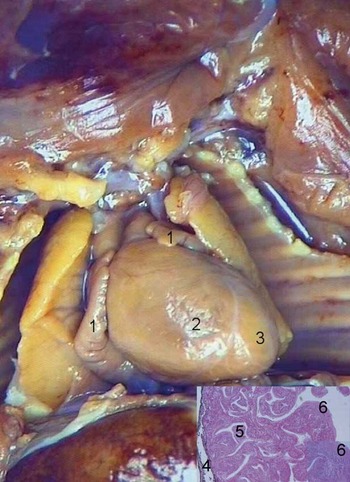
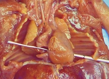
Foetal death took place at 20 weeks and 3 days. Autopsy confirmed a female foetus with a 19–20- week biometry, foetal hydrops with generalised subcutaneous oedema, a small cystic higroma, pleural and pericardial effusions, and ascites. Cardiac malformations comprised situs ambiguus with left isomerism, levoapex, levocardia, two atrial appendages with left morphology (Fig 1), and a complete unbalanced auriculoventricular septal defect, almost hypoplastic right ventricle. Inferior caval vein was on the left, and drained to a discretely dilated coronary sinus. Superior caval vein was bilateral and drained to the right and left atria, respectively. A right aortic arch with right aorta was present (Fig 2), along with a non-compacted heart (Fig 1) and endocardial fibroelastosis.
Macroscopy of the heart – left isomerism: (1) auricular appendix with left morphology, (2) right ventricle, and (3) left ventricle; microscopy of the myocardium: (4) pericardium, (5) fenestrated myocardium and non-compacted myocardium; and (6) endocardial fibroelastosis.

Macroscopy of the heart – (1) right aortic arch and (2) object through auriculoventricular septal defect – from left ventricle to right atrium.

Other associated malformations were symmetrical liver with gall bladder on the left, stomach on the right, intestinal malrotation with midline vermiform appendix, spleen at the midline and polysplenia, diaphragmatic weakness, hypoplastic and bilobulated lungs, and hypoplastic kidneys with lobulation loss.
Discussion
As soon as foetal hydrops was detected, suspicion of a cardiac malformation as the aetiological factor arised. However, other causes were considered and excluded, such as alfa-talassaemia, given the patient’s race. Aneuploidy was discarded as a possible cause, given the normal karyotype. Therefore, the foetal echocardiogram that showed a complete auriculoventricular septal defect was primordial for the suspicion that left isomerism and non-compacted myocardium might be the aetiological factors of the foetal hydrops.
Left isomerism is a congenital anomaly characterised by the presence of two atrial appendages with left morphology and bilobed lungs, with two main left bronchi. Abnormalities may affect the entire body as in total mirror-image sidedness, or involve individual organ systems.Reference Edwards, Allen, Gutgesell and Clark3, Reference Pepes, Zidere and Allan4 Non-compacted myocardium is a rare, potentially fatal, congenital anomaly, related to abnormal endomyocardial embryogenesis and characterised by a halt in the myocardial fibres’ compaction.Reference Edwards, Allen, Gutgesell and Clark3, Reference Sen-Chowdhry and McKenna5–Reference Yang, Zhou, Sunnassee and Liu8 These diagnoses were considered mainly due to the cardiac malformations that were present, for example, much enlarged left atrium, a common auriculoventricular valve with severe regurgitation, an hypoplastic and almost motionless right ventricle, and an apparently non-compacted myocardium, in association with the foetal bradycardia – a complete atrioventricular block.Reference Edwards, Allen, Gutgesell and Clark3, Reference Pepes, Zidere and Allan4, Reference Saeed, Vegsundvåg and Lode7–Reference Jaeggi and Friedberg9 An investigation of foetal bradycardia due to complete cardiac block serves to identify intrauterine left isomerism.Reference Peacock and Joseph10 The foetal bradycardia was due to the existence of two atrial appendages with left morphology, which led to an absent or atresic sinus node, typical of the left isomerism.
These diagnostic hypotheses needed confirmation that was attainable by autopsy, which confirmed the suspicions that arised in the prenatal period. The presence of hydrops is, therefore, a sign associated with a bad prognosis.Reference Cardoso1, Reference Bellini, Hennekam and Fulcheri2 This case report shows the importance of prenatal ultrasonographic scans and autopsy in the assessment of the underlying aetiology, which determines the recurrence risk.
One should note that the case presented here, with combined left isomerism and non-compacted myocardium, is rare, given the prenatal diagnosis.
Acknowledgements
The authors thank all members of Prenatal Diagnosis Unit of Centro Hospitalar de Vila Nova de Gaia/Espinho: Francisco Valente, Margarida Mesquita, Matilde Azevedo, Conceição Brito, Cristina Godinho, and Ana Olívia Sousa.