Introduction
According to Ellis and Brust (Reference Ellis and Brust1973), the Ochlerotatus communis complex consists of four species in North America, including: O. communis (De Geer, 1776), O. churchillensis (Ellis and Brust, Reference Ellis and Brust1973), Ochlerotatus nevadensis (Chapman and Barr, Reference Chapman and Barr1964), and Otahoensis tahoensis (Dyar, 1916). These species are morphologically cryptic and thus identification based on morphology is exceedingly difficult. Most characteristics are diagnostic for only one species within the complex or for a specific developmental stage or sex. For example, the shape of the larval comb scales is only diagnostic for the identification of O. nevadensis in the fourth larval instar (Ellis and Brust Reference Ellis and Brust1973). In the adult stage, male members of this complex may be distinguished by the size of genital gonocoxites (Ellis and Brust Reference Ellis and Brust1973). Females, however, are much more difficult to identify. The shape of the metatarsal claw is diagnostic for O. communis from other members of the complex, but this character is not always reliable for distinguishing the other species (Brust and Munstermann Reference Brust and Munstermann1992).
Species within the O. communis complex have interesting distributions suggesting that both allopatric and sympatric speciation have played a role in their evolution. Ochlerotatus communis has a Holarctic distribution and has been collected throughout much of North America (Wood et al. Reference Wood, Dang and Ellis1979). Ochlerotatus nevadensis has been reported from southeastern British Columbia, Canada and northwestern parts of the United States of America (Belton Reference Belton1983) but has only been collected in mountainous regions suggesting that this species might be reproductively isolated from other species within the complex (Belton Reference Belton1982; Schutz and Eldridge Reference Schutz and Eldridge1993). Ochlerotatus tahoensis has a restricted range of distribution from the Sierra Nevadas to the southern Cascades and Klamath mountains in California, United States of America but has only been collected from high elevations (1500–3000 m; Schutz and Eldridge Reference Schutz and Eldridge1993). Ochlerotatus tahoensis can be distinguished from O. nevadensis based on larval morphological characters and from all species in the complex by collection locality (Darsie Reference Darsie1995).
Ochlerotatus communis and Ochlerotatus churchillensis have a sympatric distribution in northeastern and southeastern Manitoba and western Alberta, Canada (Ellis and Brust Reference Ellis and Brust1973; Wood et al. Reference Wood, Dang and Ellis1979) and thus are particularly difficult to identify. These two species have similar larval habitats as both species can be found in snow-melt pools within or at the margins of deciduous and coniferous forests (Ellis and Brust Reference Ellis and Brust1973). However, O. churchillensis females have smaller salivary glands than O. communis due to their autogenous ovarian development. This character may be useful to separate populations of O. churchillensis from all other members of the complex (Ellis and Brust Reference Ellis and Brust1973). Morphometric characteristics of the metatarsal claws are the primary characters used for the identification of adult females, although the measurements are not discrete with considerable overlap across species (Brust and Munstermann Reference Brust and Munstermann1992).
The combination of reproductive differences in addition to small morphological differences, particularly for males, originally led Ellis and Brust (Reference Ellis and Brust1973) to describe O. churchillensis as a distinct species from O. communis. Brust and Munstermann (Reference Brust and Munstermann1992) suggested isozyme analysis as a method for distinguishing members of the O. communis complex. This method, however, lacks consistency and requires very fresh specimens, thereby limiting the use of this technique as a routine tool for species identification (Schutz and Eldridge Reference Schutz and Eldridge1993). Currently, there is no reliable method for quickly and accurately separating O. communis from O. churchillensis.
Accurate and rapid identification of pathogens and vectors is essential in epidemiological studies of mosquito-borne diseases. Ochlerotatus communis is a vector of Jamestown Canyon virus (JCV) and snow shoe hare virus (SSHV) in North America (McLean et al. Reference McLean, Judd and Shives1981; Heard et al. Reference Heard, Zhang and Grimstad1990), but the clinical significance of O. churchillensis has not yet been studied. Both viruses are widely distributed across temperate areas of North America, and several boreal Aedes Meigen, 1818 and Ochlerotatus Lynch Arribálzaga, 1891 species are the primary vectors (Grimstad Reference Grimstad1988; Andreadis et al. Reference Andreadis, Anderson, Armstrong and Main2008; Bennett et al. Reference Bennett, Gresko, Nelson, Murphy and Whitehead2012). Although JCV antibodies have been detected in moose, elk, bison, mule deer, domestic bovine and equine, white-tailed deer is the only known amplifying host in the natural transmission cycle of the virus (Grimstad Reference Grimstad1988; Rust et al. Reference Rust, Thompson, Matthews, Beauty and Chun1999). The common symptoms of JCV infection in humans ranges from nonspecific mild febrile illnesses, acute central nervous system infection, and respiratory system involvement (Grimstad Reference Grimstad1988, Reference Grimstad2001). SSHV was isolated for the first time from snowshoe hare (Lepus americanus Erxleben, 1777; Mammalia: Leporidae) in Montana, United States of America in 1959 (Burgdorfer et al. Reference Burgdorfer, Newhouse and Thomas1961). However, its antibodies also have recently been detected in several small vertebrates and domestic animals such as equine, cattle, dog, and chicken (Bennett et al. Reference Bennett, Gresko, Nelson, Murphy and Whitehead2012). The symptoms of SSHV infection in humans include fever, headache, vomiting, meningitis, and encephalitis (Fauvel et al. Reference Fauvel, Artsob, Calisher, Davignon, Chagnon and Skvorc-Ranko1980). Developing molecular markers could improve the reliability of identification of the cryptic species in the O. communis complex and greatly facilitate mosquito vector surveillance.
Unlike morphological characters, molecular tools can be applied for the identification of species in all developmental stages and for damaged specimens. Cytochrome c oxidase subunit I (COI) has been used for species identification in many insect species (Hebert et al. Reference Hebert, Cywinska, Ball and DeWaard2003; Smith et al. Reference Smith, Woodley, Janzen, Hallwachs and Hebert2006; Rivera and Currie Reference Rivera and Currie2009; Boring et al. Reference Boring, Sharanowski and Sharkey2011; Hernández-Triana et al. Reference Hernández-Triana, Crainey, Hall, Fatih, MacKenzie-Dodds and Shelley2012). Cywinska et al. (Reference Cywinska, Hunter and Hebert2006) also used DNA barcodes for the identification of several species of Aedes and Ochlerotatus in Canada; however, O. churchillensis was not included in their analysis. Polymerase chain reaction followed by restriction fragment length polymorphism (PCR-RFLP) has been successfully used as an effective molecular tool for the identification of two other cryptic Ochlerotatus species: Ochlerotatus triseriatus (Say, 1823) and Ochlerotatus hendersoni (Cockerell, 1918) (Reno et al. Reference Reno, Vodkin and Novak2000). As sequencing is not required for identification, RFLP may be a cost-effective and rapid alternative to DNA barcoding for species identification. This study was part of a larger ongoing project to develop RFLP profiles for all black-legged Ochlerotatus species in Manitoba, Canada. Due to the cryptic morphology and sympatric distribution of O. communis and O. churchillensis in Canada, the major aim of this study was to develop diagnostic RFLP patterns for these species and assess the applicability of this technique for rapid identification for use in vector surveillance programs in Manitoba. Additionally, we evaluated the efficacy of DNA barcoding for species identification of the members of the O. communis complex in North America (excluding O. nevadensis). Additional species of Ochlerotatus were added to the data matrix for comparative examination of the genetic variability in the DNA barcoding gene COI within and between species.
Materials and methods
Mosquito collection
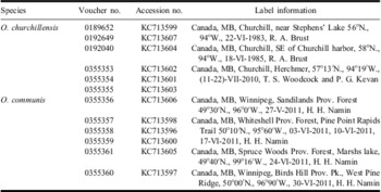
Adult mosquito specimens were collected from multiple localities in Manitoba (including known localities for Ochlerotatus churchillensis) using aspirators between May and July 2011 (Table 1). The specimens of O. churchillensis, collected by Brust (Table 1), were obtained from the Wallis–Roughley Museum of Entomology (University of Manitoba, Winnipeg, Manitoba, Canada). Additional specimens of this species were obtained from the collected materials of a project entitled “Insect biodiversity in the Churchill region and Wapusk National Park”, collected using a malaise trap in July 2010. Although malaise traps are not an appropriate method of collecting adult female specimens, the specimens were adequately preserved to make putative morphological identifications of O. churchillensis and allowed for increased sampling of this taxon. Collected female specimens were identified using the following morphological keys (Ellis and Brust Reference Ellis and Brust1973; Wood et al. Reference Wood, Dang and Ellis1979; Thielman and Hunter Reference Thielman and Hunter2007) and deposited in the Wallis–Roughley Museum of Entomology at the University of Manitoba. Collection information for all specimens is provided in Table 1.
Specimen locality, voucher, and GenBank numbers for Ochlerotatus communis and Ochlerotatus churchillensis specimens collected in this study.

DNA extraction and amplification
Genomic DNA was extracted from legs of the collected specimens and from the whole body of previously mounted museum specimens. DNA extraction was carried out using the Qiagen DNEasy extraction kit (Qiagen Inc., Valencia, California, United States of America) following the manufacturer's instructions. Universal primers LCOI490 (5′-GGTCAACAAATCATAAAGATATTGG-3′) and HCO2198 (5′-TAAACTTCAGGGTGACCAAAAAATCA-3′) (Folmer et al. Reference Folmer, Black, Hoeh, Lutz and Vrijenhoek1994) were used to amplify an ∼660 base pair (bp) fragment of COI. All PCR reaction mixtures contain Chapman d 1× Standard Taq Reaction Buffer (10 mM Tris-HCl; 50 mM KCl; 1.5 mM MgCl2 pH 8.3, New England Biolabs, Ipswich, Massachusetts, United States of America), 200 μM dNTP (Invitrogen, Carlsbad, California, United States of America), 0.4 μM of each primer, 1 unit Taq DNA polymerase (New England Biolabs), ∼1–2 μg of genomic DNA, and purified water to a final volume of 50 μL. The PCR amplification protocol consisted of an initial denaturation at 95 °C for 60 seconds, followed by 30 cycles of 60 seconds at 94 °C, 60 seconds at 50°C, 60 seconds at 72 °C, and a final extension of 72 °C for seven minutes. The PCR products were purified with ExoSAP-IT (Affymetrix, Santa Clara, California, United States of America) using 25% of the recommended reagent amount, but otherwise following the manufacturer's instructions. Sequencing was carried out using the BigDye Terminator Cycle Sequencing Kit (Applied Biosystems, Carlsbad, California, United States of America), with reaction products analysed on an Applied Biosystems 3730×l DNA Analyzer at Eurofins MWG Operon (Huntsville, Alabama, United States of America).
Data analysis
Sequence contigs were assembled, trimmed, and edited using Geneious version 5.4.5 (Drummond et al. Reference Drummond, Ashton, Buxton, Cheung, Cooper and Duran2011), and deposited in GenBank (see Table 1 for accession numbers). Additional COI sequences of Ochlerotatus species were obtained from the sequence data from Mitchell et al. (Reference Mitchell, Sperling and Hickey2002), Morlais and Severson (Reference Morlais and Severson2002), Cywinska et al. (Reference Cywinska, Hunter and Hebert2006), and Gibson et al. (Reference Gibson, Kao, Blevins and Travers2012), and are listed in Supplementary Table 1.
Multiple sequence alignments were performed using ClustalX v. 2.0.11 (Larkin et al. Reference Larkin, Blackshields, Brown, Chenna, McGettigan and McWilliam2007) with default settings and checked for reading frame accuracy. Interspecies and intraspecies genetic p-distances were calculated using MEGA 5 (Tamura et al. Reference Tamura, Peterson, Peterson, Stecher, Nei and Kumar2011). The general time-reversible model, with a parameter for invariant sites and rate heterogeneity modeled under a gamma distribution (GTR + I + G) was chosen as the best fitting model of nucleotide substitution using the Akaike Information Criterion. The model selection test was carried out using PAUP beta v. 4.0 b10 (Swofford Reference Swofford2000), ModelTest v.3.7 (Posada and Crandall Reference Posada and Crandall1998), and the ModelTest Server (Posada Reference Posada2006). Bayesian phylogenetic inference was performed using MrBayes v.3.1.2 (Huelsenbeck and Ronquist Reference Huelsenbeck and Ronquist2001; Ronquist and Huelsenbeck Reference Ronquist and Huelsenbeck2003) with two independent searches with four chains each, default priors, and run for 5 000 000 generations, sampling every 1000th generation. Culex territans Walker was chosen as the outgroup for the phylogenetic analysis. Convergence of runs and suitable mixing was ascertained by examining the likelihood plots for each run and when the average standard deviation of split frequencies was <0.2 (Huelsenbeck and Ronquist Reference Huelsenbeck and Ronquist2001; Ronquist and Huelsenbeck Reference Ronquist and Huelsenbeck2003). Trees were summarised from the two independent searches using majority rule consensus after discarding 25% of the samples for burn-in.
Restriction digestion
AluI was determined as the best restriction enzyme for producing a unique RFLP pattern for each species using NEB Cutter 2.0 (Vincze et al. Reference Vincze, Posdai and Roberts2003). The digestion mixtures consisted of 5–7 μL PCR-products (∼50–150 ng), 5 units of AluI (Invitrogen), 1× incubation buffer (1.5 mM Tris-HCl, pH 7.5; 1.5 mM MgCl2; 0.15 mM Dithiothreitol) and purified water to a final volume of 15 μL. The digested mixture was incubated at 37 °C for two hours and the digested fragments were resolved on a 1.5% agarose gel, stained with Gel Red (Biotium Inc., Hayward, California, United States of America). The restricted DNA patterns were photographed on a Gel Doc EZ imager (BioRAD Inc, Mississauga, Ontario, Canada) and the length of each restriction fragment was estimated using a 50-base DNA ladder (Novagen, Darmstadt, Germany).
Results and discussion
Sequence and phylogenic analyses
The final alignment was 658 bp in length of which 236 (35.9%) sites were variable and 224 (34%) were parsimony informative. The COI sequences showed a strong AT base composition bias ranging from 65.4% (Ochlerotatus implicatus (Vockeroth, 1954)) to 70.6% (Aedes vexans (Meigen, 1830)), with an average of 68% for all taxa. No stop codons or indel events were observed among the studied sequences. Intraspecific genetic variation within the O. communis complex ranged from 0% for O. tahoensis to 0.94% for O. churchillensis (Table 2). However, only two sequences of O. tahoensis were included in this analysis and no reference sequence for O. nevadensis was available for study. The highest interspecific genetic variation was observed between O. churchillensis and O. communis (5.1%) while the lowest value was detected between O. churchillensis and O. tahoensis (3.0%; Table 2). Although there is no widely accepted threshold of interspecific genetic variation for species identification using DNA barcoding, a sequence divergence of <2% has been observed by Cywinska et al. (Reference Cywinska, Hunter and Hebert2006) for species delineation of Ochlerotatus and Aedes species. Given the low intraspecific variability and high interspecific distances, O. churchillensis, O. communis, and O. tahoensis can be delineated using COI barcodes (Table 2). These distinct barcodes support the conclusions of Ellis and Brust (Reference Ellis and Brust1973) that O. churchillensis is a distinct species from O. communis. Ellis and Brust (Reference Ellis and Brust1973) and Brust and Munstermann (Reference Brust and Munstermann1992) suggested O. tahoensis is a valid species distinct from O. communis based on morphological and allozyme evidence. The DNA barcode data from this study also supports O. tahoensis as a genetically distinct species.
The interspecific and intraspecific p-distances among the members of the Ochlerotatus communis complex calculated using MEGA 5 (Tamura et al. Reference Tamura, Peterson, Peterson, Stecher, Nei and Kumar2011) based on a 658 bp sequence of cytochrome oxidase 1 and the number of analysed specimens for each species.

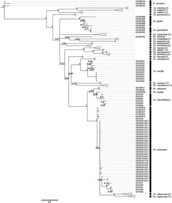
On the basis of Bayesian inference of phylogenetic relationships, Aedes and Ochlerotatus were recovered as well-supported monophyletic sister taxa (Fig. 1). These results are congruent with the results of Shepard et al. (Reference Shepard, Andreadis and Vossbrinck2006) based on 18s rDNA, and also support the proposed classification of Culicidae by Reinert (Reference Reinert2000) with Ochlerotatus as a distinct, valid genus from Aedes. Although the recognition of Ochlerotatus as a distinct genus has been controversial (Black Reference Black2004; Savage and Strickman Reference Savage and Strickman2004; Edman Reference Edman2005), the current study provides additional evidence supporting recognition of Ochlerotatus by Reinert (Reference Reinert2000). Furthermore, we agree with Black (Reference Black2004) that taxonomic stability is best achieved when taxonomy follows phylogeny.
Bayesian majority-rule consensus tree for the COI DNA sequences under the GTR + I + G model. Posterior probabilities are given for the respective nodes. Monophyletic clades of individual species were collapsed (indicated with a triangle) if they were not the focus of this study for easier visualisation. The number of analysed specimens for collapsed nodes is indicated in brackets beside the species name. GenBank and BOLD Accession numbers for specimens within collapsed clades can be found in Supplementary Table 1. For other sequences used from previously published data sets, the accession numbers are listed on the figure and in Supplementary Table 1. For specimens collected and sequenced in this study, voucher numbers are listed on the figure and accession numbers are listed in Table 1. One sequence of Ochlerotatus aurifer (GU907850) downloaded from GenBank was possibly misidentified or contaminated. COI, cytochrome c oxidase subunit I.

Ochlerotatus churchillensis was recovered as sister to O. tahoensis (Fig. 1). However, one sequence of Ochlerotatus aurifer (GU907850) was recovered within the O. churchillensis clade. Since all the other sequences of O. aurifer were recovered as a monophyletic group sister to O. cantator + O. canadensis, this sequence (GU907850) was likely contaminated or the specimen was misidentified prior to sequencing (Fig. 1). This sequence of O. aurifer (GU907850) was downloaded from GenBank and has not been published in a peer-reviewed journal, highlighting the risks with GenBank sequences, particularly from unpublished data. Ochlerotatus communis was recovered as paraphyletic with respect to Ochlerotatus abserratus + O. implicatus (Fig. 1). Although there are distinct morphological differences that separate O. abserratus and O. implicatus from members of the O. communis complex (Carpenter and LaCasse Reference Carpenter and LaCasse1955; Wood et al. Reference Wood, Dang and Ellis1979), the phylogenetic relationships recovered here suggest that the O. communis complex may include additional species. However, additional genes and taxon sampling will be necessary to establish the limits of the O. communis complex, as not all taxa within Ochlerotatus were included in this study and one gene phylogenies are typically inadequate for resolving phylogenetic relationships (Rokas et al. Reference Rokas, Williams, King and Carroll2003; Rubinoff and Holland Reference Rubinoff and Holland2005; Sharanowski et al. Reference Sharanowski, Dowling and Sharkey2011).
All specimens of O. churchillensis were collected in northern Manitoba, near Churchill. Even though efforts were made to collect this species from known localities in southern Manitoba, O. churchillensis was not collected from the known sympatric regions in Manitoba. It may be possible that this species now has a more restricted distribution, similar to O. tahoensis and O. nevadensis, which would facilitate identification of all species within the O. communis complex based on locality.
PCR-RFLP analysis
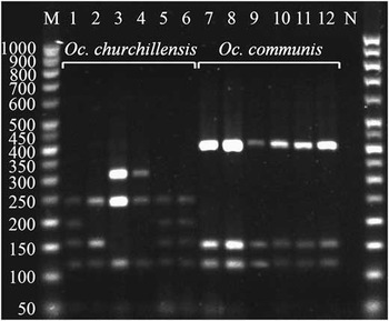
The PCR-RFLP banding patterns were developed for the identification of O. churchillensis and O. communis based on species specific restriction sites of AluI in the COI sequences. The RFLP patterns were consistent with the predicted digestion patterns of O. communis and O. churchillensis sequences from GenBank, BOLD (www.boldsystems.org), and multiple field collected specimens. Three different RFLP patterns, corresponding to those clades found in the Bayesian analysis, were observed for O. churchillensis specimens after digestion with AluI. The first RFLP pattern was characterised by fragments of ∼250, 200, 160, and 120 bp and was observed in three specimens of O. churchillensis (0355353, 0192649, and 0192040 in lanes 1, 5, and 6, respectively, Fig. 2). The second RFLP profile was typified by fragments of ∼320, 250, 120, and 50 bp, and was observed in two specimens of O. churchillensis (0355355 and 0189652 in lanes 3 and 4, respectively, Fig. 2). The third RFLP pattern included fragments of ∼250, two fragments of 150, and one fragment at 120 and 50 bp. This pattern was only observed in one specimen (0355354 in lane 2, Fig. 2). Variation at the third codon position of amino acids 139 and 175 cause the three different RFLP patterns observed in O. churchillensis. Alternatively, digestion with AluI produced one consistent diagnostic RFLP pattern for all specimens of O. communis tested in this study. The O. communis RFLP profile was characterised by fragments of ∼400, 150, 120, and 50 bp (0355356-0355361 shown in lanes 7–12, Fig. 2). This digestion RFLP pattern was also consistent with the predicted RFLP pattern for the sequences of O. communis from GenBank using NEB Cutter 2.0 (Vincze et al. Reference Vincze, Posdai and Roberts2003). Although the analysed specimens of O. communis and O. churchillensis were not collected from the sympatric areas, the RFLP pattern for O. communis (with the largest fragment at 400 bp) was distinct from all RFLP profiles for O. churchillensis (with no bands larger than 320 bp) after digestion using AluI.
Digestion patterns of COI PCR products with AluI restriction enzyme for Ochlerotatus communis and Ochlerotatus churchillensis. Gel lanes are labeled across the top as follows: M: 50 bp DNA ladder, 1–5: O. churchillensis (0355353, 0355354, 0355355, 0189652, 0192649, 0192040), 7–12: O. communis (0355356–0355361) and N: Negative control. According to the RFLP patterns, a 250 bp band was detected in all specimens of O. churchillensis (never observed in O. communis), and a 400 bp band was observed for all specimens of O. communis (never observed in O. churchillensis). COI, cytochrome c oxidase subunit I; PCR, polymerase chain reaction.

These distinctive profiles were created because the 73rd and 74th amino acids (based on the complete CO1 gene from Aedes aegypti Linnaeus, 1762, NC_010241.1) are conserved glycine and alanine residues in the O. communis complex, respectively. In all members of the complex analysed here, the codon for the glycine residue is always GGA. However, the downstream alanine residue codon is different in O. churchillensis (GCT) and O. communis (GCC), causing an additional cut at this site for O. churchillensis. Thus, it is this mutation that confers the diagnostic RFLP patterns for these species. It is predicted that O. tahoensis would not be cut by AluI at this site, as the alanine residue is encoded by GCA, based on the two downloaded sequences analysed in this (Supplementary Table 1). Thus, this species would exhibit the same 400 bp fragment as found in O. communis (Fig. 2).
The PCR-RFLP method allows for accurate and relatively inexpensive molecular identification compared with DNA-barcoding for distinguishing O. churchillensis from O. communis. RFLP-based identification is quick and requires approximately six hours to finish, including 15 minutes for DNA extraction (using fast DNA extraction kits), two hours for DNA amplification, one hour for checking the integrity of PCR products, two hours for restriction digestion, and one hour to visualise the RFLP banding patterns. This method can be used to reduce taxonomic uncertainty and can facilitate the efficient identification of these cryptic species in all life stages.
Conclusion
This work has provided the first COI barcodes for O. churchillensis and developed the first RFLP-based identification tool for two sympatric members of the O. communis complex in Canada: O. communis and O. churchillensis. At least three members of the O. communis complex have diagnostic DNA barcodes using COI, including O. churchillensis, O. communis, and O. tahoensis. Unfortunately, O. nevadensis was not included in this study. The developed RFLP profiles can be used as a quick diagnostic tool to identify O. communis from O. churchillensis. Only through accurate identification of cryptic species can the vector potential of each species be determined, and this is critical for disease epidemiology. The molecular identification tools developed in this study will help facilitate monitoring of O. communis to predict future outbreaks of JCV and SSHV and will also facilitate future research to assess the potential of O. churchillensis as a vector of these diseases.
Acknowledgements
The authors are very grateful to Reiny A. Brust and Terry D. Galloway (Department of Entomology, University of Manitoba) for comments and advice. Theythank Thomas Woodcock (Department of Biology, Wilfred Laurier University) for providing mosquito specimens from Churchill and to Phillip Snarr for technical support. They also thank two anonymous reviewers whose comments and suggestions greatly improved the manuscript. This research was supported in part by funding provided to B. Sharanowski by Manitoba Agriculture, Food, and Rural Initiatives and the University of Manitoba Research Grants Program.