Implications
Mitochondrial metabolic capacity can be manipulated by diet in utero. In particularly, maternal overnutrition have been shown to induce skeletal muscle mitochondrial dysfunction in offspring, which contributes to the development of type 2 diabetes. It is important to better understand how nutritional interventions during gestation affect fetal skeletal muscle mitochondrial biogenesis. The novel finding was that moderately increased maternal energy intake decreased mitochondrial biogenesis and antioxidative capacity in skeletal muscle of pig fetuses. Fetal growth is crucial for skeletal muscle development and therefore these results will help to gain insight into the fetal origin of metabolic syndrome such as obesity.
Introduction
Obesity and associated metabolic disorders have reached epidemic proportions in most parts of the world, even among teenagers and children. These diseases are due to multifactorial causes that include genetic and environmental factors (O’Rahilly et al., Reference O’Rahilly, Barroso and Wareham2005). Human epidemiological and animal experimental studies indicate that alteration in fetal growth and development in response to prenatal environmental conditions such as nutritional stimulus may have deleterious persistent health effects lasting for the entire life (Wu et al., Reference Wu, Bazer, Cudd, Meininger and Spencer2004; Catalano et al., Reference Catalano, Farrell, Thomas, Huston-Presley, Mencin, de Mouzon and Amini2009). Researches have demonstrated a link between maternal malnutrition and offspring risk of metabolic diseases (Barker et al., Reference Barker, Godfrey, Gluckman, Harding, Owens and Robinson1993; Tappia et al., Reference Tappia, Guzman, Dunn and Aroutiounova2013). Currently, in nutritional programming studies focused on maternal overnutrition and obesity animal models are widely used. Indeed, maternal overnutrition has been reported to perturb fetal development, leading to persistent effects on offspring, including predisposition to obesity and type 2 diabetes (T2D) (Wu et al., Reference Wu, Bazer, Cudd, Meininger and Spencer2004; Samuelsson et al., Reference Samuelsson, Matthews, Argenton, Christie, McConnell, Jansen, Piersma, Ozanne, Twinn and Remacle2008; Du et al., Reference Du, Yan, Tong, Zhao and Zhu2010). Nevertheless, it is unclear whether a defined degree of increase in maternal energy intake could produce significant effects on fetal development.
Mitochondria are the principal sites of cellular energy source ATP and reactive oxygen species (ROS) production (Starkov, Reference Starkov2008). Skeletal muscle composes the largest metabolic tissue in the body and is one of the key insulin-responsive organs (Lowell and Shulman, Reference Lowell and Shulman2005). Mitochondrial dysfunction in skeletal muscle has been associated with metabolic diseases in human and animal models (Pagel-Langenickel et al., Reference Pagel-Langenickel, Bao, Pang and Sack2010). Likewise, decreased oxidative phosphorylation, antioxidative capacity and mitochondrial electron transport chain enzyme activities in the liver and skeletal muscle have been observed in the offspring subjected to maternal overnutrition and diabetes (Bruce et al., Reference Bruce, Cagampang, Argenton, Zhang, Ethirajan, Burdge, Bateman, Clough, Poston and Hanson2009; Zhang et al., Reference Zhang, Strakovsky, Zhou, Zhang and Pan2011; Latouche et al., Reference Latouche, Heywood, Henry, Ziemann, Lazarus, El-Osta, Armitage and Kingwell2014). In mammals, muscle fibers are formed prenatally, and subsequent skeletal muscle development depends largely on muscle fiber hypertrophy (Nissen et al., Reference Nissen, Danielsen, Jorgensen and Oksbjerg2003; Zou et al., Reference Zou, He, Yu, Yu, Mao, Zheng, He, Huang, Shu and Liu2016). Therefore, skeletal muscle is particularly vulnerable during the very early stage of mammalian development, and understanding the effect of maternal overnutrition on mitochondrial biogenesis and function in skeletal muscle of the fetuses may provide useful information on the fetal origins of the metabolic syndrome.
Besides, recent studies have shown that mitochondria play a prominent role in the regulation of skeletal myogenesis (Rochard et al., Reference Rochard, Rodier, Casas, Cassar-Malek, Marchal-Victorion, Daury, Wrutniak and Cabello2000; Wagatsuma and Sakuma, Reference Wagatsuma and Sakuma2013). Increased mitochondrial mass, mitochondrial DNA (mtDNA) copy number and mitochondrial respiration have been observed during C2C12 myogenic differentiation (Remels et al., Reference Remels, Langen, Schrauwen, Schaart, Schols and Gosker2010), suggesting the increased mitochondrial function may be essential for skeletal myogenesis. Moreover, blocking mitochondrial biogenesis inhibits myogenic differentiation (Rochard et al., Reference Rochard, Rodier, Casas, Cassar-Malek, Marchal-Victorion, Daury, Wrutniak and Cabello2000). Our previous study using a pig model indicated that moderately increased maternal dietary energy intake delayed fetal skeletal muscle differentiation and maturity on day 90 of gestation through changing myogenic gene expression and muscle fiber characteristics (Zou et al., Reference Zou, He, Yu, Yu, Mao, Zheng, He, Huang, Shu and Liu2016). Therefore, we hypothesized that the impairment of mitochondrial function may contribute to maternal HE diet-induced delayed skeletal myogenesis in pig fetuses. The aim of the present study was to assess the effects of moderately increased maternal dietary energy intake on mitochondrial biogenesis and function in skeletal muscle of the pig fetuses.
Material and methods
The experimental procedures were approved by the Animal Care and Use Committee of Sichuan Agricultural University.
Animals and diets
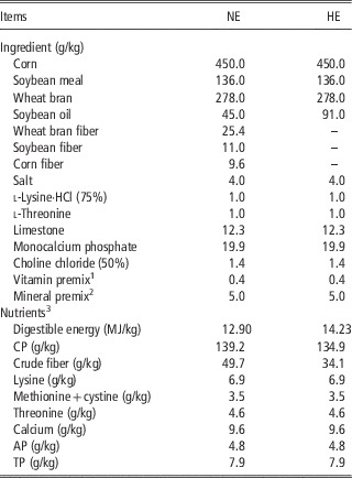
In total, 12 primiparous, purebred Large White sows (initial BW 135.5±1.6 kg) were artificially inseminated three times with the semen of purebred Large White boars at the third observation of estrus. The day of the last insemination constituted the 1st day of gestation. The pregnant sows were randomly assigned to one of two experimental groups differing in daily digestible energy (DE) intake: normal energy (NE) and high energy (HE) groups (n=6/group). The NE sows were fed 2.4 kg/day of a control diet containing 12.90 MJ of DE/kg to achieve a target energy intake of 30.96 MJ DE/day following the National Research Council (NRC) (2012) recommendation for gestating sow, and those in the HE group were fed 2.4 kg/day of a HE diet containing 14.23 MJ of DE/kg to achieve a target energy intake of 34.15 MJ DE/day. The HE diet was formulated to allow the sow to ingest daily 10% more DE than the NE group. Sows were fed isoprotein corn–wheat bran–soybean meal-based diets (Table 1). Although the additional dietary fiber was added to the NE diet to decrease energy concentration, crude fiber (CF) intake was only 1.56% higher in NE sows than HE sows. Danielsen and Vestergaard (Reference Danielsen and Vestergaard2001) reported that even excess dietary fiber intake (CF, 4.6% v. 12.7%) of pregnant sows influenced neither fetal survival nor litter size at birth. Consequently, it is quite unlikely in this experiment that the dietary fiber supplementation affected the fetal survival rate directly. The dietary treatments were introduced from mating to day 90 of gestation. During gestation, sows were housed in individual feed stalls in a breeding facility, and fed twice daily (0800 and 1400 h) with 50% of the daily ration each time. All sows had free access to drinking water. Two sows were excluded from day 30 (one NE and one HE sow) because they were non-pregnant. Another two sows had to be excluded from the experiment: one NE sows due to sudden disease and one HE sows due to had only three fetuses.
Ingredients and composition of the experimental diets (as-fed basis)

NE=normal energy intake; HE=high energy intake; AP=available phosphorus; TP=total phosphorus.
1 Provided the following per kg diet: vitamin A, 1.2 mg; vitamin D3, 0.02 mg; vitamin E, 44 mg; vitamin K3, 0.5 mg; thiamin, 1 mg; riboflavin, 3.75 mg; pyridoxine, 1 mg; vitamin B12, 0.015 mg; niacin, 10 mg; pantothenic acid, 12 mg; folic acid, 1.3 mg; biotin, 0.2 mg.
2 Provided the following per kg diet: Fe, 80 mg; Cu, 10 mg; Zn, 100 mg; Mn, 25 mg; Se, 0.15 mg; I, 0.14 mg.
3 All data were calculated values.
Sample collection
The fasted BWs of sows were determined in the morning before feeding on days 35, 55 and 90 of gestation. After a 12-h overnight fast on day 90 of gestation, four pregnant sows of both groups were anaesthetized with an intramuscular injection of Zoletil (Zoletil 50, Virbac, Carros, France; 4 mg/kg BW). The maternal abdomen was opened and the reproductive tracts were removed immediately. Blood from the umbilical vein of the fetus were collected and coagulated for 30 min, then centrifuged at 3500×g for 10 min to separate serum which was stored at −20°C until analysis. The BW of each fetus was recorded and fetal: maternal weight ratio was calculated. Two female fetuses and two male fetuses, with weights close to the average level, were selected from each sow for muscle sample collection. The longissimus (LM) muscles were collected from the selected fetuses, snap-frozen in liquid nitrogen and stored at −80°C until subsequent analysis.
Hormone and biochemical measurements
The growth hormone (GH) concentration in umbilical vein serum was measured using a commercially available radioimmunoassay kit purchased from Beijing North Institute of Biotechnology (Beijing, China). Insulin was determined with porcine ELISA kit (R&D Systems, Minneapolis, MN, USA). Sensitivities of the assays were 0.02 ng/ml and 2.15 pmol/l for GH and insulin, respectively. Intra- and inter-assay CV were 4.3% and 6.6% for GH and 3.9% and 7.4% for insulin, respectively. The concentrations of triglyceride and glucose in umbilical vein serum were determined using commercial kits (Jiancheng Institute of Bioengineering, Nanjing, Jiangsu, China) according to the provided instructions, respectively. The homeostasis model assessment (HOMA) index ((fasting insulin×fasting glucose)/22.5) was used as indicators of insulin sensitivity as previously described (Galili et al., Reference Galili, Versari, Sattler, Olson, Mannheim, McConnell, Chade, Lerman and Lerman2007).
The activities of superoxide dismutase (SOD), glutathione peroxidase (GPx) and catalase (CAT) and malondialdehyde (MDA) concentration in skeletal muscle were measured by assay kits (Jiancheng Institute of Bioengineering). NAD+/NADH ratio in skeletal muscle was measured using the BioVision NAD/NADH Quantitation kit (Milpitas, CA, USA) according to the manufacturer’s instructions.
Mitochondrial DNA copy number
Total DNA was extracted from LM muscle using QIAamp DNA extraction kit (QIAGEN, Valencia, CA, USA). The mtDNA copy number was calculated using quantitative real-time PCR by measuring the threshold cycle ratio (ΔC t ) of a mitochondrial encoded gene (mitochondrial cytochrome b) v. a nuclear-encoded gene (18S ribosomal RNA (rRNA)). The sequence of primers are presented in Supplementary Table S1.
Gene expression
Total RNA was extracted from frozen muscle tissues using RNAiso Plus reagent (Takara, Dalian, China). The concentration and purity of RNA were estimated by UV/VIS spectrophotometer (A260/A280, Beckman Coulter DU800; Beckman Coulter Inc., Fullerton, CA, USA), and the OD260 : OD280 ratio (where OD is the optical density) ranged from 1.8 to 2.0. The integrity of RNA was checked by denaturing gel electrophoresis with the 28S : 18S rRNA band ratio ⩾1.8. Following RNA isolation, total RNA was reverse transcribed into complementary DNA (cDNA) using the PrimeScriptTM RT reagent kit (Takara) according to the manufacturer’s recommendations. Real-time quantitative PCR was carried out on the ABI Prism® 7900HT Sequence Detection System (Applied Biosystems, Foster City, CA, USA) using SYBR® Premix Ex TaqTM II (Takara). The reactions were performed in a final volume of 10 μl containing 5 μl of SYBR® Premix Ex TaqTM II, 0.8 μl of the primer pair, 0.2 μl of ROX Reference Dye, 1 μl of cDNA template and 3 μl of distilled water dH2O. The thermal cycling parameters comprised an initial denaturation step at 95°C for 30 s, 40 cycles of PCR reaction at 95°C for 5 s and 60°C for 34 s, followed by a dissociation step at 95°C for 15 s, 60°C for 1 min and 95°C for 15 s. To confirm specific product amplification, melt curve analysis was conducted. All measurements were performed in triplicate. Primers for individual genes were designed using Primer Express 3.0 (Applied Biosystems) and shown in Supplementary Table S1. For normalization, β-actin was chosen as the reference gene as no variation in its expression was observed between treatments. The relative messenger RNA (mRNA) abundance of each gene was calculated using the
 $$2^{{{\minus}\Delta \Delta C_{t} }} $$
method as previously described (Livak and Schmittgen, Reference Livak and Schmittgen2001). In brief, a ΔC
t
value is the C
t
difference between target gene and the reference gene
 $$\left( {\Delta C_{t} \,{\equals}\,C_{t}^{{{\rm target}}} {\minus}C_{t}^{{{\rm reference}}} } \right)$$
. For each of the target genes, the ΔΔC
t
values of all the samples were determined by subtracting the average ΔC
t
of the corresponding NE group. The ΔΔC
t
values were then converted to fold differences by raising 2 to the power
 $${\minus}\Delta \Delta C_{t} \,\left( {2^{{{\minus}\Delta \Delta C_{t} }} } \right)$$
. The mRNA level of each target gene for NE group was set to 1.0.
Statistical analysis
All statistical analyses were performed using Statistical Analysis System (version 8.1, SAS Institute Inc.). Sow data was compared using a t test. Data obtained from the selected fetuses were analysis using the MIXED procedures of SAS according to the following model:
 $$Y_{{ijk}} \,{\equals}\,\mu {\plus}\alpha _{i} {\plus}\beta _{j} {\plus}(\alpha \beta )_{{ij}} {\plus}U_{k} {\plus}{\epsilon}_{{ijk}} $$
where μ is the mean, α i the effect of treatment (i=HE, NE), β j the effect of sex (j=male, female), αβ ij the interaction between treatment and gender, U k the litter (k=1, 2, … , 8), ε ijk represents the random error. However, none of the gender and interactions were statistically significant. Therefore, they were removed from the model, and data from female and male fetuses were pooled together for analysis. The least square means (LSM) were separated using the PDIFF option with the Tukey adjustment. Statistical significance was considered as P<0.05. Results are presented in the table as LSM±pooled SE.
Results
Gestation performance
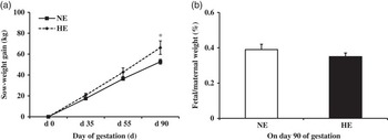
As shown in Figure 1, the weight gain of sows in HE group was significantly higher than NE sows on day 90 of gestation (P<0.05, Figure 1a). We previously reported that no significant difference was found in fetal weight between NE and HE groups (NE v. HE=737.3 v. 703.3 g, P>0.05) (Zou et al., Reference Zou, He, Yu, Yu, Mao, Zheng, He, Huang, Shu and Liu2016). Given the relationship between sow and piglets weights, the data were also analyzed as fetal: maternal weight ratio to give a more accurate description of fetal growth. However, no significant change was observed for fetal: maternal weight ratio between NE and HE groups (P>0.05, Figure 1b).
Gestation performance of sows fed normal energy (NE) or high-energy (HE) diet. (a) Changes in weight gain of sows at different days of gestation. (b) Fetal: maternal weight on day 90 of gestation. Data were expressed as least square means±SE, n=4. *P<0.05 compared with NE.

Metabolites and hormone concentrations
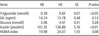
The metabolites and hormone concentrations in fetal umbilical vein serum are shown in Table 2. Concentrations of triglyceride and insulin were higher in umbilical vein serum of HE fetuses compared with NE fetuses (P<0.05). The HOMA index values also tended to be increased in HE fetuses (P=0.05). There was no significant difference in serum glucose and GH concentrations between NE and HE groups.
Concentrations of metabolites and hormones in umbilical vein serum of fetusesFootnote 1

NE=normal energy intake; HE=high energy intake; GH=growth hormone; HOMA=homeostasis model assessment.
1 Data are expressed as least square means and pooled SE, n=16/group.
Mitochondrial biogenesis in the skeletal muscle of high energy fetuses
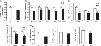
The effects of maternal HE diet on mitochondrial biogenesis and functions in skeletal muscle of the fetuses are shown in Figure 2. The mtDNA copy number was lower in skeletal muscle of HE fetuses than that in NE fetuses (P<0.05, Figure 2a). Compared with NE fetuses, the mRNA levels of genes implicated in mitochondrial biogenesis and function, including peroxisome proliferator-activated receptor-γ (PPARγ) coactivator 1α (PGC-1α), sirtuin 1 (SIRT1), nuclear respiratory factor-1 (NRF-1), mitochondrial transcription factor A (Tfam), cytochrome c oxidase 4, and cytochrome c (Cyt c) and citrate synthase (CS) were lower in HE fetuses (P<0.05, Figure 2b and c). Meanwhile, the mRNA levels of γ DNA polymerase (POLG) and single-strand DNA binding protein 1 (SSBP1) responsible for mtDNA replication were lower in HE fetuses relative to NE fetuses (P<0.05, Figure 2d). Though NADH level was not changed (P>0.05, Figure 2e), NAD+ level and NAD+/NADH ratio were remarkably decreased in HE fetuses compared with NE fetuses (P<0.05, Figure 2e).
Changes in mitochondrial biogenesis and function of the skeletal muscle of fetuses from sows fed normal energy (NE) or high-energy (HE) diet. (a) Relative abundance of mitochondrial DNA (mtDNA). (b and c) Relative messenger RNA (mRNA) expression of genes responsible for mitochondrial biogenesis (b) and function (c). (d) Relative mRNA expression of mtDNA replication genes. (e) Intracellular levels of NAD+, NADH and NAD+/NADH ratio in skeletal muscle of fetuses from sows fed NE or HE diet. Data were expressed as least square means±SE, n=16. *P<0.05 compared with NE. PGC-1α=PPARγ coactivator 1α; SIRT1=sirtuin 1; NRF-1=nuclear respiratory factor-1; Tfam=mitochondrial transcription factor A; COX4=cytochrome c oxidase 4; Cyt c=cytochrome c; CS=citrate synthase; POLG=DNA polymerase; SSBP1=single-strand DNA binding protein 1.

Malondialdehyde levels and antioxidative enzyme activities in the skeletal muscle of high energy fetuses
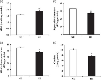
We further measured the concentration of MDA and the activities of three major antioxidative enzymes in skeletal muscle to determine the effects of maternal HE diet on ROS defense system. Compared with NE group, the MDA concentration in skeletal muscle of HE fetuses was higher (P<0.05, Figure 3a). Meanwhile, maternal HE diet significantly decreased the activities of SOD, GPx and CAT in HE fetuses (P<0.05, Figure 3b and d).
Decreased reactive oxygen species defense system in the skeletal muscle of fetuses from sows fed high-energy (HE) diet. (a) Concentration of malondialdehyde (MDA). (b to d) Activities of superoxide dismutase (b), glutathione peroxidase (c) and catalase (d). Data were expressed as least square means±SE, n=16. *P<0.05 compared with normal energy (NE).

Discussion
Most maternal nutritional intervention studies use rats or sheep as model to gain insight into the fetal origin of human metabolic syndrome. Here, we use pig as model based on the higher similarities of pig to human in physiology and genome (Merrifield et al., Reference Merrifield, Lewis, Claus, Beckonert, Dumas, Duncker, Kochhar, Rezzi, Lindon and Bailey2011). Cumulative evidence strongly indicates that reduction in mitochondrial function and oxidative metabolism in skeletal muscle during critical period of fetus growth and development was associated with the greater incidence of T2D and insulin resistance in later life (Shelley et al., Reference Shelley, Martin-Gronert, Rowlerson, Poston, Heales, Hargreaves, McConnell, Ozanne and Fernandez-Twinn2009; Latouche et al., Reference Latouche, Heywood, Henry, Ziemann, Lazarus, El-Osta, Armitage and Kingwell2014). In the present study, the intriguing findings were that HE fetuses exhibited decreased skeletal muscle mitochondrial biogenesis in response to maternal HE intake on day 90 of gestation compared with NE fetuses. Particularly, maternal HE diet led to decreased mitochondrial function in HE fetuses by reducing skeletal muscle antioxidant enzyme activities and expression of gene involved in mitochondrial function. As mitochondrial biogenesis accompanies skeletal myogenesis, the compromised mitochondrial function and decreased mitochondrial biogenesis, together with increased oxidative damage, seem to exert deleterious effects on skeletal muscle, which could partly explain our previous finding of maternal HE diet-induced delayed fetal myogenesis (Zou et al., Reference Zou, He, Yu, Yu, Mao, Zheng, He, Huang, Shu and Liu2016).
As expected, BW gain of HE sows was higher on day 90 of gestation compared with NE sows, which is consistent with previous reports (Bee, Reference Bee2004). Moreover, we found that mtDNA copy number was decreased in skeletal muscle of HE fetuses, suggesting disrupted mitochondrial biogenesis. Consistently, as CS reflects the mitochondrial enrichment of tissue (Kelley et al., Reference Kelley, He, Menshikova and Ritov2002), the CS mRNA level was decreased in HE fetuses. However, our previous results showed no difference in CS activity between NE and HE fetuses (Zou et al., Reference Zou, He, Yu, Yu, Mao, Zheng, He, Huang, Shu and Liu2016), which are similar to results obtained by Guillerm‐Regost et al. (Reference Guillerm‐Regost, Louveau, Sébert, Damon, Champ and Gondret2006) who reported that overconsumption of a high-fat diet did not affect CS activity in muscle of Yucatan minipigs. The discrepancies between mtDNA content and CS activity between groups revealed that CS activity may be influenced by other factors (Liu et al., Reference Liu, Chen, Yao, Yu, Mao, He, Huang and Zheng2012). It has been shown that CS is also rate-limiting and allosterically regulated (Cohen, Reference Cohen2014). Animal models of maternal overnutrition have shown that mitochondrial abnormalities in the progeny. Decreases in mtDNA copy number have been reported in kidney and liver of offspring of rat dams fed a high-fat diet during gestation (Taylor et al., Reference Taylor, McConnell, Khan, Holemans, Lawrence, Asare-Anane, Persaud, Jones, Petrie and Hanson2005; Burgueño et al., Reference Burgueño, Cabrerizo, Mansilla, Sookoian and Pirola2013). In addition, it has been reported that increased maternal nutrition in pigs and rats did not provide advantageous effects on offspring muscle growth and mtDNA copy number (McNamara et al., Reference McNamara, Giblin, Markham, Stickland, Berry, O’Reilly, Lynch, Kerry and Lawlor2011; Latouche et al., Reference Latouche, Heywood, Henry, Ziemann, Lazarus, El-Osta, Armitage and Kingwell2014).
The decrease in skeletal muscle mtDNA copy number indicates that the control of mitochondrial biogenesis and mtDNA replication is altered. PGC-1α is the master regulator of mitochondrial biogenesis and energy expenditure (Fernandez-Marcos and Auwerx, Reference Fernandez-Marcos and Auwerx2011). The previous study using rat model reported that maternal high-fat intake led to a significant reduction in offspring hepatic PGC-1α mRNA expression (Burgueño et al., Reference Burgueño, Cabrerizo, Mansilla, Sookoian and Pirola2013). In agreement, we found that maternal HE diet caused a decrease in the expression levels of PGC-1α in skeletal muscle of the fetuses. Furthermore, expression of PGC-1α downstream target genes, including NRF-1 and Tfam, were also markedly down-regulated in HE fetuses. Recently, SIRT1 has been known to regulate myogenesis and mitochondrial biogenesis in skeletal muscle (Hong et al., Reference Hong, Kim, Choo, Park, Yi, Yu, Lee, Yoon, Lee and Ko2014). SIRT1 expression level and its deacetylase activity are also modulated by the NAD+/NADH ratio (Nemoto et al., Reference Nemoto, Fergusson and Finkel2005). In the current study, we detected significantly lower SIRT1 mRNA level in HE fetuses, supporting the potential role of SIRT1 as a direct regulator of PGC-1α (Nemoto et al., Reference Nemoto, Fergusson and Finkel2005). The intracellular NAD+/NADH ratio, a marker of enhanced cellular oxidative capacity, was significantly decreased in HE fetuses, which is consistent with previous reports (Fainberg et al., Reference Fainberg, Almond, Li, Rauch, Bikker, Symonds and Mostyn2014). POLG and SSBP1 play a key role in mtDNA replication and repair (Scarpulla, Reference Scarpulla2006). Similar to a previous study in high-fat diet-induced obese mice (Mohamed et al., Reference Mohamed, Hajira, Pardo and Boriek2014), we also found that mRNA expression of POLG and SSBP1 in skeletal muscle of HE fetuses were decreased. Taken together, these data may account for the differences in mtDNA copy number.
Mitochondrial dysfunction was an important event in the development of T2D and insulin resistance in adult life (Pagel-Langenickel et al., Reference Pagel-Langenickel, Bao, Pang and Sack2010). It has been shown that maternal high-fat diet-induced changes in mitochondrial biogenesis in offspring are associated with decreased mitochondrial functions (Mohamed et al., Reference Mohamed, Hajira, Pardo and Boriek2014). In the present study, we found that expression levels of several genes responsible for mitochondrial function were markedly decreased in HE fetuses, implying decreased skeletal muscle mitochondrial functions. Furthermore, mitochondrial dysfunction leads to lipid peroxide accumulation and increased production of ROS (Kim et al., Reference Kim, Wei and Sowers2008). Previous studies in rodent model systems indicate that a maternal high-fat diet decreases mitochondrial electron transport chain enzymes activities and antioxidant defense capacity in skeletal muscle and liver of offspring (Bruce et al., Reference Bruce, Cagampang, Argenton, Zhang, Ethirajan, Burdge, Bateman, Clough, Poston and Hanson2009; Shelley et al., Reference Shelley, Martin-Gronert, Rowlerson, Poston, Heales, Hargreaves, McConnell, Ozanne and Fernandez-Twinn2009; Zhang et al., Reference Zhang, Strakovsky, Zhou, Zhang and Pan2011). Consistently, decreased activities of antioxidative enzymes (SOD, GPx and CAT) and increased MDA levels were also observed in skeletal muscle of HE fetuses, suggesting the failure of ROS defense system and more, which is probably one of the major determinants of mitochondrial function alterations (Bonnard et al., Reference Bonnard, Durand, Peyrol, Chanseaume, Chauvin, Morio, Vidal and Rieusset2008). In addition, several studies have shown mitochondrial dysfunction can affect whole-body metabolism and play an important in the pathophysiology of insulin resistance in offspring of high-fat diet-fed dams (Bonnard et al., Reference Bonnard, Durand, Peyrol, Chanseaume, Chauvin, Morio, Vidal and Rieusset2008; Latouche et al., Reference Latouche, Heywood, Henry, Ziemann, Lazarus, El-Osta, Armitage and Kingwell2014). In accordance with previous work (Raipuria et al., Reference Raipuria, Bahari and Morris2015), in the current study the fetuses from HE sows had a potential risk for metabolic disorder with higher insulin and triglyceride concentrations in umbilical vein serum compared with fetuses from NE sows.
Conclusion
In conclusion, our results suggest that a HE supply during gestation decreases mitochondrial biogenesis, function and antioxidant defense capacity in skeletal muscle of the fetuses on day 90 of gestation. Prenatal changes in skeletal muscle mitochondrial function may manifest in later life, causing long-term consequences on energy homeostasis.
Acknowledgments
This work was supported by the National Basic Research Program of China (2012CB124701) and the National Natural Science Foundation (31372323). The authors are grateful to the ongoing support from Zhenguo Yang and Long Che during the animal experiment.
Supplementary material
To view supplementary material for this article, please visit http://dx.doi.org/10.1017/S1751731116001269