Introduction
Fertility in female mammals depends on close communication and interaction between the somatic and germ cell lineages, from early stages of ovarian morphogenesis. This communication is bidirectional as both lineages contribute to the development and maintenance of the ovary. Oocytes require granulosa cells to grow and accumulate essential molecules to initiate and continue meiosis, a process known as oogenesis; whereas oocytes in turn influence the differentiation and proliferation of granulosa cells, promoting the formation and maturation of follicles (Eppig and Wigglesworth, Reference Eppig and Wigglesworth2000; Gilchrist et al., Reference Gilchrist, Ritter and Armstrong2004). There is a theory that the fundamental mechanisms that dictate the development of oocytes, the interactions with somatic cells and progress towards folliculogenesis, are evolutionarily conserved (Eppig and Wigglesworth, Reference Eppig and Wigglesworth2000). In this regard, it has been widely reported that the oocyte plays a crucial role in ovarian morphogenesis because, in the absence of primordial germ cells, follicular cells fail to assemble, therefore negating the folliculogenesis process, resulting in a sterile organism. This fact sets in motion the idea that a close interaction exists between the oocyte and follicular cells from very early stages of embryonic development (Eppig, Reference Eppig1991). It was therefore established that the oocyte plays a fundamental role in ovarian morphogenesis, directing the formation and growth of ovarian follicles (Erickson and Shimasaki, Reference Erickson and Shimasaki2000).
Folliculogenesis in mammals initiates with the formation of primordial follicles. These structures consist of an oocyte arrested during the diplotene stage of the first meiotic division and by granulosa cells with flat morphology that surround the oocyte (Monniaux, Reference Monniaux2016). Together, the primordial follicles constitute the follicular reserve from which all other stages of folliculogenesis and oogenesis will take place, both prenatally and postnatally (Zuckerman, Reference Zuckerman1951). When the primordial follicles leave the reserve, the oocyte grows and the cells of the granulosa, now cuboidal in shape, proliferate to surround the oocyte, forming the primary follicle. Several of the primary follicles that have started to grow are recruited as candidates to achieve ovulation, whereas those that are not selected will die of atresia. Subsequently, a cavity is formed from a re-arrangement of the granulosa cells that fills with fluid called antrum. From this moment, the follicles, now known as antral, depend on gonadotropins for their growth and development (Thomas and Vanderhyden, Reference Thomas and Vanderhyden2006). In this way, the oocyte will restart meiosis, mature and become ovulated.
Evidently, the germ cell is an essential regulator of folliculogenesis, establishing an important axis of bidirectional communication between the female gamete and the somatic cells that surround it, from the early stages of gonadal development, leading to the development of preovulatory follicles (Gougeon and Chainey, Reference Gougeon and Chainey1987; Picton, Reference Picton2001; Gilchrist et al., Reference Gilchrist, Ritter and Armstrong2004). During this bidirectional communication, there are factors that regulate the number of follicles that will mature. These, together with the recruitment of primordial follicles, have been proposed as constituting a finite number in mammals (Zuckerman, Reference Zuckerman1951). In this regard, until a few years ago it had been established that once the follicular reserve has been established, oocytes in the ovaries of mammalian species cannot be renewed, so both the number of primordial follicles and the dynamics of follicular development restrict the fertility of the ovary (Picton, Reference Picton2001; Zhang et al., Reference Zhang, Fouad, Zoma, Salama, Wentz and Al-Hendy2008). However, there are some accounts that describe nests of oogonia, prior to the establishment of the reserve of primordial follicles, which subsequently fragment, leading on the one hand to massive apoptosis and on the other hand to the formation of primordial follicles. These observations have led to the consideration of new perspectives in follicular dynamics, suggesting the existence of progenitor cells from the primordial germ cell (PGC) germ line, which may be renewing the follicular reserve and therefore maintaining the reproductive life of females (Johnson et al., Reference Johnson, Canning, Kaneko, Pru and Tilly2004). Currently, several research groups have described the existence of PGC in some mammal models such as the mouse (Esmaeilian et al., Reference Esmaeilian, Dedeoglu, Atalay and Erdemli2012), human (Stimpfel et al., Reference Stimpfel, Skutella, Cvjeticanin, Meznaric, Dovc, Novakovic, Cerkovnik, Vrtacnik-Bokal and Virant-Klun2013), pig (Bui et al., Reference Bui, Van Thuan, Kwon, Choi, Kang, Han, Kim and Kim2014) and bat (Antonio-Rubio et al., Reference Antonio-Rubio, Porras-Gómez and Moreno-Mendoza2013). In bats, the presence of adult cortical germ cells (ACGC) has been particularly described in chiropteran ovaries, which have affinity with totipotential cell markers from the germ line and from cell proliferation (Antonio-Rubio et al., Reference Antonio-Rubio, Porras-Gómez and Moreno-Mendoza2013).
Rodents and bats represent the two largest orders of mammals (Rodentia and Chiroptera), in terms of number of existing species (Murphy et al., Reference Murphy, Pevzner and O’Brien2004). As these two groups diverged approximately 90 million years ago (Cretekos et al., Reference Cretekos, Wang, Green, Martin, Rasweiler and Behringer2008), certain differences in the morphology and physiology of their ovaries have emerged. Generally, bats have polarized ovaries, in which two zones can be clearly identified: one in which the developing follicles are found and the corpus luteum known as the medullar and another region where primordial follicles and ACGC groups are located, known as the cortical region (Antonio-Rubio et al., Reference Antonio-Rubio, Porras-Gómez and Moreno-Mendoza2013). Correspondingly, these two regions have been described in the ovary of rodents, but they are not as evident (Jiménez, Reference Jiménez2009) and the presence of stem cells in the germ line is contentious. Therefore, these organisms represent good models for studying cell interactions during ovarian morphogenesis, as well as the role of possible progenitor cells for oocytes in the ovaries of these mammals.
One of the strategies for studying the interactions between the different cell lineages involved in ovarian development, as well as their postnatal maintenance, is the intercommunication that these lineages can establish after being dissociated and then maintained in culture for re-aggregation (Moscona, Reference Moscona1957; Zenzes and Engel, Reference Zenzes and Engel1981; Eppig and Wigglesworth, Reference Eppig and Wigglesworth2000; Ol et al., Reference Ol, Tasaki, Munakata, Shirasuna, Kuwayama and Iwata2015). Using this experimental procedure, it is possible to detect the ability of the oocyte and/or granulosa cells of adult ovaries to recapitulate the events of ovarian morphogenesis. Likewise, it is possible to study the ability of PGC to remain as quiescent cells or develop follicles. Therefore, in the present study, we used mice ovaries from the B6B5/EGFP strain that expressed green fluorescent protein (GFP), as well as ovaries from the Artibeus jamaicensis bat species, in which ACGC have been observed. In this way, we evaluated the role of the somatic cells of the ovary, when interacting with oocytes from another species and vice versa. Similarly, we determined the behaviour of ACGC from bat ovaries, when interacting in vitro with somatic cells from unrelated ovaries.
Materials and methods
Animals
Adult female mice between 3 and 5 months of age from the C57BL/6 strain were used in which GFP was transferred from crosses between B5/EGFP males (Ikawa et al., Reference Ikawa, Kominami, Yoshimura, Tanaka, Nishimune and Okabe1995) and C57BL/6 strain of females. In this way, the ovarian cells from the B6B5/EGFP mouse can be distinguished from those with non-fluorescent ovaries.
For bats, adult females from the A. jamaicensis species were collected in the municipality of Yautepec, Morelos, Mexico, with permits granted by the Undersecretary of Management for Environmental Protection, General Directorate of Wildlife (SEMARNAT, SGPA/DGVS/12332/15). The Yautepec area is located to the north of the state of Morelos, at an altitude of 1210 m above sea level and has a subhumid warm climate with summer rains and low deciduous forest vegetation. Bats were captured during the night using mist nets placed among vegetation and were transported in cloth bags for experimental work. Three collections were made during 2016 and 10 specimens were collected. The A. jamaicensis species was identified using the field code for this species in Mexico, described by Medellín et al. (Reference Medellín, Arita and Sanchez2008). Sexually mature females were used, identifying them by the presence of complete ossification of the growth plates of the epiphysis of the fourth phalangeal metacarpal joint (Anthony, Reference Anthony and Kunz1988). The bats were transported to the Instituto de Investigaciones Biomédicas, UNAM, in which they were maintained until sacrifice.
Conservation status
The A. jamaicensis bat species used in this study does not appear in any conservation list in Mexico. Its status is considered to be of least concern and the population is considered stable in its natural habitat (Arita and Ceballos Reference Arita and Ceballos1997; IUCN 2012).
Obtaining biological samples
All laboratory procedures were carried out in accordance with the ethical standards defining animal experiments in the Instituto de Investigaciones Biomédicas (IIB) from the UNAM, in accordance with the Guide for the Care and Use of Laboratory Animals of the Ethical Committee of the IIB (National Research Council, 1996). To sacrifice the animals, an overdose of sodium pentobarbital (0.7 ml/20 g, SEDAL-VET, Lyfsa Laboratorios, Tulancingo, Hgo., México) was applied to each female and the ovaries were removed for processing and analysis.
Disaggregated–re-aggregated cells
Mouse and bat ovaries were placed in L-15 medium (Gibco, Grand Island NY, USA) and subsequently incubated in a solution for disaggregation composed of: 0.25% trypsin (Difco, Sparks MD, USA), 0.1 mM ethylene diamine tetraacetic acid (EDTA) (Sigma Chemical Co., St. Louis MO, USA), 1.6 mg/ml of hyaluronidase (Sigma) and 20 μg/ml DNase (Thermo Scientific, CA, USA) in Rinaldini solution (Rinaldini, Reference Rinaldini1959). In this way, the ovaries were incubated for 20 min at 37°C under humid conditions, with 5% carbon dioxide. Enzymes were inactivated with fetal bovine serum (FBS; Sigma). The dissociated cells were washed twice with normal mouse serum (Calbiochem, Darmstadt, Germany), in McCoy’s 5a modified culture medium (Microlab, Mexico). Subsequently, the cells from both mouse and bat were mixed and centrifuged for 5 min at 3000 rpm in a 400 μl Eppendorf tube and the pellet was carefully recovered by cutting off the tip of the tube.
Magnetic separation of adult cortical germ cells
To facilitate the separation of ACGC from bat ovaries, the cortical region of the ovary was isolated, as this is clearly distinguishable from the medullary region and is the location for this type of cell. (Antonio-Rubio et al., Reference Antonio-Rubio, Porras-Gómez and Moreno-Mendoza2013). The cortical region was collected and incubated in 0.25% trypsin (Difco) in Rinaldini solution and 1 mM of EDTA (Sigma). After 10 min of incubation at 37°C, enzymes were removed and their activity arrested with 10% FBS in McCoy culture medium, containing 20 μg/ml DNase. A suspension of dissociated cells was obtained by repeated mixing with the aid of a Pasteur pipette. Subsequently, the dissociated cells were incubated for 1 h at 4°C with 40 μl of anti-alkaline phosphatase (AP) primary antibody (Sigma-Aldrich A9811) diluted in 160 μl of McCoy/DNase medium. After washing the cells with McCoy/DNase medium, they were incubated in 190 μl McCoy/DNase medium, containing 10 μl of anti-rabbit IgG Microbeads (Miltenyi Biotec GmbH, Bergisch Gladbach, Germany) following the manufacturer’s protocol. The cells obtained in this way were mixed in an Eppendorf tube with dissociated cells from ovaries from B6B5/EGFP mice and centrifuged. Pellets were recovered by cutting off the tip of the tubes and then placed in organ culture.
Organ culture
Pellets were cultured floating on a 0.4-μm transparent filter of low-protein-binding Biopore membrane (Millipore Corp., BRL, USA), in 0.5 ml of McCoy’s 5a modified culture medium, containing 10% normal mouse serum (Calbiochem, Darmstadt, Germany), 200 IU/ml penicillin G and 200 μg/ml of streptomycin (In Vitro, Mexico). Cell-aggregated pellets were cultured for up to 96 h at 37°C in a humid atmosphere with 5% CO2 and 95% air.
Immunofluorescence
Cell aggregates were fixed in paraformaldehyde [Sigma-Aldrich, 4% in 1× phosphate-buffered saline (PBS)] for 20 min at room temperature, washed in PBS and incubated in sucrose (Sigma-Aldrich; 30% in PBS) overnight at 4°C. Samples were then embedded in Tissue-Tek® optimum cutting temperature (O.C.T.) medium (Sakura Finetek, Torrance, CA, USA) and frozen at −70°C (dry ice/hexane). Serial sections (20 μm) were permeabilized with Triton X-100 (Sigma-Aldrich) at 0.1% in PBS for 10 min and blocked with 1% bovine serum albumin (BSA; Gibco) in PBS for 2 h at room temperature. Slices were then incubated overnight at 4°C with primary antibodies: Laminin (ab11575 Abcam; 1:200), connexin 43 (Cx43, C6219 Sigma-Aldrich; 1:200), Ddx4 (ab13840 Abcam; 1:250) and phospho-histone mitosis marker (pH3, 06–570; Upstate, Lake Placid, NY, USA; 1:200) diluted in BSA. Subsequently, they were washed four times with PBS and incubated with the secondary antibody goat anti-rabbit IgG, cyanine 3 (Cy3) (A10520 Life Technologies, USA; 1:100) for 1 h at room temperature. Finally, slices were mounted in a permanent aqueous medium (Dako Cytomation, Dako, Carpinteria, CA, USA) and stored at 4°C. The sections were observed under a confocal microscope (LSM5 Pascal, Carl Zeiss, Jena, Germany), equipped with argon–krypton and helium–neon lasers, using BP 450–490 and 546/12 filters.
Transmission electron microscopy
Following purification from the adult bat ovary, ACGC were fixed in Karnovsky’s solution (Karnovsky, Reference Karnovsky1965) for 24 h at 4°C and washed with sodium cacodylate buffer (0.1 M, pH 7.4). Subsequently, cells were post-fixed with osmium tetroxide (OsO4; Sigma-Aldrich) for 1 h. Cells were dehydrated using a series with increasing percentage concentrations of 70% to 100% alcohol (JT Baker, Edo. Mex., Mexico), placed in acetonitrile (JT Baker) for 20 min and resin infiltration was performed using an Epon solution:acetonitrile, (EMS, Hatfield, PA, USA; Amresco, Solon, OH, USA: JT Baker) 1:1 and then 2:1, for 1 h each. Finally, the isolated ACGC were embedded in pure Epon (EMS) and the resin was polymerized at 60°C for 24 h. Next, 1-μm sections were obtained using an ultramicrotome (Leica, Wetzlar, Germany), stained with toluidine blue (0.1% in bidistilled water, EMS) and observed under an optical microscope (Nikon, Melville, NY, USA). For the ultrastructural analysis, thin sections (60–100 nm) were obtained and then mounted on 3-mm diameter copper grids and contrasted with 2.5% uranyl acetate and 0.3% lead citrate, for observation under the transmission electron microscope (JEM-1010, JEOL, Tokyo, Japan).
Western blot
Adult ovaries from the A. jamaicensis phyllostomid bat and B6B5/EGFP mouse were dissected and placed in 1.5 ml Eppendorf tubes to be frozen with dry ice. For protein extraction, 500 μl lysis buffer (50 mM Tris–HCl, pH 7.4, 1% Nonidet P-40, 0.5% sodium deoxycholate and 0.1% sodium dodecyl sulfate) were added to a cocktail of protease inhibitors (Roche, 11836 170001, Mannheim, Germany). The tissue was homogenized using a manual homogenizer (disposable pellet pestle; Sigma-Aldrich Z3559971-1EA), and centrifuged at 16,000 g for 20 min at 4°C; then the supernatant was removed and stored at −80°C until use. Protein quantification was performed using a Sigma-Aldrich A2058 BSA curve, carrying out absorbance readings with an Eppendorf Biophotometer spectrophotometer (Germany) at 260 nm wavelength and the total concentration of the protein extract of each sample was determined by linear regression. A volume was taken that corresponded to 60 μm of protein diluted in Laemmli 2× loading buffer, containing 1% β-mercaptoethanol (Sigma-Aldrich, S3401) at a 1: 1 ratio, and the mixture boiled for 5 min.
A total amount of 60 μm of protein was taken that had been separated based on size by electrophoresis on 12% SDS-PAGE gels, running at 100 V for 90 min. Proteins were transferred to nitrocellulose membranes (0.45μm; Bio-Rad 162-0115, Hercules, CA, USA) using the semi-dry transfer system (Bio-Rad Transblot SD semi-dry transfer cells) at 25 V for 60 min. Membranes were blocked with non-fat dried skimmed milk (Svellty, Nestlé Mexico) and incubated overnight at 4°C. Then the membranes were incubated with a range of primary antibodies against: β-actin (1:500; Sigma A2066), Ddx4 (1:500; ab5535), Cx43 (1:500; C6219), alkaline phosphatase (AP) (1:500; Sigma A9811) Foxl2 (1:250; Santa Cruz sc68348), pH3 (1:500; Millipore 06–570), Fragillis (1:500; ab15592) and Stella (1: 500; ab19878) in 2% skimmed milk with 0.2% Tween 20 (Sigma-Aldrich P-7949) with constant stirring overnight at 4°C. The following day, three washes with 1× PBS with 0.2% Tween 20 were performed and the membranes were incubated with goat anti-rabbit horseradish peroxidase (HRP) IgG secondary antibody (1:2500, Invitrogen, A 16104, USA) for 90 min at room temperature. The immunoreactive bands were detected by chemiluminescence using the Super Signal West Dura Extended Duration Substrate kit (Thermo Scientific-34075, Rockford, IL USA), according to the manufacturer’s protocol. Finally, the membranes were exposed on films (Amersham Hyperfilm™ ECL, Buckinghamshire, UK) for 1–5 min.
Results
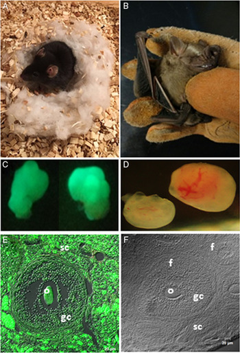
Ovaries from B6B5/EGFP mice were distinguished from those of bats by expression of GFP. The GFP expression pattern was analyzed in sections obtained by freezing, in which it was apparent that all somatic lineages that make up the ovary, and also the oocytes, stained positive for GFP (Fig. 1).
Study models, ovaries and histological sections of ovaries. (A) Transgenic mouse from B6B5/EGFP strain that expresses green fluorescent protein (GFP). (B) Artibeus jamaicensis phyllostomid bat. (C) Ovaries from the B6B5/EGFP mouse appear fluorescent under ultraviolet light. (D) Ovaries from the A. jamaicensis bat. (E) Histological section obtained from the B6B5/EGFP mouse ovary, in which all the cells that make up the organ such as the oocyte are present (o), granulosa cells (gc), and cells from the stromal compartment (sc) are positive for GFP. (F) Histology of the ovary of A. jamaicensis showing follicles (f) at different stages of development.

Cell dissociation
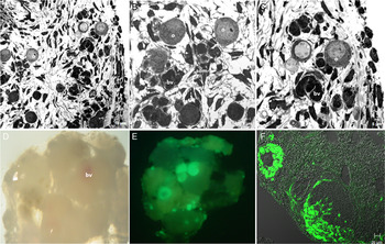
In the fixed cell pellets after bat ovaries were disaggregated and before placing them in organ culture, cells appeared to be separated and randomly distributed. At times, some cells appeared to be forming groups, but most were found in isolation, indicating that cell dissociation was almost total. The main cell types that made up the organ, such as oocytes, somatic cells and blood vessels, were observed (Fig. 2A–C). Isolated oocytes, characterized by having a spherical morphology with a prominent nucleus, were observed to be randomly distributed over loose tissue made up of granulosa cells, blood vessels and components of the extracellular matrix.
Disaggregated and disaggregated–re-aggregated (DR) ovarian cells from Artibeus jamaicensis bat and B6B6/EGFP mouse. (A–C) Disaggregated cell from the A. jamaicensis ovary, in which it can be observed that the different cell lineages that form the ovary such as oocytes (o), somatic cells (som) and blood vessels (bv) are separated and randomly distributed. (D) Disaggregated–re-aggregated after 96 h of cultivation and observed floating on a filter in the culture medium. A compact structure is apparent, in which some blood vessels (bv) are visible. (E) Same DR shown in (D), but exposed to ultraviolet light, in which zones corresponding to cells from the B6B5/EGFP mouse can be observed. (F) Histological section obtained from the DR shown in (D) and (E) in which the reconstitution of xenogeneic follicles (f) is apparent, formed by cells that are both positive (green) and negative to GFP.

Cell aggregation
After 96 h of culture, in the disaggregated–re-aggregated (DR) ovaries from the bat A. jamaicensis and the B6B5/EGFP mouse, cells re-aggregated forming a compact structure, in which ovarian lineages from both species could be seen to interact, mixing and reorganizing their ovarian morphology (Fig. 2D–F). In this way, we witnessed a recapitulation of the morphogenetic events that led to the formation of the ovary. Both cells from the bat A. jamaicensis ovary and those from the B6B5/EGFP mouse interacted and reconstituted the main structures that make up the mammal ovary: the follicles. Cellular lineages of the ovaries of both species reorganized, recapitulating folliculogenesis events, for example the formation of a basal membrane, cell proliferation and the formation of links between granulosa cells to encapsulate the oocyte and contribute to its development (Fig. 2).
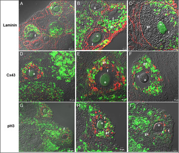
The dissociation–re-aggregation of ovarian cells from bat and mouse revealed that the oocytes from both mammalian species were capable of recruiting granulosa cells independently of the species, therefore forming xenogeneic chimeric follicles at different stages of follicular development (Fig. 3). From this, it followed that, from the initial stages of culture, cell displacement occurred that seemed to recapitulate the morphogenesis of the ovarian tissue. The granulosa cells and oocytes segregated to form the follicles, which were delimited by an evident basal lamina that separated them from the stromal compartment (Fig. 3A–C). The differentiation of cells from the theca, myoid cells and blood vessels occurred to form part of the stromatic tissue. Gap junctions, evidenced by the expression of Cx43 directly reconnected to neighbouring granulosa cells in both model species, forming the cytoplasmic compartments of the follicles (Fig. 3D, F). The proliferative activity of the dissociated and re-aggregated cells was evidenced by the expression of histone H3 (pH3), with the activity of this protein principally being found in granulosa cells from both bat and mouse ovaries (Fig. 3G, I).
Immunofluorescence showing the localization of Laminin, Cx43 and pH3 protein expression, in chimeric xenogeneic bat and mouse disaggregated–re-aggregated (DR) ovaries. (A) Expression of Laminin (red) delimiting the follicles (f) that are at different stages of growth, in the stromatic compartment. (B, C) Expression of Laminin makes it clear that the reconstituted follicles can be made up of oocytes (o) from the bat or by oocytes coming from mice (GFP positive). In the same way, the granulosa cells (gc) that surround the oocytes can come from the mouse (green, GFP positive) or from the bat. (D–F) Expression of the cellular intercommunication marker Cx43 (red), which is expressed mainly between the junctions that form granulosa cells (gc), and between these and the oocyte (o). (G–I) Location of pH3 expression (red), which is detected mainly in proliferating granulosa cells (gc).

Expression of the protein from the Ddx4 gene
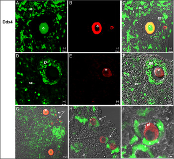
The identification and viability of the germ line in the DR ovaries was evaluated by analyzing the expression of protein (DEAD-Box Helicase 4) from the Ddx4 gene, which is expressed specifically in the germline of vertebrates and invertebrates. In chimeric DR ovaries, Ddx4 was detected in the cytoplasm of both bat and mouse oocytes, at different stages of follicular development (Fig. 4). The expression of the Ddx4 protein was also observed in smaller cells located at the periphery of the re-aggregates, which began to be surrounded by follicular cells and appeared to be organizing in primordial follicles. This type of cell corresponded exclusively to cells from bats (Fig. 4G–I).
Detection of the protein for the Ddx4 gene in chimeric DR ovaries of mouse and bat ovaries. (A) Expression of GFP in an oocyte, as well as in some granulosa cells and cells that make up the stromal compartment (sc). (B) Expression of the protein for the Ddx4 gene (red) in the cytoplasm of an oocyte (o). (C) Merge of Ddx4 and GFP in an oocyte within a follicle, formed from granulosa cells (gc), both positive and negative to GFP. (D–F) A xenogeneic chimeric follicle is presented consisting of a bat oocyte and granulosa cells (gc), mainly from mouse, are shown. (G) Detection of Ddx4 in the cytoplasm of oocytes (o) and in some cells (arrows) located in the cortical region of the DR. (H) Greater amplification of (G) in which cells positive for Ddx4 from bat can be observed as they begin to be surrounded by mouse granulosa cells (arrows). (I) Primordial follicle (pf), formed from a bat oocyte, positive to Ddx4 and some granulosa cells from the transgenic mouse ovary (*).

Isolation and re-aggregation of adult cortical germ cells
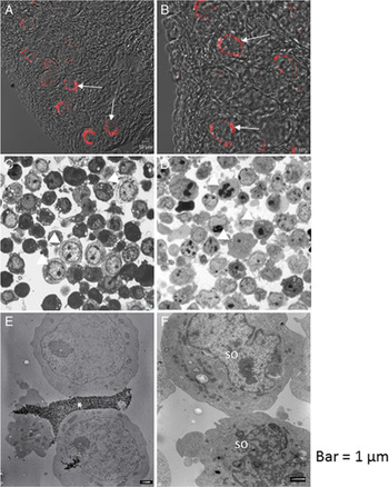
Germ cells in mammals can be labelled and distinguished from other cells with reference to their high activity in response to AP. In sections obtained by freezing bat ovaries, the AP reaction was observed at the cell surface of cells located in the cortical region, therefore these cells correspond to ACGC (Fig. 5A, B). Subsequently, using an antibody against AP and magnetic beads, two cell fractions were obtained: an enriched fraction from somatic cells (erythrocytes, granulosa cells, myoid cells, theca cells and fibroblasts) and an enriched population of ACGC from bat (Fig. 5C, D). Both cell populations were characterized by electron microscopy, revealing that the somatic cells had an irregular nucleus and that their surface presented great activity, as they manifested formation of cytoplasmic lobopodium-type prolongations (Fig. 5E, F). Regarding the ACGC population at the ultrastructural level, the presence of magnetic beads adhering to the cell membrane was apparent. These cells were observed to be larger than the somatic cells, presenting a more circular and prominent nucleus (Fig. 5E).
Isolation of adult cortical germ cells (ACGC). (A, B) In situ expression of alkaline phosphatase at the cell surface of the ACGC (arrows). (C) Enriched cell fraction of ACGC (arrowheads). (D) Enriched fraction of ovarian somatic cells (arrowheads). (E) Electron microscopy of two cells related to alkaline phosphatase with ACGC characteristics that have magnetic beads, used for their isolation, attached to their cell surface (*). (F) Electron microscopy of cells shown in (D), in which two cells with typical characteristics of somatic ovarian cells (so) can be observed. Bars, 1 μm.

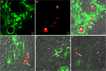
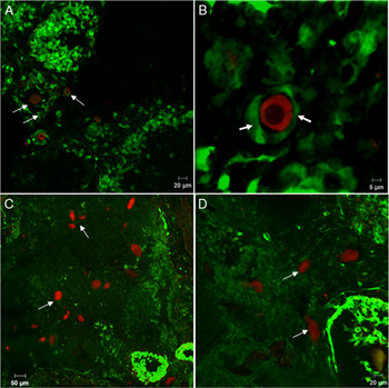
When isolated ACGC were mixed with dissociated ovarian cells from the B6B5/EGFP transgenic mouse, it was apparent that the majority of ACGC were incorporated to form primary follicles surrounded by murine granulosa cells, whereas a few others were randomly distributed in the cortical region of the ovarian chimeric re-aggregate. The distribution of the ACGC was determined by the cytoplasmic expression of Ddx4 (Fig. 6). This distribution corroborated that some isolated ACGC from bats were incorporated into follicles and interacted in this follicle with granulosa cells from both mouse and bat, whereas other ACGC remained isolated and located in the cortical region. The identity of ACGC as part of the GC lineage was evidenced by the expression of positive cells for Stella. When Stella-positive cells were found forming primordial follicles, this staining was usually observed to be cytoplasmic (Fig. 7A, B). However, if the Stella-positive cells were randomly distributed without observing follicle formation, protein expression was apparent in the nucleus (Fig. 7C, D).
Disaggregation–re-aggregation (DR) of isolated adult cortical germ cells (ACGC) together with ovarian cells from B6B6/EGFP transgenic mouse. (A) GFP expression pattern (green). (B) Protein expression pattern of the Ddx4 gene (red). (C) Merge of GFP and Ddx4 shown in (A) and (B) and combined with Nomarski microscopy. Ddx4 is observed in the cytoplasm of both oocytes (o) forming follicles and cells located in the periphery of the DR (arrows). (D) Two Ddx4 positive cells (red) are shown at the periphery of the DR, in which one of these begins to be surrounded by granulosa cells (*) and the other is kept isolated (arrow). (E, F) ACGC positive to Ddx4 (red), evidently forming primordial follicles, which are randomly located in the DR (arrows). These ACGC are surrounded by both granulosa cells positive for GFP and granulosa cells from bat.

Detection of Stella gene protein in DR of isolated adult cortical germ cells (ACGC), together with ovarian cells from B6B6/EGFP transgenic mouse. (A) Stella-positive cells (red) can be observed distributed randomly in the disaggregation–re-aggregation (DR) (arrows). (B) Cytoplasmic expression of the Stella protein in an ACGC (red) from a bat surrounded by follicular cells from the B6B5/GFP mouse (arrows). (C, D) Stella-positive cells (red) from bat that are randomly distributed in a somatic cell environment consisting mainly of cells from the B6B5/GFP mouse (green) with no evident follicle formation, in which expression of the protein can be observed in the nucleus (arrows).

Western blot
We detected proteins Ddx4, Cx43, AP, Foxl2, pH3, Fragillis and Stella by western blot analysis in homogenates of adult bat and mouse ovary. These results concurred with immunofluorescence observations. Notably, Stella expression was differential, as it was only detected in the bat ovary. The remaining proteins (Ddx4, Cx43, AP, Foxl2, pH3 and Fragillis) were identified in the ovaries of both animalmodels (bat and mouse). The reported proteins had an estimated molecular weight of: β-Actina (42 kDa), Ddx4 (76 kDa), Cx43 (43 kDa), AP (50 kDa) Foxl2 (50 kDa), pH3 (15 kDa), Fragillis (13 kDa) and Stella (17 kDa) (Fig. 8).
Validation of the primary antibodies in protein homogenates of adult A. jamaicensis bat and B6B5/GFP mouse ovary by western blot. Bands corresponding to the reported weight were identified for each of the proteins analyzed. (A) β-Actina (42 kDa), Ddx4 (76 kDa), Cx43 (43 kDa), alkaline phosphatase (AP; 50 kDa) and Foxl2 (50 kDa). (B) pH3 (15 kDa), Fragillis (13 kDa) and Stella (17 kDa).

Discussion
The experiments discussed here were performed to evaluate the cellular interactions between ovarian cells from mouse and from our A. jamaicensis bat model.
Ovarian reconstruction in vitro from cells dissociated from postnatal ovaries of mammals has been documented previously in the rat (Zenzes and Engel, Reference Zenzes and Engel1981), pig (Ol et al., Reference Ol, Tasaki, Munakata, Shirasuna, Kuwayama and Iwata2015), mouse (Young et al., Reference Young, Drummond, Akers, Bartle, Kennedy and Asaduzzaman2017) and in re-aggregated chimeric mice and rats (Eppig and Wigglesworth, Reference Eppig and Wigglesworth2000). However, among re-aggregated ovaries, the expression of markers that promote the histogenetic recapitulation of organs such as Cx43 and Laminin has not been reported, including the viability of the oocytes (Ddx4) and their proliferation (pH3). The identification of these markers could corroborate the theory that there could be differences between mammals, with respect to factors that mediate communication between the oocyte and granulosa cells. This type of study among wild species is scarce and, for bats, non-existent.
Bat species from the Phyllostomidae family are of particular interest because they exhibit reproductive traits that are similar to those of primates and rodents (Rasweiler and Badwaik, Reference Rasweiler, Badwaik, Crichton and Krutzsch2000). The most recent common ancestor shared by rodents and bats, dates back to about 90 million years ago (Murphy et al., Reference Murphy, Pevzner and O’Brien2004). Despite their divergence, both species of mammals share reproductive characteristics, especially concerning the morphology of the reproductive process. The ovarian morphology of the A. jamaicensis phyllostomid bat manifests a pattern that is similar to that of mammals, in which the ovary is divided into a cortex and a medulla, with the follicles distributed throughout the cortical region. However, the functionality of bat ovaries is different, as one of the ovaries appears to be polarized, with the primordial follicles and ACGC located in the ovarian cortex, near to the basal membrane of the tunica albuginea (Antonio-Rubio et al., Reference Antonio-Rubio, Porras-Gómez and Moreno-Mendoza2013). In contrast, in many mammalian species, such as mice, this polarization is not evident, indicating a tendency to avoid regionalization during gonadal development among this group (Jiménez, Reference Jiménez2009).
Under our experimental conditions for the dissociated-re-aggregation of ovarian cells, after 96 h of culture, the main characteristics that serve to identify the ovary under normal development conditions were reconstituted. From this, it is apparent that from the initial stages of culture, cell transfer takes place that seems to recapitulate the morphogenesis of the ovarian tissue. The granulosa and oocyte cells appear re-aggregated to form the follicles, which are delimited by an evident basal lamina that separates them from the stromatic compartment. The differentiation of cells from the theca, myoid and blood vessels takes place to form part of the stromatic tissue. This typical reconstruction of an ovary raises the possibility that an intrinsic property of the oocytes is that cellular communication is possible between these species. This means that bat oocytes can regenerate follicles using somatic mouse cells and vice versa, despite years of evolutionary divergence. Similarly, the follicular cells that are responsible for the integrity of the follicle maintain their proliferative capacity, evidenced by their activity at pH3. It is therefore feasible that, for the follicular organization that occurs during the culture period, cell recognition and cellular interactions take place between the granulosa cells and the oocyte. It has been proposed that cellular recognition may be due to a memory mechanism present at the surface of cells, in which the components at the cell surface become essential during a sequence of events that lead to the recapitulation of ovarian morphogenesis (Zenzes and Engel, Reference Zenzes and Engel1981). In this regard, the fact that oocytes are capable of recruiting granulosa cells from other species may be because the oocyte controls the rate of mammalian ovarian follicle development, as alluded to in studies, in which oocytes from secondary-stage follicles are interchanged with granulosa cells from primordial follicles of newborn ovaries to produce re-aggregated ovaries (Eppig et al., Reference Eppig, Wigglesworth and Pendola2002). These experiments suggested that a developmental programme, intrinsic to the oocyte, controls the rate of follicle development in mice (Matzuk et al., Reference Matzuk, Burns, Viveiros and Eppig2002). This possibility has been attributed to mammals, so the fact that both mouse and bat oocytes are capable of recruiting granulosa cells indistinctly from the species corroborates the pivotal function of the oocyte in mammalian folliculogenesis.
Expression of the Ddx4 protein was used for to detect the germ line in the xenogeneic re-aggregates, observing its expression in the cytoplasm of developing oocytes at different stages of folliculogenesis in both mouse and bat and in ACGC; this expression suggested that the role of the Ddx4 gene in bats is maintained. High levels of Ddx4 protein in primordial, primary and secondary follicles and its decrease in the antral follicles suggested a role for this protein prior to the formation of the antrum. The oocyte acquired the ability to resume meiosis at approximately the moment when the antrum is formed and the expression of Ddx4 decreases considerably (Song et al., Reference Song, Ma, Huang, Ding, Cui and Zhang2016).
Not only are oocytes able to re-establish complex communications between granulosa cells, but ACGC isolated from the bat reconstituted primordial follicular structures. ACGC were isolated using an anti-AP antibody, generated by a soluble complex of calf intestinal AP and rabbit antibodies against AP. As for the other primary antibodies used in this study, AP specificity in both bat and mice ovaries was validated by western blot. At the cellular level, detection of the protein by immunofluorescence was observed in the cell membrane of ACGC. These observations are of interest as AP, besides being a surface marker, has been shown to be a marker of pluripotent cells (cells of the internal cell mass) and of primordial germ cells (Štefková et al., Reference Štefková, Procházková and Pacherník2015). This fact corroborates the idea that ACGC may correspond to precursor cells of the germ line in the adult ovary of A. jamaicensis bat. In previous studies, these ACGC have been characterized as oocyte precursor cells due to their affinity for germline, stem cell and proliferation markers in the A. jamaicensis bat (Antonio-Rubio et al., Reference Antonio-Rubio, Porras-Gómez and Moreno-Mendoza2013). In the present study, when ACGC from the bat ovary were placed in a functional ovarian environment such as that of adult mice, they formed primordial follicles, using mouse follicular cells; in the same way mouse oocytes reconstructed follicles using bat follicular cells. This suggests that the somatic lineage from both mammalian species, in particular the granulosa or follicular cells, interacted to form complex communicating junctions between themselves and between the ACGC. This involved gap junctions and paracrine factors that were required to promote oocyte growth and regulate meiosis. This finding is of great interest, as in recent years it has been proposed that mammalian postnatal ovaries contain germline stem cells, suggesting a mechanism of neo-oogenesis in which self-renewal of oocyte precursor cells persists during postnatal development. The fact that isolated ACGC form primordial follicles corroborates the possibility that this is an early germinal lineage present in the cortical region of A. jamaicensis ovary that renews the follicular pool during the postnatal life of the organism. Granulosa cells also interact with cells from the stromal compartment, such as myoid cells and fibroblasts, to secrete components from the extracellular matrix for ovarian reorganization. It is therefore possible that the fundamental mechanisms that govern the development of the oocyte and the interactions of the oocyte with its somatic cells are evolutionarily conserved between mice and bats.
Stella/Dppa3/PGC7 gene is expressed during the PGC specification period. Stella was originally identified in mouse preimplantation embryos, PGC and developing germ cells, in which it localizes in both the nucleus and cytoplasm (Saitou et al., Reference Saitou, Barton and Surani2002; Sato et al., Reference Sato, Jimura, Kurokawa, Fujita, Abe, Masuhara, Yasunaga, Ryo, Yamamoto and Nakano2002; Wongtrakoongate et al., Reference Wongtrakoongate, Jones, Gokhale and Andrews2013). In mouse ovaries, Stella gene was mainly identified, localized in both the nucleus and cytoplasm of oocytes at primary and primordial stages (Saitou et al., Reference Saitou, Barton and Surani2002). Therefore, in the present study, when this factor was detected it was used as a marker of germ cells in the early stages. In this sense, our work presents the first evidence of Stella expression in adult germ cells detected in a wildlife species. In our DR ovaries, the expression of Ddx4, as well as that of Stella in ACGC, suggested that cells isolated from the bat belonged to the germline and were capable of forming primordial follicles. Alternatively, in bat, they could remain as germ line progenitor cells that may renew folliculogenesis in the adult bat ovary. These findings corroborate those of previous studies, in which ACGC were characterized as germline progenitor cells that renew the oocyte pool in adult bat ovary (Antonio-Rubio et al., Reference Antonio-Rubio, Porras-Gómez and Moreno-Mendoza2013).
Acknowledgements
Thanks to Programa de Becas Posdoctorales de la UNAM, DGAPA for the scholarship granted to Tania Janeth Porras-Gómez. This work was supported by UNAM-PAPIIT IN204119.
Conflicts of interest
The authors declare no conflicts of interest.